

doi: 10.1016/S1569-2590(05)10005-6.
Ciliary Body and Ciliary Epithelium
Affiliations
- PMID: 21234280
- PMCID: PMC3018825
- DOI: 10.1016/S1569-2590(05)10005-6
Item in Clipboard
Ciliary Body and Ciliary Epithelium
Adv Organ Biol.
.
No abstract available
Figures
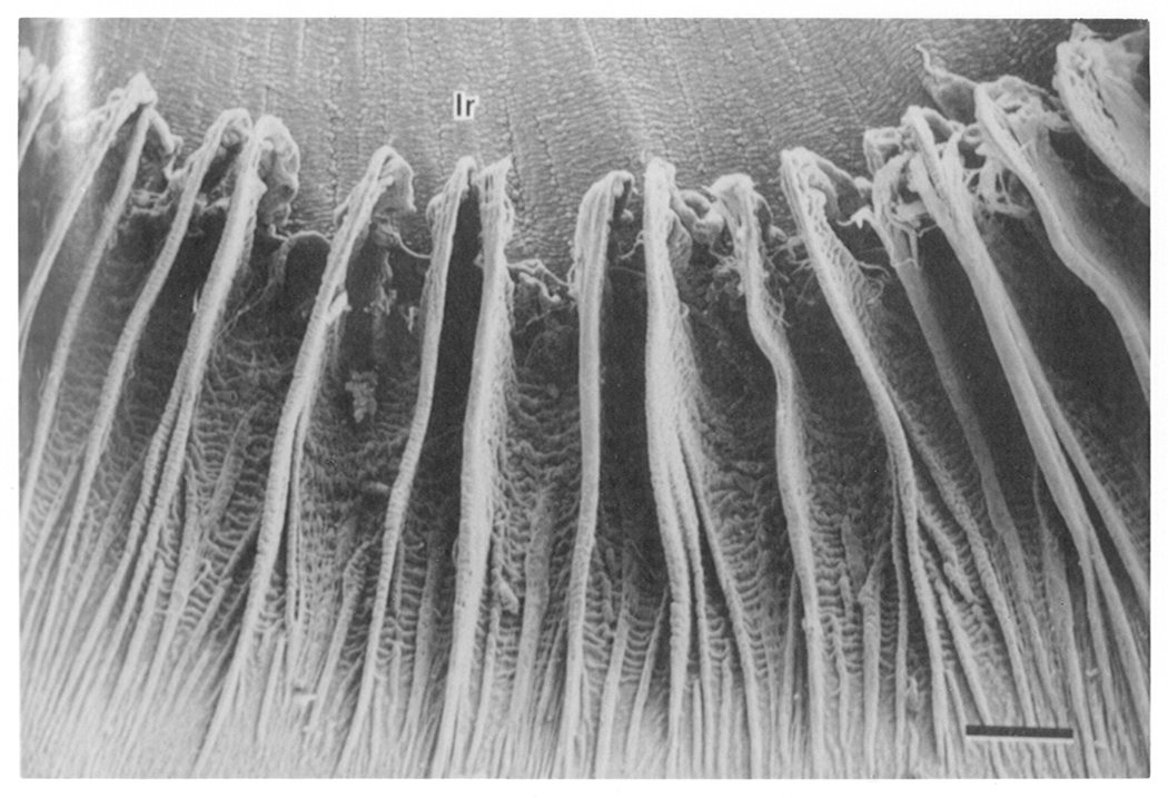
Shown in this scanning electromicrograph, the ciliary processes resemble fins that are arranged radially at the root of the iris on its posterior surface. Ir = iris. Bar = 0.5mm. Taken from Morrison et al. Invest Opthalmol Vis Sci. 28, 1325–1340, 1987, J.B. Lippincott Company. Used with permission.
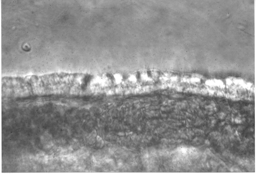
Figure 2a. Light photomicrograph showing the edge of a ciliary process from an albino rabbit. The bilayer of epithelial cells (indicated by an arrow) can be seen covering the blood capillaries within the ciliary process. Figure 2b. Scanning electromicrograph of arterioles (A) and veins (V) entering and exiting the goat ciliary process. These blood vessels serve the very elaborate capillary bed (C). Bar = 0.15mm. From Morrison et al. Invest Opthalmol Vis Sci. 28,1325–1340, 1987, J.B. Lippincott Company. Used with permission.
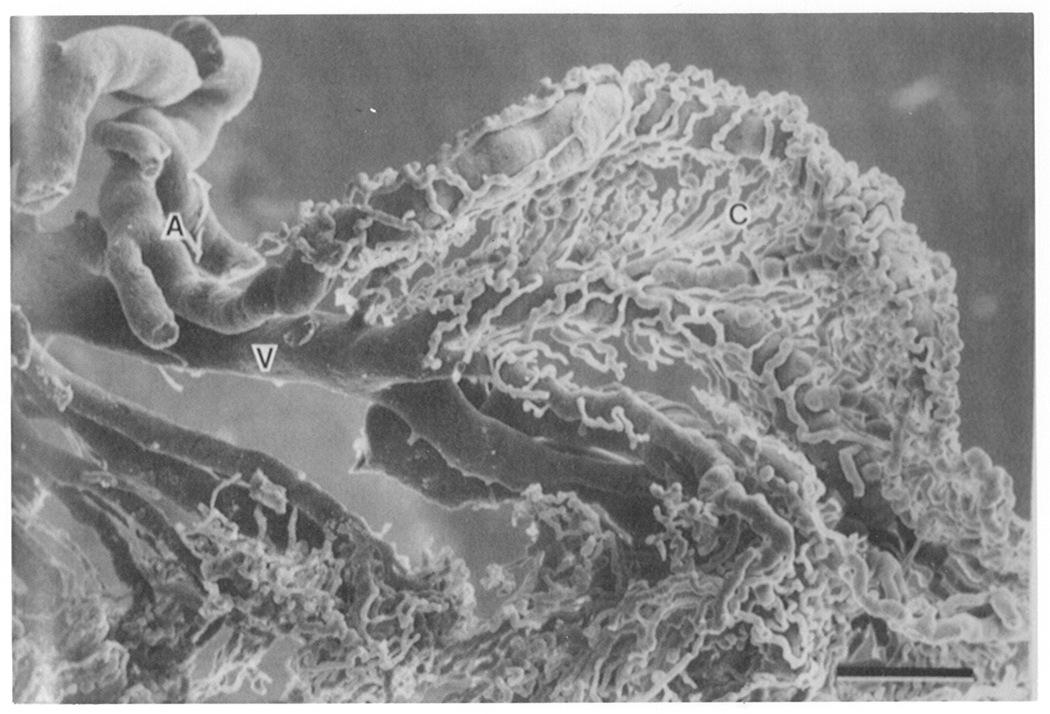
Figure 2a. Light photomicrograph showing the edge of a ciliary process from an albino rabbit. The bilayer of epithelial cells (indicated by an arrow) can be seen covering the blood capillaries within the ciliary process. Figure 2b. Scanning electromicrograph of arterioles (A) and veins (V) entering and exiting the goat ciliary process. These blood vessels serve the very elaborate capillary bed (C). Bar = 0.15mm. From Morrison et al. Invest Opthalmol Vis Sci. 28,1325–1340, 1987, J.B. Lippincott Company. Used with permission.
The pigmented ciliary epithelium is shown at the top with the nonpigmented ciliary epithelium beneath. Note that tight junctions are located in the nonpigmented ciliary epithelium cell layer, the layer which faces the posterior chamber of the eye. From Raviola, 1977, Exp. Eye Res., 25(Suppl), 25–63, Academic Press, Inc. Used with permission.
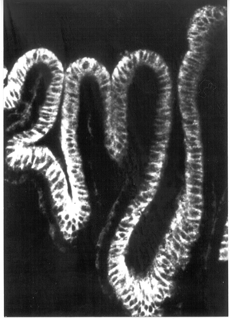
The confocal microscopic image shows the result of an immunolocalization study carried out using an antibody directed against the beta-1 subunit of Na,K-ATPase. As judged by fluorescence intensity, Na,K-ATPase beta-1 polypeptide is abundant in the nonpigmented ciliary epithelium layer, particularly at the basal and lateral surfaces (the NPE layer points upward in this photograph). The Na,K-ATPase beta-1 signal is much more faint in the pigmented cell layer (downward-facing cells in this picture) where it is seen at the basal surface. Taken from an unpublished collaborative study by the author, Amy E. Moseley (University of Cincinnati, Cincinnati, OH), and Steven Bassnett (Washington University, St. Louis, MO).
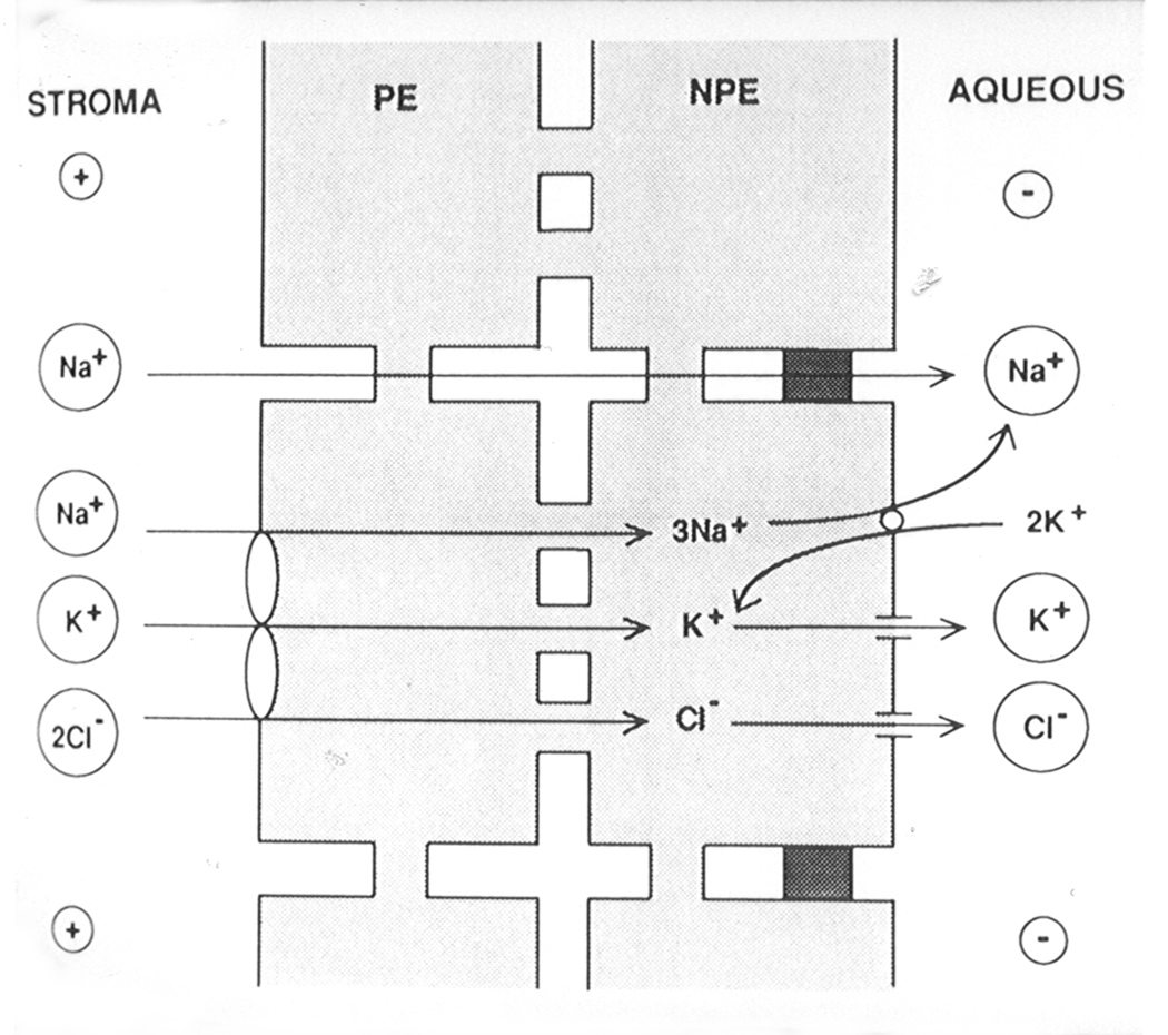
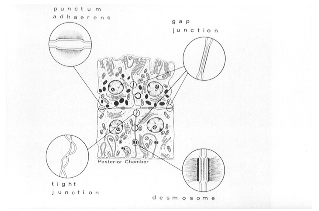
Nonpigmented cells (NPE) and pigmented cells (PE) communicate via gap junctions. Densely shaded areas between NPE cells represent tight junctions. The model shows sodium, potassium and chloride entering the PE via a Na/K/2Cl cotransporter and passing via gap junctions to the NPE. There, sodium is exported via Na,K-ATPase (the sodium pump). In the NPE, potassium imported by Na,K-ATPase is recycled out of the cell via potassium channels. Chloride exits the NPE via chloride channels. Taken from Coca-Prados et al. 1995, Am. J. Physiol. 268, C572–C579, The American Physiological Society. Used with permission.
References
-
- Avila MY, Seidler RW, Stone RA, Civan MM. Inhibitors of NHE-1 Na+/H+ exchange reduce mouse intraocular pressure. Invest. Ophthalmol. Vis. Sci. 2002;43:1897–1902. - PubMed
-
- Chen S, Sears M. A low conductance chloride channel in the basolateral membranes of the non-pigmented ciliary epithelium of the rabbit eye. Curr. Eye Res. 1997;16:710–718. - PubMed
-
- Chu TC, Candia OA. Active transport of ascorbate across the isolated rabbit ciliary epithelium. Invest. Ophthalmol. Vis. Sci. 1988;29:594–599. - PubMed
-
- Coca-Prados M, Anguita J, Chalfant MS, Civan MM. PKC-sensitive Cl− channels associated with ciliary epithelial homologue of pICln−. Am. J. Physiol. 1995;268:C572–C579. - PubMed
-
- Coca-Prados M, Escribano J, Ortego J. Differential gene expression in the human ciliary epithelium. Progress. Ret. Eye Res. 1999;18:403–4729. - PubMed
Grants and funding
LinkOut - more resources
Full Text Sources