Review
. 2011 Feb;21(1):12-20.
doi: 10.1016/j.gde.2010.12.001. Epub 2011 Jan 13.

Apoptosis and oncogenesis: give and take in the BCL-2 family

Affiliations
Review

Apoptosis and oncogenesis: give and take in the BCL-2 family

Fabien Llambi et al. Curr Opin Genet Dev. 2011 Feb.

Abstract

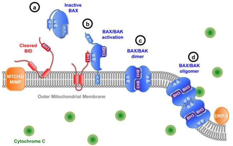
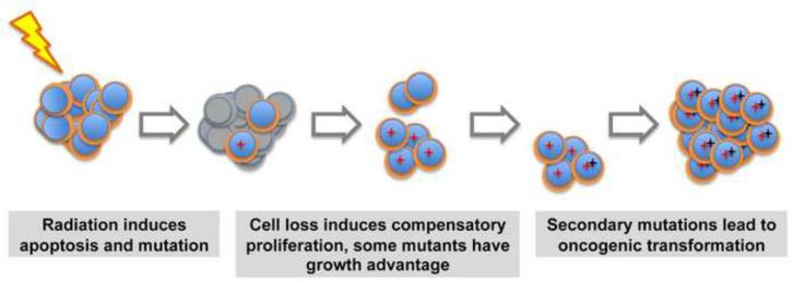
The mitochondrial pathway of apoptosis constitutes one of the main safeguards against tumorigenesis. The BCL-2 family includes the central players of this pathway that regulate cell fate through the control of mitochondrial outer membrane permeabilization (MOMP), and important progress has been made in understanding the dynamic interactions between pro-apoptotic and anti-apoptotic BCL-2 proteins. In particular, recent studies have delineated a stepwise model for the induction of MOMP. BCL-2 proteins are often dysregulated in cancer, leading to increased survival of abnormal cells; however, recent studies have paradoxically shown that apoptosis induction can under some circumstances drive tumor formation, perhaps by inducing compensatory proliferation under conditions of cellular stress. These observations underline the complexity of BCL-2 protein function in oncogenesis.


Figures

FIGURE 1
FIGURE 1
Stepwise model for BAX and BAK activation. (a) In healthy cells, BAX is a cytosolic protein, its C-terminal α-helice (CT) being sequestered in a hydrophobic pocket, while BAK is constitutively anchored into the outer mitochondrial membrane (OMM). Following apoptotic stimulation, BH3-only proteins are produced and/or activated. In the particular case of cleaved BID, targeting to the OMM is facilitated by mitochondrial proteins such as MTCH2/MIMP. (b) Binding of BAX or BAK by BH3-only activator through an interface close to the α-helice 6 (α6) triggers a series of extensive conformational changes including exposure of the N-terminal α-helice (NT) and transient exposure of the BH3-domain (BH3). Additionally, BAX activation induces displacement of the C-terminus which targets it to the OMM and frees the hydrophobic groove (c) Next, BAX and BAK further insert into the membrane and dimerize “nose to nose” though reciprocal BH3-domain-groove interaction. (d) Further oligomerization is then achieved though α-helix 6 interactions in two dimers (“back to back”). Induction of MOMP by formation of higher order oligomers is facilitated by DRP-1 induced membrane curvature.
FIGURE 2
FIGURE 2
Regulation of MCL-1 anti-apoptotic activity. MCL-1 pro-survival function is regulated by two mechanisms: control of protein level and inhibition by BH3-only proteins. The PI3K/AKT signaling pathway increases MCL-1 translation through mTOR signaling complex 1. MCL-1 protein level is also tightly regulated by proteasomal degradation. Three E3 ubiquitin ligase complexes MULE, β-TrCP, APC/C and one deubiquitinase USP9X, have been indentified as direct regulators of MCL-1 ubiquitylation. Alternatively, MCL-1 stability is also controlled by ubiquitin-independent proteasomal degradation. The second mode of regulation of MCL-1 pro-survival function is by occlusion of MCL-1 hydrophobic groove by BH3-only proteins. Additionally, shorter versions of MCL-1, generated by alternative splicing, could have a similar function by engaging MCL-1L through their BH3 domains.
FIGURE 3
FIGURE 3
Model for apoptosis driven tumor formation. In wild type mice, γ-irradiation simultaneously induces massive apoptotic cell death in the haematopoietic compartment and oncogenic mutations in the surviving population. Leukocyte depletion, and possibly surrounding apoptotic cells, induces compensatory proliferation of the hematopoietic stem/progenitor cells (HSCs). Some mutant HSCs clones, such as the one with disrupted p53 function, may display a proliferative advantage and outcompete non-mutated cells. This could provide favorable conditions for accumulation of secondary mutations eventually leading to malignant transformation. In the absence of PUMA, cell loss via apoptosis does not occur, and no compensatory proliferation follows. This model suggests that compensatory proliferation and growth advantage of some mutations are necessary for oncogenesis in this system of radiation-induced tumorigenesis.

References

    1. Armitage P, Doll R. The age distribution of cancer and a multi-stage theory of carcinogenesis. Br J Cancer. 1954;8:1–12. - PMC - PubMed
    1. Hanahan D, Weinberg RA. The hallmarks of cancer. Cell. 2000;100:57–70. - PubMed
    1. Luo J, Solimini NL, Elledge SJ. Principles of cancer therapy: oncogene and non-oncogene addiction. Cell. 2009;136:823–837. - PMC - PubMed
    1. Green DR, Evan GI. A matter of life and death. Cancer Cell. 2002;1:19–30. - PubMed
    1. Taylor RC, Cullen SP, Martin SJ. Apoptosis: controlled demolition at the cellular level. Nat Rev Mol Cell Biol. 2008;9:231–241. - PubMed

Substances