. 2011 Mar 1;411(1):87-102.
doi: 10.1016/j.virol.2010.12.033. Epub 2011 Jan 14.

Rapid SIV Env-specific mucosal and serum antibody induction augments cellular immunity in protecting immunized, elite-controller macaques against high dose heterologous SIV challenge

Affiliations

Rapid SIV Env-specific mucosal and serum antibody induction augments cellular immunity in protecting immunized, elite-controller macaques against high dose heterologous SIV challenge

L Jean Patterson et al. Virology. .

Abstract

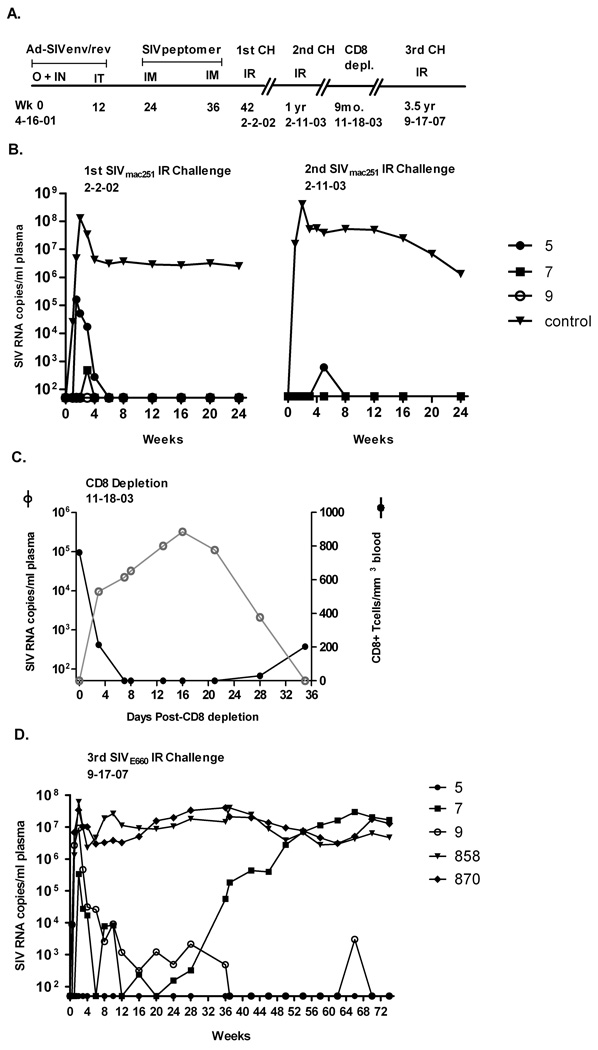
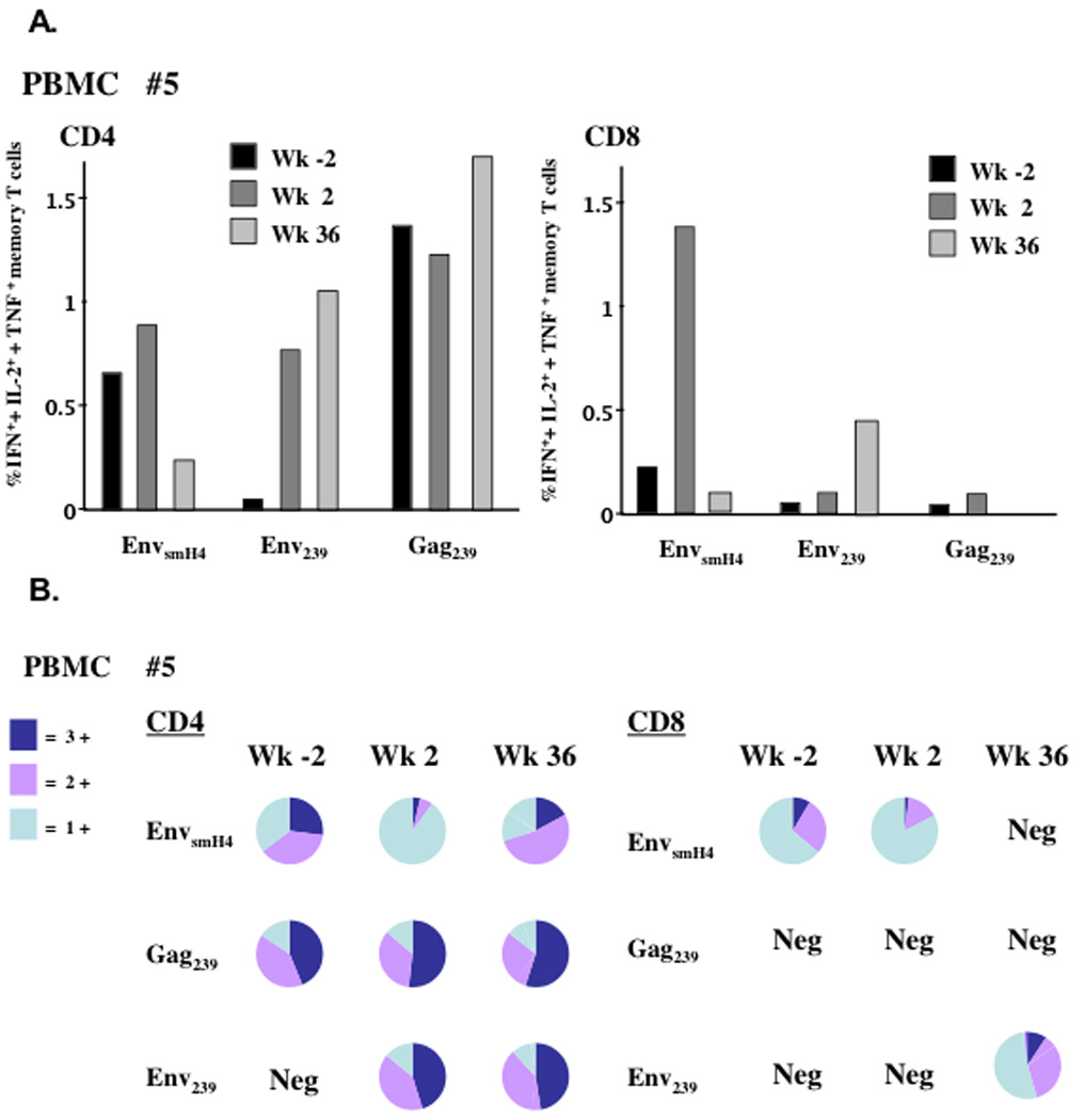
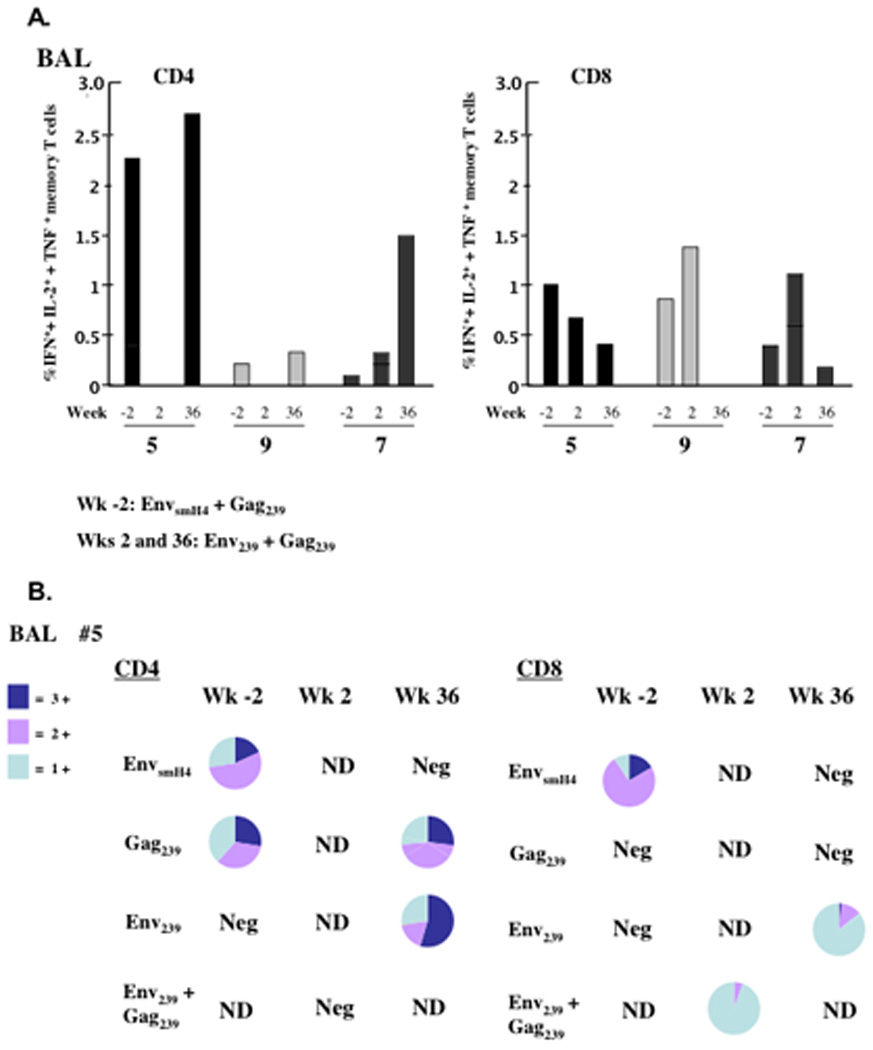
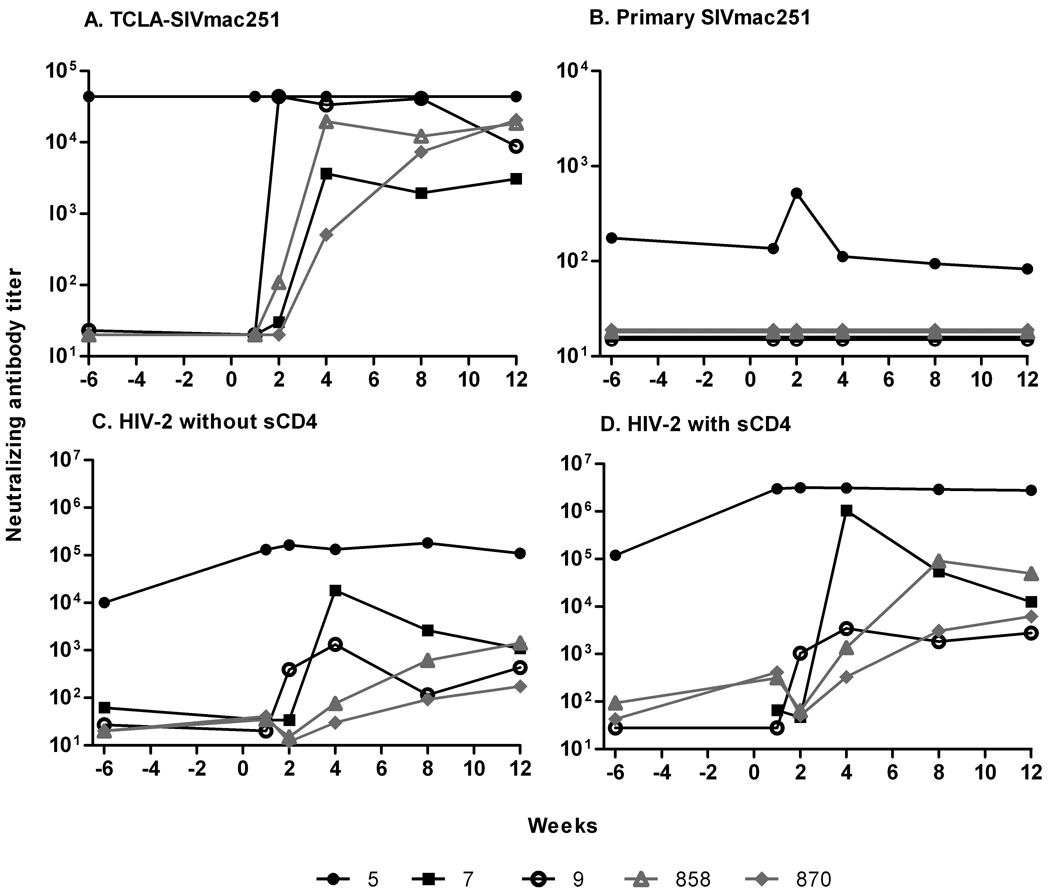
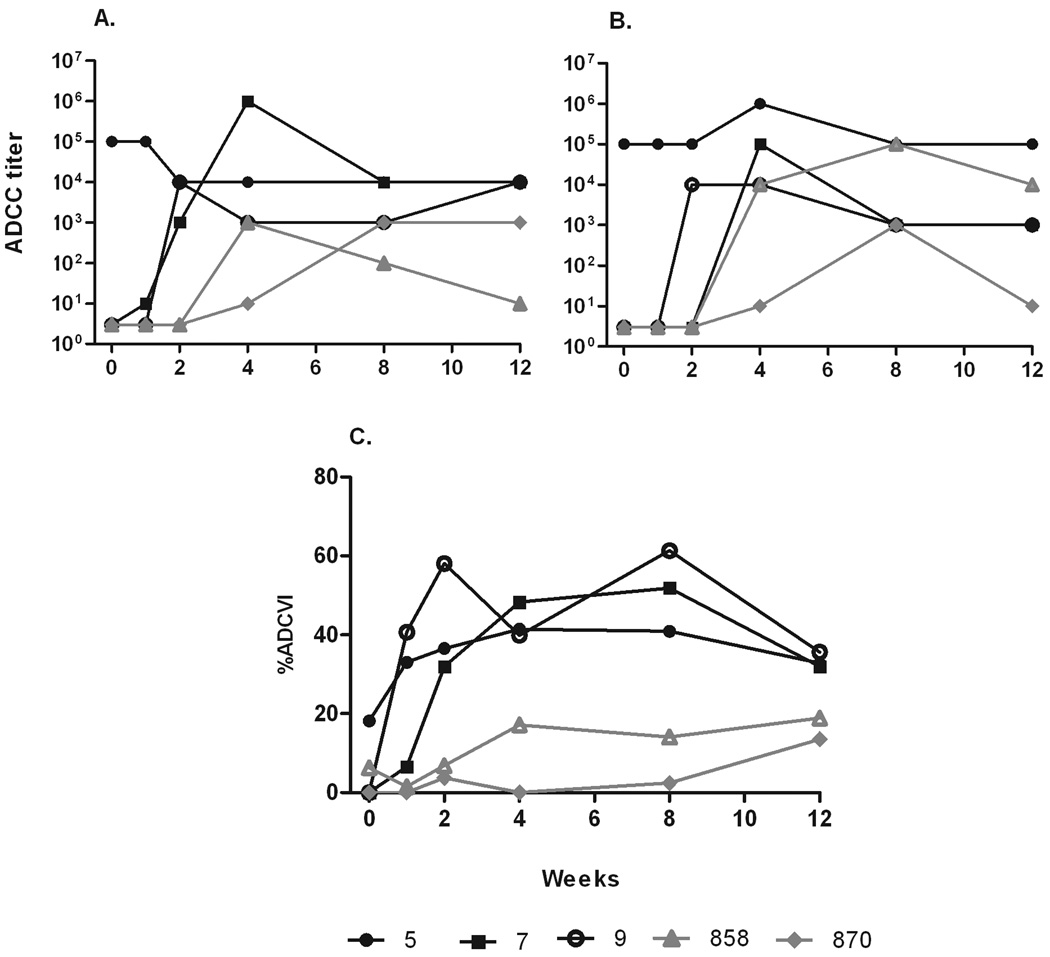
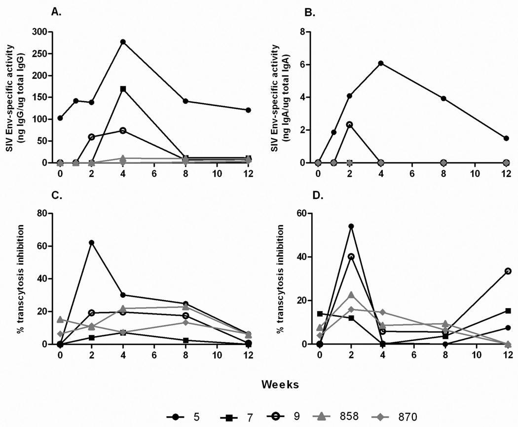
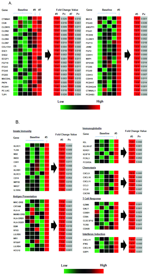
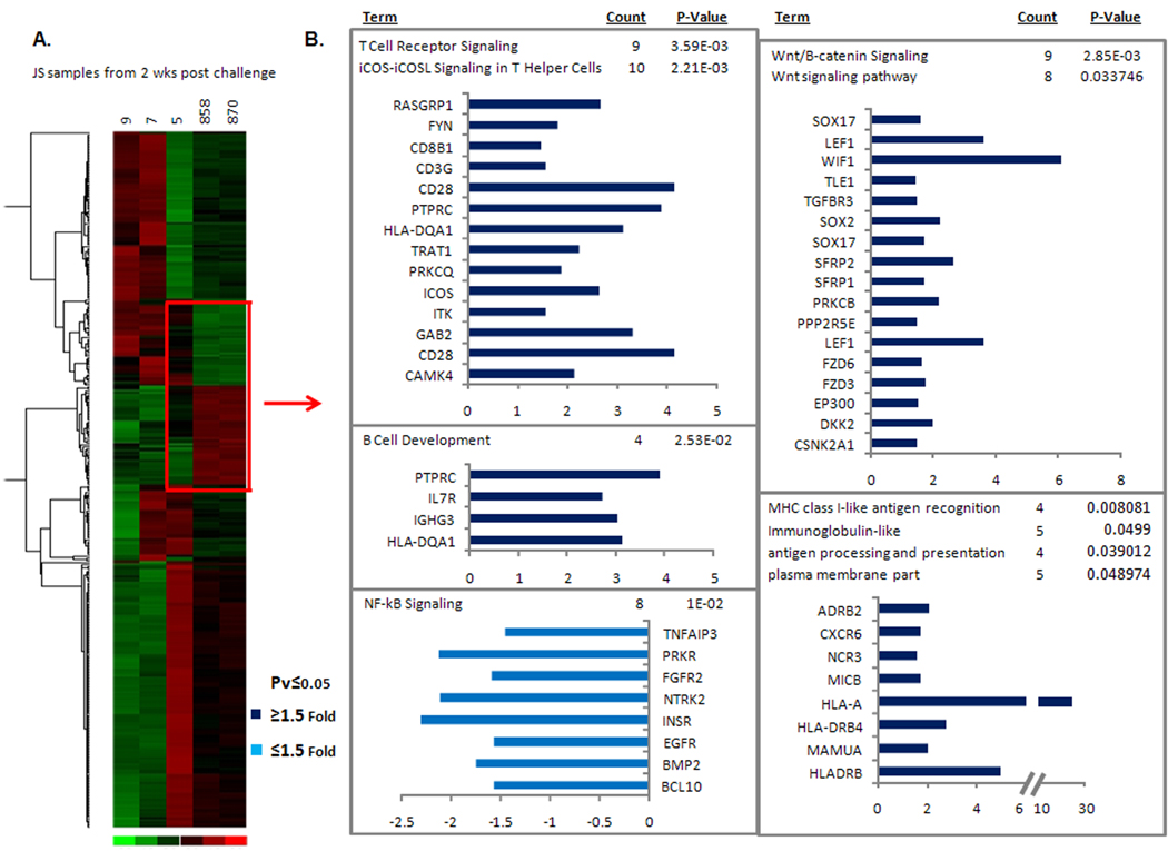
Three Indian rhesus macaques, Ad-SIV primed/protein boosted and exposed twice to high-dose mucosal SIV(mac251) challenges, exhibited elite control of viremia over 6.5 years. They were negative for host factors associated with control of SIV infection. After a third intrarectal challenge with SIV(smE660), all controlled viremia, with one (macaque #5) maintaining undetectable viremia in blood. Acquisition was not blocked, but virus was contained in the jejunum and draining lymph nodes. Polyfunctional memory T cell responses and high-titered neutralizing and non-neutralizing serum and mucosal antibodies were present before and maintained post-challenge. The level of protection seen for animal #5 was predicted from analyses of gene transcription in jejunum 2 weeks post-challenge. Macaques #7 and #9, exhibiting lower pre-challenge cellular and humoral immunity, partially controlled the SIV(smE660) challenge. Initial vaccine-induced control by macaque #5 extended to the SIV(smE660) challenge due to multiple immune mechanisms that were boosted and augmented by cryptic SIV exposure.


Figures

Figure 1
Figure 1
Immunization and challenge history of elite controller macaques (31, 40). (A) Macaques #5, #7, and #9 were originally immunized with an Ad-SIVsmH4 env/rev recombinant vector and boosted with a conformational CD4 binding site peptomer. Two priming immunizations given first orally and intranasally and second intratracheally were followed by two intramuscular boosts of peptomer in PBS. A high dose SIVmac251 rectal challenge was administered 6 weeks later. The times of second and third intrarectal challenges with SIVmac251 and SIVsmE660, respectively, and an intermediate in vivo CD8 depletion are indicated. (B) SIV RNA copies/ml plasma are shown for all three macaques, exhibiting elite control of the first high dose SIVmac251 rectal challenge and durable protection from a identical, second SIVmac251 rectal challenge one year later. Control group represents the geometric mean of 8 and 2 control macaques for the first and second challenges, respectively. (C) In vivo peripheral CD8+ T cell depletion resulted in reemergence of virus for macaque #5. Virus remained undetectable for #7 and #9 (not shown). (D) Outcome of a third heterologous rectal SIVE660 challenge, 3.5 years after the CD8 depletion study and 8 years since initial immunization. Macaques 858 and 870 are naïve controls.
Figure 2
Figure 2
The sum percent of IFN-γ+, + IL-2+, + TNF-α+ total memory T cells in PBMCs for macaque #5. (A) Shown are the total cytokine positive cells among total memory CD4 or CD8 T cells at weeks −2, 2 and 36 post SIVsmE660 challenge after stimulation with three separate peptide pools. (B) Pie charts for each flow sample from panel A, representing the percent triple, double and single cytokine secreting cells. Neg = negative response.
Figure 3
Figure 3
The sum percent of IFN-γ+, + IL-2+, + TNF-α+ total memory T cells in BAL samples for macaques #5, #7 and #9 over time. (A) Shown are the total cytokine positive cells among total memory CD4 or CD8 T cells at weeks -2, 2 and 36 post SIVsmE660 challenge after stimulation with peptide pools noted. Cell numbers were lower at week 2 post-challenge, so peptide pools were combined for stimulations. For ease in interpretation, responses were summed at weeks -2 and 36 as well. (B) Pie charts for each flow sample from panel A, representing the percent triple, double and single cytokine secreting cells. Neg = negative response. ND = denotes not done.
Figure 4
Figure 4
SIVmac251 serum binding antibody titers (log10) throughout the three SIV challenges.
Figure 5
Figure 5
Neutralizing antibody titers in plasma after SIVsmE660 challenge. Neutralization titers to (A) TCLA-SIV mac251 and (B) primary SIVmac251. CD4-induced antibody neutralization of HIV-2 without sCD4 added (C) and with sCD4 added (D). In (A), neutralizing titers for macaque #5 were >4.4×10⁴ at all time points.
Figure 6
Figure 6
Non-neutralizing antibody titers in plasma after SIVsmE660 challenge. ADCC titers of plasma using SIV251 gp120- (A) and SIVsmE660 gp120-coated target cells (B). (C) % ADCVI of SIVsmE660 infection of rhesus PBMC using plasma diluted 1:200.
Figure 7
Figure 7
Mucosal antibody binding titers and transcytosis inhibition by rectal secretions post SIVsmE660 challenge. SIVmac251 Env specific IgG (A) and IgA (B) titers shown as specific activity values (ratio of specific divided by total IgG or IgA in each sample). Transcytosis inhibition of SIVmac251 (C) and SIVsmE660 (D) across a tight epithelial cell barrier.
Figure 8
Figure 8
Transcriptional profile of jejunal biopsies week 2 post challenge. (A) Up-regulated genes involved in barrier integrity, epithelial repair and regeneration. (B) Up-regulated immune response genes for macaque #5.
Figure 9
Figure 9
Changes in gene expression levels in jejunal tissue in vaccinated and control macaques 2 weeks post SIVsmE660 challenge. (A) Heat diagram of changes in gene expression in each vaccinated animal as compared to control animals (#858 and #870) following SIVsmE660 challenge. Relative increase (red) or decrease (green) of mRNA levels are shown. (B) Following hierarchical clustering, gene expression levels for the elite controller macaque #5 were then compared to the two control macaques (genes within red box). Pathways statistically over represented in the gene lists are shown to the right of the red arrow with the genes and P value indicated.

References

    1. Baker BM, Block BL, Rothchild AC, Walker BD. Elite control of HIV infection: implications for vaccine design. Expert Opin. Biol. Ther. 2009;9:55–69. - PMC - PubMed
    1. Bertley FM, Kozlowski PA, Wang S-W, Chappelle J, Patel J, Sonuyi O, Mazzara G, Montefiori D, Carville A, Mansfield KG, Aldovini A. Control of simian/human immunodeficiency virus viremia and disease progression after IL-2-augmented DNA-modified vaccinia virus Ankara nasal vaccination in nonhuman primates. J. Immunol. 2004;172:3745–3757. - PubMed
    1. Bomsel M. Transcytosis of infectious human immunodeficiency virus across a tight human epithelial cell line barrier. Nat. Med. 1997;3:42–47. - PubMed
    1. Bosinger SE, Li Q, Gordon SN, Klatt NR, Duan L, Xu L, Francella N, Sidahmed A, Smith AJ, Cramer EM, Zeng M, Masopust D, Carlis JV, Ran L, Vanderford TH, Paiardini M, Isett RB, Baldwin DA, Else JG, Staprans SI, Silvestri G, Haase AT, Kelvin DJ. Global genomic analysis reveals rapid control of a robust innate response in SIV-infected sooty mangabeys. J. Clin. Invest. 2009;119:3556–3572. - PMC - PubMed
    1. Bostik P, Kobkitjaroen J, Tang W, Villinger F, Pereira LE, Little DM, Stephenson ST, Bouzyk B, Ansari AA. Decreased NK cell frequency and function is associated with increased risk of KIR3DL allele polymorphism in simian immunodeficiency virus-infected rhesus macaques with high viral loads. J. Immunol. 2009;182:3638–3649. - PMC - PubMed

Publication types

MeSH terms