Radial diffusivity predicts demyelination in ex vivo multiple sclerosis spinal cords
- PMID: 21238597
- PMCID: PMC3062747
- DOI: 10.1016/j.neuroimage.2011.01.007
Radial diffusivity predicts demyelination in ex vivo multiple sclerosis spinal cords
Abstract
Objective: Correlation of diffusion tensor imaging (DTI) with histochemical staining for demyelination and axonal damage in multiple sclerosis (MS) ex vivo human cervical spinal cords.
Background: In MS, demyelination, axonal degeneration, and inflammation contribute to disease pathogenesis to variable degrees. Based upon in vivo animal studies with acute injury and histopathologic correlation, we hypothesized that DTI can differentiate between axonal and myelin pathologies within humans.
Methods: DTI was performed at 4.7 T on 9 MS and 5 normal control fixed cervical spinal cord blocks following autopsy. Sections were then stained for Luxol fast blue (LFB), Bielschowsky silver, and hematoxylin and eosin (H&E). Regions of interest (ROIs) were graded semi-quantitatively as normal myelination, mild (<50%) demyelination, or moderate-severe (>50%) demyelination. Corresponding axonal counts were manually determined on Bielschowsky silver. ROIs were mapped to co-registered DTI parameter slices. DTI parameters evaluated included standard quantitative assessments of apparent diffusion coefficient (ADC), relative anisotropy (RA), axial diffusivity and radial diffusivity. Statistical correlations were made between histochemical gradings and DTI parameters using linear mixed models.
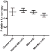
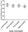
Results: Within ROIs in MS subjects, increased radial diffusivity distinguished worsening severities of demyelination. Relative anisotropy was decreased in the setting of moderate-severe demyelination compared to normal areas and areas of mild demyelination. Radial diffusivity, ADC, and RA became increasingly altered within quartiles of worsening axonal counts. Axial diffusivity did not correlate with axonal density (p=0.091).
Conclusions: Increased radial diffusivity can serve as a surrogate for demyelination. However, radial diffusivity was also altered with axon injury, suggesting that this measure is not pathologically specific within chronic human MS tissue. We propose that radial diffusivity can serve as a marker of overall tissue integrity within chronic MS lesions. This study provides pathologic foundation for on-going in vivo DTI studies in MS.
Copyright © 2011 Elsevier Inc. All rights reserved.
Figures


References
-
- Agosta F, Absinta M, Sormani MP, Ghezzi A, Bertolotto A, Montanari E, Comi G, Filippi M. In vivo assessment of cervical cord damage in MS patients: a longitudinal diffusion tensor MRI study. Brain. 2007;130:2211–2219. - PubMed
-
- Barkhof F. MRI in multiple sclerosis: correlation with expanded disability status scale (EDSS) Mult Scler. 1999;5:283–286. - PubMed
-
- Barnes D, Munro PM, Youl BD, Prineas JW, McDonald WI. The longstanding MS lesion. A quantitative MRI and electron microscopic study. Brain. 1991;114 ( Pt 3):1271–1280. - PubMed
-
- Benedetti B, Rocca MA, Rovaris M, Caputo D, Zaffaroni M, Capra R, Bertolotto A, Martinelli V, Comi G, Filippi M. A diffusion tensor MRI study of cervical cord damage in benign and secondary progressive multiple sclerosis patients. J Neurol Neurosurg Psychiatry. 2010;81:26–30. - PubMed
-
- Bergers E, Bot JC, De Groot CJ, Polman CH, Lycklama a Nijeholt GJ, Castelijns JA, van der Valk P, Barkhof F. Axonal damage in the spinal cord of MS patients occurs largely independent of T2 MRI lesions. Neurology. 2002;59:1766–1771. - PubMed
Publication types
MeSH terms
Grants and funding
LinkOut - more resources
Full Text Sources
Other Literature Sources
Medical

