. 2011 Jan 20;9(1):32-45.
doi: 10.1016/j.chom.2010.12.002.

Recruitment and activation of a lipid kinase by hepatitis C virus NS5A is essential for integrity of the membranous replication compartment

Affiliations

Recruitment and activation of a lipid kinase by hepatitis C virus NS5A is essential for integrity of the membranous replication compartment

Simon Reiss et al. Cell Host Microbe. .

Abstract

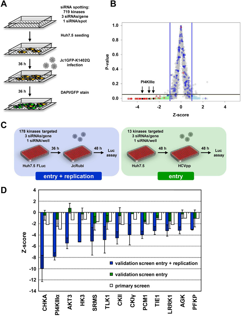
Hepatitis C virus (HCV) is a major causative agent of chronic liver disease in humans. To gain insight into host factor requirements for HCV replication, we performed a siRNA screen of the human kinome and identified 13 different kinases, including phosphatidylinositol-4 kinase III alpha (PI4KIIIα), as being required for HCV replication. Consistent with elevated levels of the PI4KIIIα product phosphatidylinositol-4-phosphate (PI4P) detected in HCV-infected cultured hepatocytes and liver tissue from chronic hepatitis C patients, the enzymatic activity of PI4KIIIα was critical for HCV replication. Viral nonstructural protein 5A (NS5A) was found to interact with PI4KIIIα and stimulate its kinase activity. The absence of PI4KIIIα activity induced a dramatic change in the ultrastructural morphology of the membranous HCV replication complex. Our analysis suggests that the direct activation of a lipid kinase by HCV NS5A contributes critically to the integrity of the membranous viral replication complex.


Figures

Fig. 1
Fig. 1. High throughput siRNA screen to identify kinases involved in HCV entry and replication
(A) Assay setup of the primary siRNA screen. Each chamber slide was coated with 384 different siRNA spots. Huh7.5 cells were infected 36 h after seeding and another 36 h later, cells were fixed and stained. (B) Virus specific signal intensities obtained in the primary screen were quantified and values were normalized intra- and inter-slide wise as well as between the 12 repetitions. Mean z-scores for all siRNAs tested are depicted in grey. Z-scores of negative and positive controls are highlighted in blue and red, respectively. With a hit criterion of −1 > z > 1 and a p-value < 0.05 indicated by the blue and black bars, respectively, 83 potential dependency and 95 potential restriction factors were identified (light green and light blue, respectively). Positions of PI4KIIIα hits are indicated by arrowheads. (C) Assay setup of the entry and replication validation screen (left panel). For each candidate 3 unique siRNAs were tested in a 96-well plate format. Huh7.5 FLuc cells were infected with JcR-ubi to measure effects of the silencing on HCV entry and replication (4 repetitions). To measure effects of the silencing of validated hits on entry (right panel), Huh7.5 cells were infected with HCVpp containing a FLuc reporter gene and luciferase activity was determined 48 h later (7 repetitions). (D) Summary of mean z-scores of hits determined in the two independent validation screens. Z-scores determined in the primary screen (white bars) are shown for comparison.
Fig. 2
Fig. 2. Network of HCV dependency factors
The depicted network highlights pathways in which dependency factors identified in our study (hexagonal nodes) and in previous HCV screens (ellipsoid nodes) are involved. Squares represent kinases that were found in our study and have already been known as dependency factors. The mean z-score of the two most inhibitory siRNAs per gene in the entry and replication validation screen is indicated by the color intensity of the nodes. Factors in white nodes were either not studied in this work or below the hit criterion in the primary screen; red-shaded nodes display candidates analyzed in the validation screen. Solid lines indicate proteins that are known to interact with each other or to participate in the same protein complex. Pathways for which we found a significant enrichment of HCV dependency factors are linked by colored areas and labeled at the top and bottom.
Fig. 3
Fig. 3. Effect of PI4KIIIα and PI4KIIIβ silencing on HCV replication and colocalization of these lipid kinases with NS5A
(A, B) Cells containing a genotype (gt) 2a or 1b luciferase reporter replicon specified in the top of each panel were transfected with siRNAs targeting PI4KIIIα or PI4KIIIβ (three individual siRNAs each) or with the positive control siRNA (HCV321) or the negative control siRNAs (DV-3’NTR or NegC#1). SiRNAs were transfected twice and RNA replication was determined 48 h after the second transfection by luciferase assay. RLUs were normalized to the mean of the negative controls. Data (mean +/− SD; n = 3 in triplicates) were analyzed using a one-way t-test. (C) Lysates prepared from Con1ET replicon cells 48 h after the second transfection were analyzed by immunobloting for PI4KIIIα, PI4KIIIβ, Transferrin receptor (TfR) and NS5A. Levels of NS5A were quantified and normalized to the corresponding TfR levels. Values are given in % relative to the mean of the NS5A levels of the negative control treated cells. (D, E) Huh7-Lunet cells (mock) or cells containing a selectable JFH-1 replicon (neo-sgJFH-1) were stained with a NS5A- (red) and either a PI4KIIIα- (D) or a PI4KIIIβ-specific antibody (green) (E). Nuclear DNA was stained with DAPI (blue). An enlargement of the sections indicated by a white square in each of the merged images is shown in the corresponding crop panel in the bottom. Images were acquired with a confocal microscope.
Fig. 4
Fig. 4. Impact of PI4KIIIα silencing on HCV production and integrity of the membranous web
(A) Expression of PI4KIIIα in Huh7.5 or LH86 cells was silenced by using individual siRNAs and cells were infected with JcR-2a. SiRNA HCV321 served as positive control, siRNAs DV-3’NTR and NegC#1 were used as negative controls. Virus replication was determined by luciferase assay. Data (mean +/− SD; n = 2 in duplicates) were analyzed using a one-way t-test. (B) Same as in (A) but measuring Dengue virus replication by using a Renilla reporter virus which will be described elsewhere. Data (mean +/− SD; n = 2 in duplicates) were analyzed using a one-way t-test. P-values below 0.05 or 0.001 are indicated by one or three asterisks, respectively. (C) Huh7.5 cells with stable knock-down of PI4KIIIα expression (sh-PI4KIIIα) or expressing a non-targeting shRNA (sh-NT) were stably transduced with shRNA-resistant PI4KIIIα wild type (wt) or D1957A mutant (inactive) expression constructs. For comparison, the PI4KIIIα knock-down cell line was transduced with the empty expression vector in parallel (empty vector). Cell lines were additionally transfected with siRNA #3 targeting PI4KIIIα (corresponding to the sequence of the shRNA used for stable silencing) prior to infection with JcR-2A with a MOI of 0.5 TCID50/cell. Virus replication was determined by luciferase assay 48 h post infection. As negative control, sh-NT cells were infected and analyzed in parallel. (D) Huh7-Lunet/T7 cells stably expressing a PI4KIIIα-specific shRNA (sh-PI4KIIIα) or a non-targeting shRNA (sh-NT) (as described in (C)) were transfected with a T7 promoter-based NS3 to NS5B expression construct. Cells were stained with a NS5A-specific antibody (red) and nuclear DNA was stained with DAPI (blue). Note the formation of profound NS5A “clusters” in PI4KIIIα knock-down cells, but not in sh-NT cells. (E) Cell lines described in panel C were transfected with a NS3 to NS5B polyprotein expression construct and the percentage of “wildtype” and “cluster” phenotype (D) was determined for 250 HCV-positive cells per condition. (F) Control (upper panels) and PI4KIIIα-silenced cells (lower panels) were transfected with a NS3 to NS5B expression construct containing a functional NS5A with a GFP insertion in domain III (Schaller et al., 2007) and prepared for EM analysis. The GFP insertion had no impact on the morphology of membrane alterations induced by the HCV NS-proteins in IF or EM (not shown) and does not interfere with RNA replication in a replicon context (Schaller et al., 2007). Consecutive enlargements of the boxed areas are shown from left to right. Note the very heterogeneous membranous web (MW) in control cells and the clusters of small double membrane vesicles (DMVs) (indicated by yellow arrows) in sh-PI4KIIIα cells. Size markers are given in the lower right of each panel. N, nucleus; LD, lipid droplet; MMVs, multi-membrane vesicles; rER, rough endoplasmic reticulum; m, mitochondrium.
Fig. 5
Fig. 5. PI4P and PI4KIIIα levels in HCV containing cells
(A) HCV permissive Lunet/CD81 cells were mock treated or infected with Jc1. Cells were fixed at given time points and stained with a NS5A- (red) and a PI4P-specific antibody (green). Nuclear DNA was stained with DAPI (blue). An enlargement of the section indicated by the white square is shown in the crop panel in the bottom. Images were acquired with a confocal microscope. (B) Quantification of PI4P levels by immunofluorescence analysis (green bars) and immunoblot (black bars). For immunofluorescence of mock- or Jc1-transfected cells mean values and standard error of the mean (SEM) of 80 cells per condition are shown. For immunoblot, cells transfected with Jc1 or subgenomic replicons specified in the bottom were lysed after 48 h. Lipids were extracted, spotted onto membranes and PI4P was quantified using the PI4P Mass strip kit (Echelon). Signals were quantified by using the Quantity One software (Bio-Rad). Error bars represent the SEM of three independent experiments. IF, immunofluorescence; IB, immunoblotting; n.t., not tested (C) Lysates used in (B) were prepared from mock- or Jc1-transfected cells at time points post transfection given in the top and analyzed by immunobloting for PI4KIIIα, NS5A and Calnexin (loading control). (D) Immunohistochemistry on snap-frozen liver tissues from a HCV negative person (10H6; Table S4, S5) or a patient with chronic HCV infection (3E6; Table S4, S5). Consecutive liver sections were stained for NS5A, PI4P and core. Note that cells staining positive for viral proteins and PI4P reside in the same region of each section.
Fig. 6
Fig. 6. Role of NS5A domain I in recruitment of PI4KIIIα and induction of PI4P
(A) Huh7-Lunet/T7 cells were cotransfected with HA-tagged PI4KIIIα and expression constructs encoding individual viral proteins specified in the top. After 8 h cell proteins were radiolabeled for 16 h with [35S] methionine/cysteine-containing medium. Cells were lysed and immunoprecipitation was performed using antibodies indicated in the bottom. Filled arrowheads point to HCV proteins; the open arrowhead marks PI4KIIIα. (B) Huh7-Lunet/T7 cells were cotransfected with HA-tagged PI4KIIIα and pTM-based NS3 to NS5B polyprotein constructs containing given deletions of individual NS5A domains. Immunoprecipitation was performed as described in (A) using an NS5A-specific antibody. Filled arrowheads on the right refer to NS5A variants; the open arrowhead marks PI4KIIIα. (C) Huh7-Lunet/T7 cells were transfected with constructs encoding NS3 to NS5B harboring wildtype NS5A or NS5A deletions mutants specified in the top. Twenty four hours after transfection NS5A (red) and PI4P (green) were detected by immunofluorescence. Nuclear DNA was stained with DAPI (blue). Images were acquired with a confocal microscope. Numbers in the bottom refer to percentage and SEM of NS5A positive dot-like structures costaining with PI4P. Values are derived from analysis of 20 individual cells.
Fig. 7
Fig. 7. Activation of PI4KIIIα by NS5A
(A) Analysis of recombinantly expressed and purified HCV nonstructural proteins and PI4KIIIα. NS3 and NS5A were expressed as full-length proteins with N-terminal hexahistidine tag, NS5B was C-terminally tagged and lacked the C-terminal membrane insertion sequence. One µg of each protein was loaded onto a SDS 7 % polyacrylamide gel and proteins were visualized by Coomassie blue staining. (B) Purified PI4KIIIα was incubated with a phosphatidylinositol containing substrate and radiolabeled γ-[32P] ATP in the presence or absence of the indicated purified viral proteins. Molar ratios of PI4KIIIα and a given viral protein are indicated in the bottom. PI4KIIIα activity was determined by measuring the incorporation of [32P] into the phosphatidylinositol substrate. As controls, the PI4KIIIα inhibitor Wortmannin (WM; 100 µM) was added to the reaction. Data (mean +/− SD; n = 3 in duplicates) were analyzed using a two-way t-test. P-values below 0.05 or 0.001 are indicated by one or three asterisks, respectively. n.d., not detectable (C) Naïve Huh7-Lunet/T7 cells or cells with stable knock-down of PI4KIIIα expression (sh-PI4KIIIα) or expressing a non-targeting shRNA (sh-NT) were transfected with a NS3 to NS5B polyprotein expression construct under control of the T7 RNA polymerase promoter (pTM-NS3-5B) and analyzed for NS5A (red) and PI4P (green) distribution. A fraction of the cells were treated with 30 µM PIK93 for 16 h. Mock transfected Huh7-Lunet/T7 cells (left column of panels) are shown for reference. Nuclear DNA was stained with DAPI (blue). Images were acquired with a confocal microscope. White arrows point to a regular Golgi-like distribution of PI4P.

Comment in

References

    1. Amako Y, Sarkeshik A, Hotta H, Yates J, III, Siddiqui A. Role of oxysterol binding protein in hepatitis C virus infection. J. Virol. 2009;83:9237–9246. - PMC - PubMed
    1. Appel N, Zayas M, Miller S, Krijnse-Locker J, Schaller T, Friebe P, Kallis S, Engel U, Bartenschlager R. Essential role of domain III of nonstructural protein 5A for hepatitis C virus infectious particle assembly. PLoS. Pathog. 2008;4 e1000035. - PMC - PubMed
    1. Backes P, Quinkert D, Reiss S, Binder M, Zayas M, Rescher U, Gerke V, Bartenschlager R, Lohmann V. Role of annexin A2 in the production of infectious hepatitis C virus particles. J. Virol. 2010;84:5775–5789. - PMC - PubMed
    1. Balla A, Balla T. Phosphatidylinositol 4-kinases: old enzymes with emerging functions. Trends Cell Biol. 2006;16:351–361. - PubMed
    1. Balla T, Downing GJ, Jaffe H, Kim S, Zolyomi A, Catt KJ. Isolation and molecular cloning of wortmannin-sensitive bovine type III phosphatidylinositol 4-kinases. J. Biol. Chem. 1997;272:18358–18366. - PubMed

Publication types

MeSH terms

Substances