. 2011 Apr 1;286(13):11569-78.
doi: 10.1074/jbc.M110.201442. Epub 2011 Jan 14.

Arfaptins are localized to the trans-Golgi by interaction with Arl1, but not Arfs

Affiliations

Arfaptins are localized to the trans-Golgi by interaction with Arl1, but not Arfs

Zhiqiu Man et al. J Biol Chem. .

Abstract

Arfaptins (arfaptin-1 and arfaptin-2/POR1) were originally identified as binding partners of the Arf small GTPases. Both proteins contain a BAR (Bin/Amphiphysin/Rvs) domain, which participates in membrane deformation. Here we show that arfaptins associate with trans-Golgi membranes. Unexpectedly, Arl1 (Arf-like 1), but not Arfs, determines the trans-Golgi association of arfaptins. We also demonstrate that arfaptins interact with Arl1 through their BAR domain-containing region and compete for Arl1 binding with golgin-97 and golgin-245/p230, both of which also bind to Arl1 through their GRIP (golgin-97/RanBP2/Imh1p/p230) domains. However, arfaptins and these golgins show only limited colocalization at the trans-Golgi. Time-lapse imaging of cells overexpressing fluorescent protein-tagged arfaptins and golgin-97 reveals that arfaptins, but not golgin-97, are included in vesicular and tubular structures emanating from the Golgi region. These observations indicate that arfaptins are recruited onto trans-Golgi membranes by interacting with Arl1, and capable of inducing membrane deformation via their BAR domains.


Figures
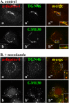
FIGURE 1.
FIGURE 1.
Association of arfaptin-1 with the trans-Golgi. HeLa cells left untreated (A) or treated with 5 μg/ml nocodazole for 2 h (B) were doubly stained for arfaptin-1 (a and b) and either TGN46 (a′) or GM130 (b′).
FIGURE 2.
FIGURE 2.
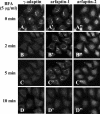
BFA-insensitive, Arf-independent Golgi-association of arfaptins. HeLa cells were incubated with 5 μg/ml BFA for the indicated time periods and triply stained for γ-adaptin (A–D), arfaptin-1 (A′–D′) and arfaptin-2 (A″–D″).
FIGURE 3.
FIGURE 3.
Binding of arfaptins to Arl1. A, binding of arfaptins to GTP-bound Arl1. Lysates of HeLa cells expressing C-terminally HA-tagged Arl1(WT), Arl1(Q71L), or Arl1(T31N) were pulled down with GST, GST-GGA1(GAT), GST-arfaptin-1, or GST-arfaptin-2 as indicated, and analyzed by immunoblot for HA as described under “Materials and Methods.” B, BAR domain-containing C-terminal region of arfaptins is responsible for Arl1 binding. Lysates of HeLa cells expressing Arl1(Q71L)-HA were pulled down with a GST fusion protein of full-length, N-terminal portion or C-terminal portion of arfaptin-1 or arfaptin-2 and analyzed by immunoblot for HA. C, schematic representation of the structures of human arfaptin-1 and arfaptin-2. Regions of constructs used in the experiment shown in B are indicated.
FIGURE 4.
FIGURE 4.
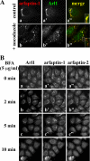
Colocalization of arfaptin-1 with Arl1. A, HeLa cells left untreated (a-a″) or treated with 5 μg/ml nocodazole for 2 h (b-b″) were doubly stained for arfaptin-1 (a and b) and Arl1 (a′ and b′). B, HeLa cells were incubated with 5 μg/ml BFA for the indicated time periods and triply stained for Arl1 (a–d), arfaptin-1 (a′–d′), and arfaptin-2 (a″–d).
FIGURE 5.
FIGURE 5.
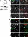
Arl1-dependent Golgi-association of arfaptins. A, HeLa cells were treated with a pool of siRNAs for Arl1, ARFRP1, or LacZ and processed for Western blot analysis with antibody to Arl1, ARFRP1, arfaptin-1, or arfaptin-2 as indicated. B, Arl1-dependent Golgi-localization of arfaptins. HeLa cells were treated with a pool of siRNAs for Arl1 (b-b′′′), ARFRP1 (d-d′′′), or LacZ (a-a′′′ and c-c′′′) and triply stained for arfaptin-1 (a–d), arfaptin-2 (a′–d′), and either Arl1 (a″ and b″) or ARFRP1 (c″ and d″). C, rescue experiment. HeLa cells treated with a pool of siRNAs for Arl1 (b-b′′′ and d-d′′′) or LacZ (a-a′′′ and c-c′′′) were transiently transfected with an expression vector for Arl1-HA and triply stained for HA (a′–d′), golgin-245 (a″–d″), and either arfaptin-1 (a and b) or arfaptin-2 (c and d).
FIGURE 6.
FIGURE 6.
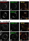
Comparison of localization of arfaptin-1 with that of golgins. HeLa cells left untreated (A) or treated with 5 μg/ml nocodazole for 2 h (B) were doubly stained for arfaptin-1 (a and b) and either golgin-245 (a′) or golgin-97 (b′).
FIGURE 7.
FIGURE 7.
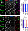
Exogenous expression of arfaptins displaces golgin-245 from the Golgi. HeLa cells were transfected with a control EGFP vector (A and D), or an expression vector for EGFP-arfaptin-1 (B and E), or EGFP-arfaptin-2 (C and F) and processed for immunofluorescence analysis with antibodies against golgin-245 (A′–C′) and TGN46 (A″–C″), or against Arl1 (D′–F′) and golgin-245 (D″–F″). G, numbers of cells with the golgin-245 and TGN46 staining in the Golgi region were counted in the population of cells expressing EGFP, EGFP-arfaptin-1, and EGFP-arfaptin-2 (n = 100), and represented as a bar graph. H, numbers of cells with the Arl1 and golgin-245 staining in the Golgi region were counted in the population of cells expressing EGFP, EGFP-arfaptin-1, and EGFP-arfaptin-2 (n = 100), and represented as a bar graph.
FIGURE 8.
FIGURE 8.
Exogenous expression of arfaptins inhibits StxB transport to the Golgi. HeLa cells were transfected with a control EGFP vector (A′ and D′), or an expression vector for EGFP-arfaptin-1 (B′ and E′), or EGFP-arfaptin-2 (C′ and F′), incubated with StxB for 50 min at 4 °C and then internalized it for 10 min (A–C) or 30 min (D–F) at 37 °C. The cells were then stained with anti-StxB antibody. G, numbers of cells (30 min uptake) with the StxB staining in the Golgi region were counted in the population of cells expressing EGFP, EGFP-arfaptin-1, and EGFP-arfaptin-2 (n = 100), and represented as a bar graph.
FIGURE 9.
FIGURE 9.
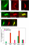
Localization of arfaptin-2 on Golgi-derived tubules. HeLa cells transiently expressing Arl1-EGFP (A), mCherry-arfaptin-2 (B), Arl1-EGFP and mCherry-arfaptin-2 (C), mCherry-golgin-97 (D), or Arl1-EGFP and mCherry-golgin-97 (E) were subjected to time-lapse recording. Single frames derived from supplemental Videos S1, S2, S3, S4, and S5, respectively, are shown. F, numbers of vesicles and tubules positive for Arl1-EGFP, mCherry-arfaptin-2, and mCherry-golgin-97 that were formed de novo from the Golgi region within 300 s in single cells. The values represent means ± S.D. of 10 cells observed for those expressing Arl1-EGFP alone, mCherry-arfaptin-2 alone, mCherry-golgin-97 alone, Arl1-EGFP, and mCherry-arfaptin-2, and Arl1-EGFP and mCherry-golgin-97.
FIGURE 10.
FIGURE 10.
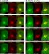
Enhanced Golgi tubulation by inhibition of myosin II activation. HeLa cells transiently expressing Arl1-EGFP and mCherry-arfaptin-2 were mock-treated with DMSO (A) or treated with 30 μm ML7 and 10 μm Y-27632 for 90 min (B), and subjected to time-lapse recording. Representative frames from supplemental Videos S6 and S7, respectively, are shown.

References

    1. Moss J., Vaughan M. (1995) J. Biol. Chem. 270, 12327–12330 - PubMed
    1. D'Souza-Schorey C., Chavrier P. (2006) Nat. Rev. Mol. Cell Biol. 7, 347–358 - PubMed
    1. Kahn R. A., Cherfils J., Elias M., Lovering R. C., Munro S., Schurmann A. (2006) J. Cell Biol. 172, 645–650 - PMC - PubMed
    1. Gillingham A. K., Munro S. (2007) Annu. Rev. Cell Dev. Biol. 23, 579–611 - PubMed
    1. Kanoh H., Williger B. T., Exton J. H. (1997) J. Biol. Chem. 272, 5421–5429 - PubMed

Publication types

MeSH terms