. 2011 Mar 25;286(12):10265-75.
doi: 10.1074/jbc.M110.188268. Epub 2011 Jan 14.

Cortisol synthesis in epidermis is induced by IL-1 and tissue injury

Affiliations

Cortisol synthesis in epidermis is induced by IL-1 and tissue injury

Sasa Vukelic et al. J Biol Chem. .

Abstract

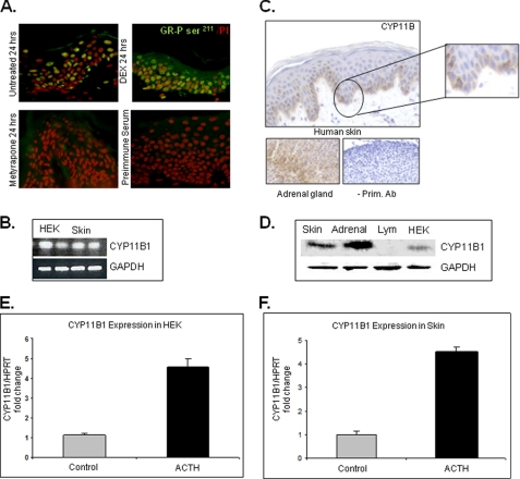
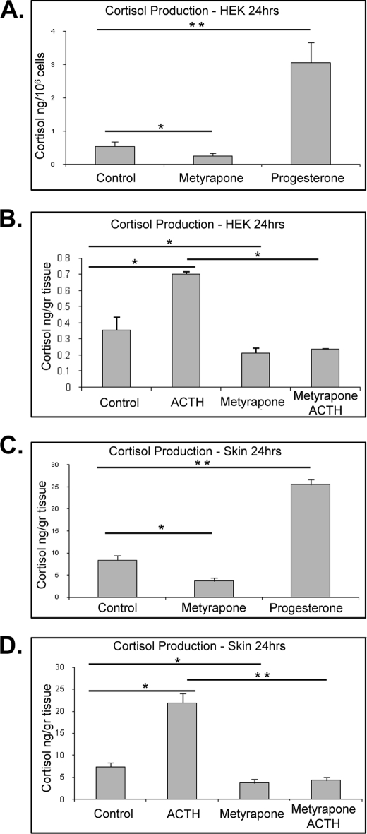
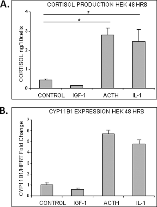
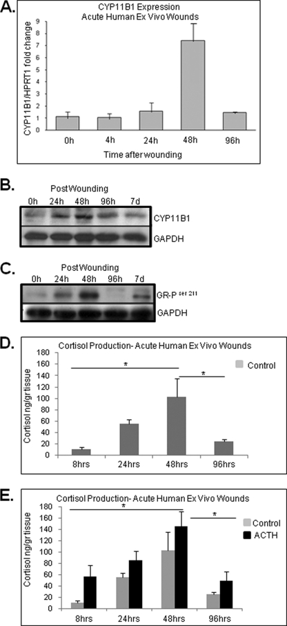
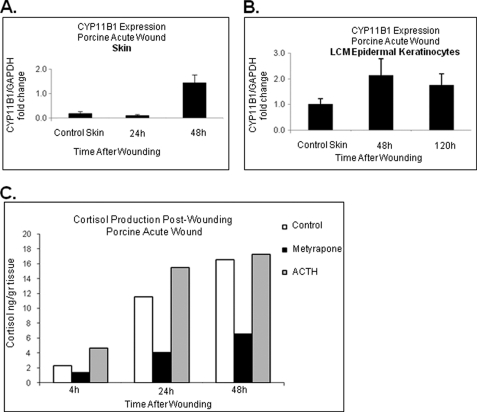
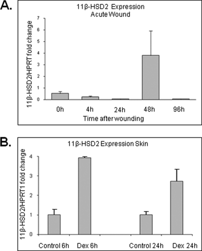
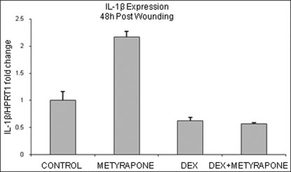
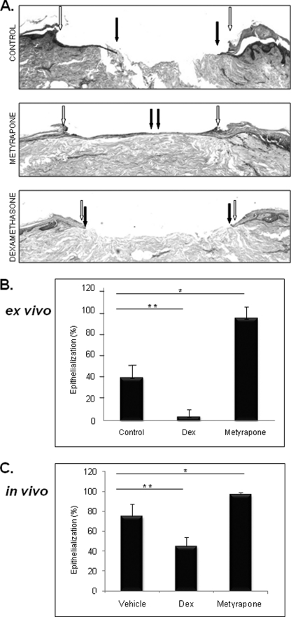
Glucocorticoids (GCs) are known inhibitors of wound healing. In this study we report the novel finding that both keratinocytes in vitro and epidermis in vivo synthesize cortisol and how this synthesis regulates wound healing. We show that epidermis expresses enzymes essential for cortisol synthesis, including steroid 11 β-hydroxylase (CYP11B1), and an enzyme that controls negative feedback mechanism, 11β-hydroxysteroid dehydrogenase 2 (11βHSD2). We also found that cortisol synthesis in keratinocytes and skin can be stimulated by ACTH and inhibited by metyrapone (CYP11B1 enzyme inhibitor). Interestingly, IL-1β, the first epidermal signal of tissue injury, induces the expression of CYP11B1 and increases cortisol production by keratinocytes. Additionally, we found induction of CYP11B1 increased production of cortisol and activation of GR pathway during wound healing ex vivo and in vivo using human and porcine wound models, respectively. Conversely, inhibition of cortisol synthesis during wound healing increases IL-1β production, suggesting that cortisol synthesis in epidermis may serve as a local negative feedback to proinflammatory cytokines. Local GCs synthesis, therefore, may provide control of the initial proinflammatory response, preventing excessive inflammation upon tissue injury. Inhibition of GC synthesis accelerated wound closure in vivo, providing the evidence that modulation of cortisol synthesis in epidermis may be an important regulatory mechanism during wound healing.


Figures

FIGURE 1.
FIGURE 1.
GR receptor is activated in human epidermis. A, immunofluorescence staining with GR- phosphorylated Ser211 antibody of human skin explants maintained in the absence of GCs for 24 h revealed the nuclear presence of the receptor in keratinocytes. Dexamethasone (DEX)-treated skin, a positive control, confirmed nuclear translocation of GR after topical corticosteroid treatment. No staining is found in the absence of primary antibody. B, qRT- PCR confirms expression of CYP11B1 in HEK and skin. Expression levels were normalized to GAPDH. C, immunoperoxidase staining of human skin with CYP11B antibody (Ab) shows increased staining of basal and first suprabasal keratinocytes, whereas staining intensity decreases in upper epidermal layers. No staining was observed in sections incubated without primary antibody. As expected, signal was present in zona fasciculata of mouse adrenal gland. D, Western blot with CYP11B1 antibody and protein extracts from adrenal gland, skin, and HEK and lymphocytes (Lym) is shown. Both keratinocytes and skin showed significant protein levels of CYP11B1 in comparison to adrenal gland (positive control), whereas no CYP11B1 was detected in lymphocytes, which do not express CYP11B1 (52) and served as negative control. E, ACTH stimulates CYP11B1 expression in keratinocytes. HEK were incubated with/without ACTH (10−7 m) for 24 h (n = 4). Quantitative real time PCR for the expression of CYP11B1 is shown. Expression levels were normalized to HPRT1. Error bars represent S.D. F, ACTH stimulates CYP11B1 expression in skin. Skin specimens were incubated with/without ACTH (10−7 m) for 24 h (n = 4). Quantitative real time PCR for the expression of CYP11B1 is shown. Error bars represent S.D.
FIGURE 2.
FIGURE 2.
Cortisol levels in culture medium. A, effects of progesterone and metyrapone on cortisol synthesis in HEK are shown. Cells were incubated with progesterone (10−6 m) and/or metyrapone (10−3 m) for 24 h (n = 4). Progesterone as a substrate stimulates cortisol production (p < 0.01), and metyrapone inhibits it (p < 0.05). Data represent the mean ± S.D. and were analyzed by one-way ANOVA (F = 463.89; p < 0.0001) and Dunnett's post hoc test (*, p < 0.05; **, p < 0.01). B, ACTH stimulates cortisol production, and metyrapone inhibits it (p < 0.01). HEK were incubated with/without ACTH (10−7 m) for 24 h. Metyrapone co-treatment reverses the effect of ACTH (p < 0.01). Data represent the mean ± S.D. (n = 4) and were analyzed by one-way ANOVA (F = 73.85; p < 0.0001) and Tukey's post hoc test (*, p < 0.01). C, effects of progesterone and metyrapone on cortisol synthesis in skin are shown. Skin explants were incubated with progesterone (10−6 m) and/or metyrapone (10−3 m) for 24 h (n = 3). Progesterone as a substrate stimulates cortisol production (p < 0.01), and metyrapone inhibits it (p < 0.05). Data represent the mean ± S.D. and were analyzed by one-way ANOVA (F = 142.66; p < 0.0001) and Dunnett's post hoc test (*, p < 0.05; **, p < 0.01). D, ACTH stimulates cortisol production. Skin punches were incubated with/without ACTH (10−7 m) for 6 h (p < 0.01). Metyrapone co-treatment reverses the effect of ACTH (p < 0.001). Data represent the mean ± S.D. (n = 4) and were analyzed by one-way ANOVA (F = 176.27; p < 0.0001) and Tukey's post hoc test (*, p < 0.01; **, p < 0.001).
FIGURE 3.
FIGURE 3.
IL-1β and IGF-1 modulate CYP11B1 expression and cortisol production in HEK. A, IL-1 and ACTH stimulate cortisol production in HEK (p < 0.01), whereas IGF-1 inhibits it. Cells were incubated with IL-1β (10 ng/ml), IGF-1 (100 ng/ml), or ACTH (10−7 m) for 24 h (n = 5). Data represent the mean ± S.D. and were analyzed by one-way ANOVA (F = 75.32; p < 0.0001) and Dunnett's post hoc test (*, p < 0.01). B, IL-1 stimulates CYP11B1 expression in HEK, and IGF-1 inhibits it. Cells were incubated with IL-1β (10 ng/ml), IGF-1(100 ng/ml), or ACTH (10−7 m) for 24 h (n = 3). Quantitative real time PCR for the expression of CYP11B1 is shown. Expression levels were normalized to HPRT1. Error bars represent S.D.
FIGURE 4.
FIGURE 4.
Expression of CYP11B1 and cortisol production during acute wound healing. A, expression of CYP11B1 during acute wound healing is shown. Acute wounds were maintained for 0, 4, 24, 48, and 96 h post wounding at the air-liquid interface, and the expression levels of CYP11B1 were determined using quantitative real time PCR (n = 4). Expression levels were normalized to HPRT1. Error bars represent S.D. B, immunoblotting shows the gradual increase in CYP11B1 protein levels during acute wound healing, with a peak at 48 h post wounding. Protein extracts from acute wound explants were tested with anti-CYP11B1 antibody. Loading control with anti-GAPDH antibody is shown. C, immunoblotting is shown. Protein extracts from acute wound explants show an increase in GR-phosphorylated Ser211 48 h after the wounding. GAPDH was used as a loading control. D and E, cortisol levels in culture medium are shown. Media from acute wounds explants with/without ACTH was collected at 8, 24, 48, and 96 h post wounding and assessed with ELISA for presence of cortisol (n = 4). Characteristic induction of cortisol production was detected during acute wound healing peaking at 48 h post wounding (p < 0.05). Data represent the mean ± S.D. (n = 4) and were analyzed by one-way ANOVA (F = 5.4; p < 0.05) and Tukey's post hoc test (*, p < 0.05).
FIGURE 5.
FIGURE 5.
Expression of CYP11B1 and cortisol synthesis during acute wound healing in vivo. A, CYP11B1 gene expression determined using real-time qPCR showed induction at 48 h post wounding as compared with normal porcine skin (n = 3). Error bars represent S.D. B, laser microdissected epidermal keratinocytes demonstrate induction of CYP11B1 enzyme 48 h post wounding. At the later time points CYP11B1 expression decreased back to the level of normal, unwounded skin (n = 3). Error bars represent S.D. C, porcine wound explants show the highest levels of cortisol production 48 h after the wounding (n = 2). Cortisol production is inhibited with metyrapone (10−3 m) and induced by ACTH (10−7 m).
FIGURE 6.
FIGURE 6.
11β-HSD2 (negative feedback enzyme) is induced by glucocorticoids. A, expression of 11β-HSD2 during ex vivo human acute wound healing (n = 3) is shown. Induction of 11β-HSD2 48 h post wounding is shown. Quantitative real time PCR for the expression of 11β-HSD2 is shown. Expression levels were normalized to HPRT1. Error bars represent S.D. B, dexamethasone (Dex, 10−3 m) treatment for 6 h of human skin leads to a 4-fold induction of 11β-HSD2 (n = 3). Quantitative real time PCR for the expression of 11β-HSD2 is shown. Expression levels were normalized to HPRT1. Error bars represent S.D.
FIGURE 7.
FIGURE 7.
Inhibition of cortisol production during wound healing leads to induction of IL-1β expression. Acute wound explants were treated with metyrapone (10−3 m), dexamethasone (DEX, 10−3 m), or combination for 48 h (n = 3). Metyrapone treatment induces expression of IL-1β, whereas dexamethasone reverses that effect. Error bars represent S.D.
FIGURE 8.
FIGURE 8.
Metyrapone promotes wound healing in ex vivo human and in vivo porcine wound models. A, hematoxylin and eosin staining demonstrates complete epithelialization after treatment of acute ex vivo wounds with metyrapone. Open arrows indicate wound edges after initial wounding, whereas solid arrows point to the epithelialized edges of the migrating fronts 4 days after the wounding. B, quantification of the rate of epithelialization 4 days after wounding is shown. Metyrapone (10−3 m) promotes epithelialization in a human skin organ culture model (*, p < 0.05). Topical dexamethasone (Dex, 10−3 m) treatment delayed epithelialization when compared with control untreated skin (**, p < 0.03) (n = 3). C, quantification of the epithelialization rate in a porcine deep partial thickness model is shown. Metyrapone treatment promotes epithelialization (*, p < 0.01), whereas dexamethasone inhibits it (**, p < 0.006) (n = 4). Error bars represent S.D.

References

    1. Brem H., Tomic-Canic M. (2007) J. Clin. Invest. 117, 1219–1222 - PMC - PubMed
    1. Braun S., Hanselmann C., Gassmann M. G., auf dem Keller U., Born-Berclaz C., Chan K., Kan Y. W., Werner S. (2002) Mol. Cell. Biol. 22, 5492–5505 - PMC - PubMed
    1. Loots M. A., Lamme E. N., Zeegelaar J., Mekkes J. R., Bos J. D., Middelkoop E. (1998) J. Invest. Dermatol. 111, 850–857 - PubMed
    1. Mast B. A., Schultz G. S. (1996) Wound Repair Regen. 4, 411–420 - PubMed
    1. Robson M. C. (1997) Wound Repair Regen. 5, 12–17 - PubMed

Publication types

MeSH terms