

A comparison between bitewing radiographs taken with rectangular and circular collimators in UK military dental practices: a retrospective study
- PMID: 21239573
- PMCID: PMC3520300
- DOI: 10.1259/dmfr/86968802
A comparison between bitewing radiographs taken with rectangular and circular collimators in UK military dental practices: a retrospective study
Abstract
Objectives: The aim of this study was to determine any increase in the incidence of cone cut errors that adversely affected diagnostic yield resulting in more retakes using rectangular collimation with film holders in bitewing radiography. Comparisons were also made with other positioning errors that occurred when bitewings were taken with circular collimation, with and without film holders.
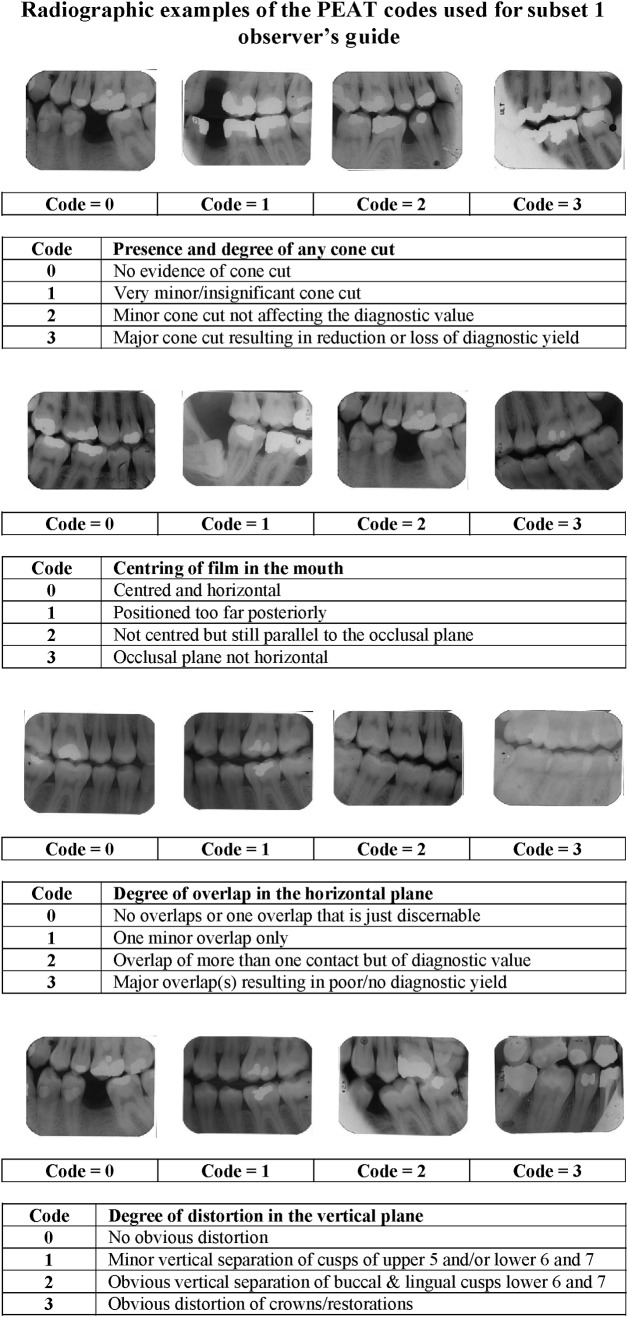
Methods: A preliminary questionnaire was used to determine the year that rectangular collimation was adopted by military dental practice. 3 time-framed subsets, each of 1000 bitewing radiographs, were identified: subset 1, films taken with circular collimators without film holders; subset 2, films taken with circular collimators with film holders; and subset 3, films taken with rectangular collimators with film holders. Each subset was assessed for positioning errors of cone cut, horizontal overlap, vertical distortion and film centring. The χ(2) test was used to test significant differences amongst the three subsets.
Results: The use of film holders with circular collimation significantly reduced the incidence of cone cut errors from 21.7% to 3.3%. There was an increase in the incidence of cone cut errors from 3.3% to 20.9% when rectangular collimation was used, but the actual number considered "rejects" was very small, only 0.1% (1 in 1000 films) in subset 2 and 0.3% (3 of 1000 films) in subset 3, when assessed for diagnostic yield.
Conclusions: This study provides evidence that rectangular collimation did not significantly affect the diagnostic yield of bitewing radiographs despite the presence of cone cut. Therefore, all practitioners should adopt rectangular collimation.
Figures

Comment in
-
Letter to the editor concerning Parrott LA, Ng SY. A comparison between bitewing radiographs taken with rectangular and circular collimators in UK military dental practices: a retrospective study published in Dentomaxillofacial Radiology (2001;40:102-109).Dentomaxillofac Radiol. 2011 May;40(4):262-3. doi: 10.1259/dmfr/38497493. Dentomaxillofac Radiol. 2011. PMID: 21493884 Free PMC article. No abstract available.
Similar articles
-
Evidence on radiation dose reduction using rectangular collimation: a systematic review.Int Dent J. 2019 Apr;69(2):84-97. doi: 10.1111/idj.12411. Epub 2018 Jun 29. Int Dent J. 2019. PMID: 29959778 Free PMC article.
-
Letter to the editor concerning Parrott LA, Ng SY. A comparison between bitewing radiographs taken with rectangular and circular collimators in UK military dental practices: a retrospective study published in Dentomaxillofacial Radiology (2001;40:102-109).Dentomaxillofac Radiol. 2011 May;40(4):262-3. doi: 10.1259/dmfr/38497493. Dentomaxillofac Radiol. 2011. PMID: 21493884 Free PMC article. No abstract available.
-
Rectangular collimation and radiographic efficacy in eight general dental practices in the West Midlands.Prim Dent Care. 2004 Jul;11(3):81-6. doi: 10.1308/1355761041208539. Prim Dent Care. 2004. PMID: 15242564
-
Comparison of technical errors in pediatric bitewing radiographs acquired with round vs rectangular collimation.Oral Surg Oral Med Oral Pathol Oral Radiol. 2022 Mar;133(3):333-342. doi: 10.1016/j.oooo.2021.09.002. Epub 2021 Sep 14. Oral Surg Oral Med Oral Pathol Oral Radiol. 2022. PMID: 34627711
-
Factors affecting the diagnostic quality of bitewing radiographs: a review.Br Dent J. 1998 Jan 24;184(2):80-4; discussion 77. doi: 10.1038/sj.bdj.4809549. Br Dent J. 1998. PMID: 9489215 Review.
Cited by
-
Proximal caries detection accuracy using intraoral bitewing radiography, extraoral bitewing radiography and panoramic radiography.Dentomaxillofac Radiol. 2012 Sep;41(6):450-9. doi: 10.1259/dmfr/30526171. Dentomaxillofac Radiol. 2012. PMID: 22868296 Free PMC article.
-
Evidence on radiation dose reduction using rectangular collimation: a systematic review.Int Dent J. 2019 Apr;69(2):84-97. doi: 10.1111/idj.12411. Epub 2018 Jun 29. Int Dent J. 2019. PMID: 29959778 Free PMC article.
-
Pathogenesis, Clinical Features, Diagnosis, and Management of Radiation Hazards in Dentistry.Open Dent J. 2018 Sep 28;12:742-752. doi: 10.2174/1745017901814010742. eCollection 2018. Open Dent J. 2018. PMID: 30369984 Free PMC article. Review.
-
Oblique lateral radiographs and bitewings; estimation of organ doses in head and neck region with Monte Carlo calculations.Dentomaxillofac Radiol. 2014;43(6):20130419. doi: 10.1259/dmfr.20130419. Epub 2014 May 16. Dentomaxillofac Radiol. 2014. PMID: 24834483 Free PMC article.
-
Development of an open project rectangular collimator for use with intraoral dental X-ray unit.Radiol Phys Technol. 2024 Mar;17(1):315-321. doi: 10.1007/s12194-023-00772-9. Epub 2024 Jan 24. Radiol Phys Technol. 2024. PMID: 38265510
References
-
- Recommendations on radiographic procedures technical report No 8 (Revision) Int Dent J 1989;39:147–148 - PubMed
-
- European guidelines on radiation protection in dental radiology. The safe use of radiographs in dental practice. Issue No 126. Office of Official Publications of the European Communities 2004.
-
- Araki K, Kanda S. Radiological characteristics of lead foils in dental packets: analysis of components and shielding effect. Dentomaxillofac Radiol 1992;21:21–25 - PubMed
-
- Mauriello SM, Overman VP, Jansen L. Current techniques and principles in dental radiology: part II. J Pract Hyg 2002:Nov/Dec 22–25
-
- Langland OE, Langlais RP, Preece JW. Principles of dental imaging (2nd edn). Lippincott Williams & Wilkins 2002 Philadelphia, PA.
