

Case Reports
doi: 10.1259/dmfr/55423624.
A case of desmoplastic ameloblastoma arising in the maxillary alveolus: the origin and time-course changes in the early stage of tumour development observed on dental radiographs
Affiliations
- PMID: 21239577
- PMCID: PMC3520299
- DOI: 10.1259/dmfr/55423624
Item in Clipboard
Case Reports
A case of desmoplastic ameloblastoma arising in the maxillary alveolus: the origin and time-course changes in the early stage of tumour development observed on dental radiographs
Dentomaxillofac Radiol.
2011 Feb.
Abstract
In this article we report a case of desmoplastic ameloblastoma in which chronological changes in the early development can be observed on dental radiographs. The tumour grew very slowly and did not appear to have a strong potential for local extension like typical ameloblastomas. Radiological findings of our case suggest the tumour arose from the periodontal membrane. However, it was not possible to obtain conclusive histopathological evidence.
Figures

Panoramic radiograph from the initial examination. An ill-defined multilocular mixed radiopaque/radiolucent lesion between the canine and the first premolar of the right maxilla is shown. The roots of the canine and the second molar are displaced
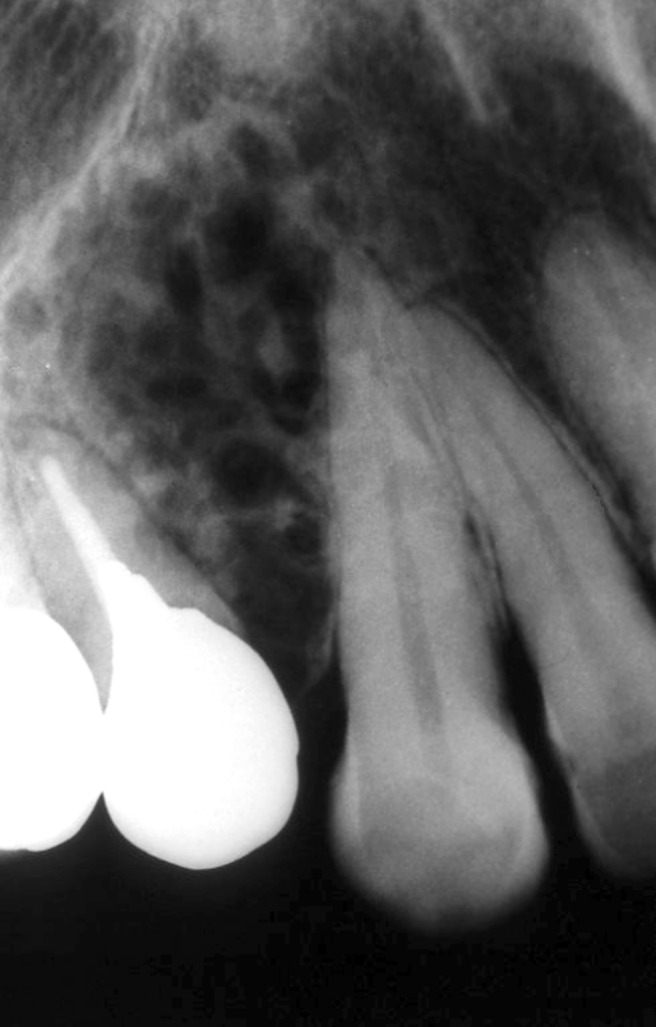
A dental radiograph from the initial examination. Note disappearance of lamina dura without root resorption of the canine and the first premolar

Axial CT in bone window at two different locations in the maxilla showing buccolingual expansion of the cortical plate of the maxillary alveolus

Multiplanar reconstructed CT image parallel to the dental arch. Note the disappearance of the lamina dura of the canine adjacent to the lesion

Histopathological specimen (haematoxylin–eosin stain, ×100). (a) The specimen shows that the tumour is composed of rich fibrous connective tissue with a scattering of ameloblastomatous foci. (b) The ameloblastomatous foci are small and complicated in shape. The ameloblastomatous foci are often atrophic or hyperkeratotic owing to enhanced fibrosis around them

(a) Dental radiograph 8 years before the first visit to our hospital. Note the radiolucent lesion between the canine and the first premolar near the alveolar crest (arrow). The lamina dura and the periodontal ligament space next to the lesion are unclear. (b) The dental radiograph 3 years before the first visit to our hospital. The lesion has developed in comparison with Figure 6a. The lamina dura and the periodontal ligament space of the canine have become increasingly unclear
References
-
- Eversole LR, Leider AS, Hansen LS. Ameloblastomas with pronounced desmoplasia. J Oral Maxillofac Surg 1984;42:735–740 - PubMed
-
- Louis PJ, Fugler RC, August M. Mixed radiolucent/radiopaque lesion of the maxilla. J Oral Maxillofac Surg 2000;58:86–90 - PubMed
-
- Lam KY, Chan AC, Wu PC, Chau KY, Tideman H, Wei W. Desmoplastic variant of ameloblastoma in Chinese patients. Br J Oral Maxillofac Surg 1998;36:129–134 - PubMed
-
- Manuel S, Simon D, Rajendran R, Naik BR. Desmoplastic ameloblastoma: a case report. J Oral Maxillofac Surg 2002;60:1186–1188 - PubMed
-
- Pillai RS, Ongole R, Ahsan A, Radhakrishnan RA, Pai KM. Recurrent desmoplastic ameloblastoma of the maxilla: a case report. J Can Dent Assoc 2004;70:100–104 - PubMed
