. 2011 Apr;300(4):H1518-29.
doi: 10.1152/ajpheart.00928.2010. Epub 2011 Jan 14.

Nicotine aggravates the brain postischemic inflammatory response

Affiliations

Nicotine aggravates the brain postischemic inflammatory response

Shayna T Bradford et al. Am J Physiol Heart Circ Physiol. 2011 Apr.

Abstract

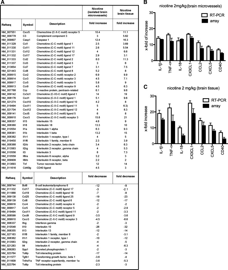
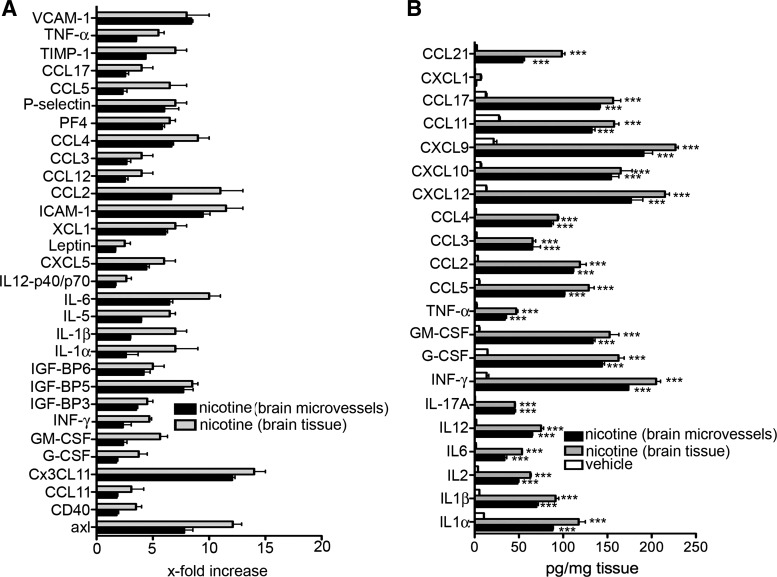
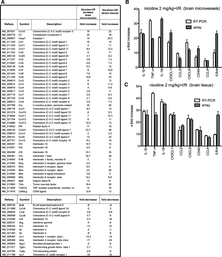
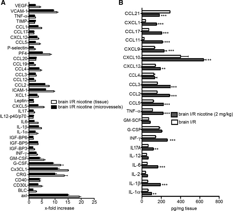
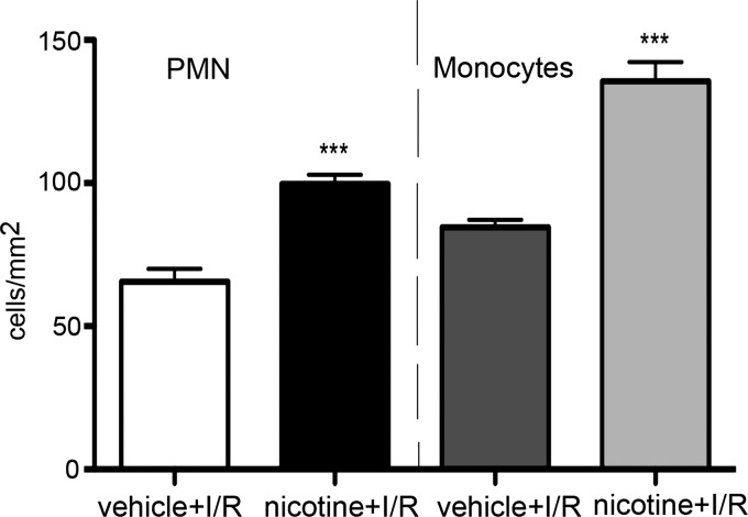
A substantial body of evidence suggests that nicotine adversely affects cerebral blood flow and the blood-brain barrier and is a risk factor for stroke. The present study investigated the effect of nicotine on cerebrovascular endothelium under basal and ischemia/reperfusion injury under in vivo condition. Nicotine (2 mg/kg sc) was administered to mice over 14 days, which resulted in plasma nicotine levels of ∼100 ng/ml, reflecting plasma concentrations in average to heavy smokers. An analysis of the phenotype of isolated brain microvessels after nicotine exposure indicated higher expression of inflammatory mediators, cytokines (IL-1β, TNF-α, and IL-18), chemokines (CCL2 and CX(3)CL1), and adhesion molecules (ICAM-1, VCAM-1, and P-selectins), and this was accompanied by enhanced leukocyte infiltration into brain during ischemia/reperfusion (P < 0.01). Nicotine had a profound effect on ischemia/reperfusion injury; i.e., increased brain infarct size (P < 0.01), worse neurological deficits, and a higher mortality rate. These experiments illuminate, for the first time, how nicotine regulates brain endothelial cell phenotype and postischemic inflammatory response at the brain-vascular interface.


Figures

Fig. 1.
Fig. 1.
A: list of the transcripts modulated by nicotine in brain tissue and isolated brain microvessels from mice treated with nicotine (2 mg/kg) for 14 days. Fold increase/decrease indicate the level of up- or downregulation of transcripts compared with control (vehicle-treated mice). Three independent samples were analyzed by RT2 real-time PCR array. B and C: quantitative real-time PCR for IL-1β, TNF-α, IL-18, CX3CL1, CCL2, CXCL5, and CD40 was carried out on RNA from isolated brain microvessels (B) and brain tissue (C) from nicotine-treated (n = 3) and vehicle-treated mice (n = 3). Expression of target genes was normalized to control vehicle-treated mice. Values are presented as means ± SD. The fold changes obtained with RT-PCR were similar to those obtained by the PCR array.
Fig. 2.
Fig. 2.
Cytokine antibody array (A) and ELISA (B) analysis of isolated brain microvessels and brain. For the cytokine array, 62 cytokines, adhesion molecules, and chemokines were assayed in 3 samples. Values after nicotine treatment (2 mg/kg; 14 days) were normalized to brain microvessels or brain tissue isolated from vehicle-treated mice. For the ELISA, samples (brain microvessels or brain tissue) were taken after mice had been treated with nicotine (2 mg/kg) or vehicle for 14 days. Values represent means ± SD from 3 independent brain samples. ***P < 0.001. GM-CSF, granulocyte/macrophage colony-stimulating factor; TIMP, tissue inhibitor of matrix metalloproteinase; INF, interferon.
Fig. 3.
Fig. 3.
A: Kaplan-Meier survival curve in mice exposed to either nicotine (2 mg/kg) or vehicle (0.9% NaCl) for 14 days followed by induction of middle cerebral artery occlusion (MCAO). B: summary of neurological scores in nicotine- (2 mg/kg) and vehicle-treated mice at day 3 after transient MCAO. No neurological deficit scores are 0; maximal deficit score is 4. Vehicle (n = 15) and nicotine (n = 15), *P < 0.05 by Chi-squared test. C: 2,3,5-triphenyltetrazolium chloride-stained coronal sections of brain illustrating typical infarcts (arrows) 3 days after reperfusion in nicotine- and vehicle-treated mice. Bar graph showing infarct volumes at day 3 after transient MCAO in vehicle (n = 10) and nicotine (0.5, 2.0, and 5.0 mg/kg) (n = 7) mice. D: cortical and striatal infarct volume in same experimental animals as in C. Values are means ± SD. ***P < 0.001. E: brain edema formation after MCAO was evaluated by measuring the brain water content in ischemic and contralateral hemispheres at day 3 after transient MCAO. Values are means ± SD for nicotine-treated mice (0.5, 2.0, and 5.0 mg/kg; n = 7) vehicle-treated mice (n = 7). F: indirect measure (to correct for edema/infarct resolution) of total infarct volume at day 3 after transient MCAO in vehicle- and nicotine-treated (0.5, 2.0, and 5.0 mg/kg) mice. **P < 0.01.
Fig. 4.
Fig. 4.
A: list of the genes modulated by nicotine in isolated brain microvessels and brain tissue from around the ischemic area (penumbra) of mice treated with nicotine (2 mg/kg) for 14 days followed by transient MCAO and reperfusion for 3 days. Fold increase/decrease indicates level of up- or downregulation of genes compared with control (mice treated with vehicle for 14 days, followed by transient MCAO and reperfusion for 3 days). Three independent samples were analyzed by real-time RT-PCR array. B and C: quantitative real-time PCR for IL-1β, TNF-α, IL-18, CX3CL1, CCL2, CXCL5, CD40, CCL4, and IL-6ra was carried out on RNA of isolated brain microvessels (B) or brain tissue (C) from nicotine-treated mice with brain ischemia/reperfusion (I/R) injury (n = 3) or vehicle-treated mice with brain I/R injury (n = 3). Expression of target genes was normalized to control (vehicle-treated mice with brain I/R injury). Values are presented as means ± SD. The real-time PCR confirmed the changes in these genes detected by the PCR array although there were some differences in the absolute level of upregulation with the two techniques.
Fig. 5.
Fig. 5.
A: cytokine antibody array of brain microvessels and brain tissue from mice exposed to nicotine (2 mg/kg) for 14 days followed by a MCAO for 3 days. For the cytokine array, 62 cytokines, adhesion molecules, and chemokines were assayed in 3 samples. The values were normalized to brain microvessels or brain tissue from the ischemic penumbra from vehicle-treated mice that underwent MCAO with reperfusion. B: ELISA analysis of penumbral brain samples collected from mice treated with nicotine or vehicle for 14 days followed by a MCAO for 3 days. Values represent means ± SD from n = 3 independent experiments of brain samples or brain microvessels. **P < 0.01, *** P < 0.001 comparing vehicle-treated and nicotine-treated mice that underwent MCAO with reperfusion. BMEC, brain microvascular endothelial cell.
Fig. 6.
Fig. 6.
Immunohistochemical analysis of polymorphonuclear leukocytes (PMNs) and monocytes infiltration into brain of mice exposed to nicotine (2 mg/kg) or vehicle for 14 days before MCAO. Analysis was performed at day 3 of reperfusion. Anti-MPO (PMN) and anti-Ly6G (monocytes) antibodies were used. ***P < 0.001 vs. vehicle-treated group.

References

    1. Abbruscato TJ, Lopez SP, Roder K, Paulson JR. Regulation of blood-brain barrier Na,K,2Cl-cotransporter through phosphorylation during in vitro stroke conditions and nicotine exposure. J Pharmacol Exp Ther 310: 459– 468, 2004. - PubMed
    1. Albaugh G, Bellavance E, Strande L, Heinburger S, Hewitt CW, Alexander JB. Nicotine induces mononuclear leukocyte adhesion and expression of adhesion molecules, VCAM and ICAM, in endothelial cells in vitro. Ann Vasc Surg 18: 302– 307, 2004. - PubMed
    1. Albaugh G, Kann B, Strande L, Vemulapalli P, Hewitt C, Alexander JB. Nicotine induces endothelial TNF-alpha expression, which mediates growth retardation in vitro. J Surg Res 99: 381– 384, 2001. - PubMed
    1. Andjelkovic AV, Stamatovic SM, Keep RF. The protective effects of preconditioning on cerebral endothelial cells in vitro. J Cereb Blood Flow Metab 23: 1348– 1355, 2003. - PubMed
    1. Apelberg BJ, Onicescu G, Avila-Tang E, Samet JM. Estimating the risks and benefits of nicotine replacement therapy for smoking cessation in the United States. Am J Public Health 100: 341– 348, 2010. - PMC - PubMed

Publication types

MeSH terms