. 2011 Feb 16;30(4):665-78.
doi: 10.1038/emboj.2010.353. Epub 2011 Jan 14.

Cdc42-dependent formation of the ZO-1/MRCKβ complex at the leading edge controls cell migration

Affiliations

Cdc42-dependent formation of the ZO-1/MRCKβ complex at the leading edge controls cell migration

Lin Huo et al. EMBO J. .

Abstract

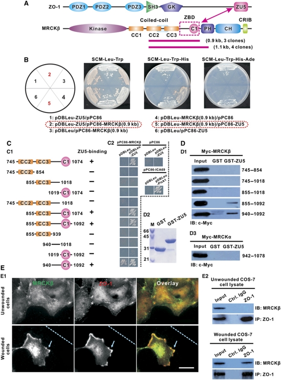
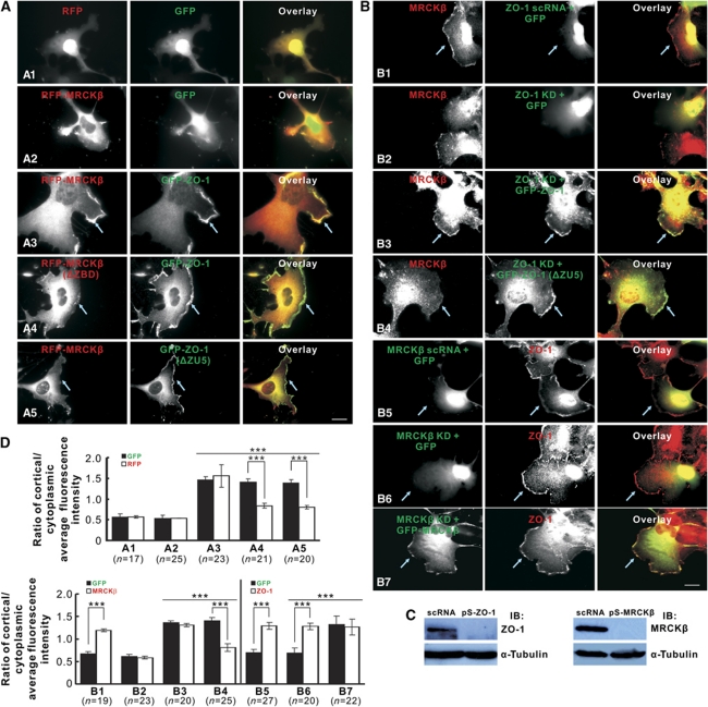
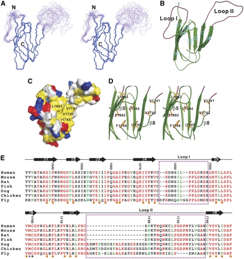
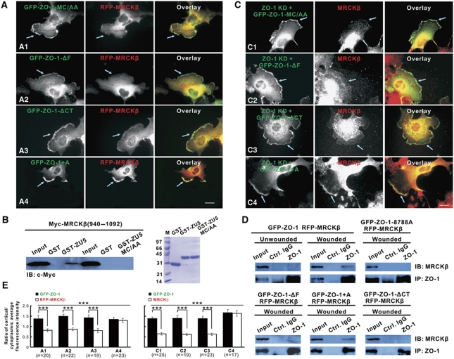
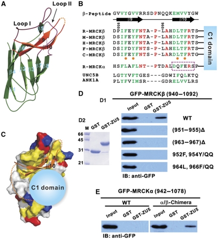
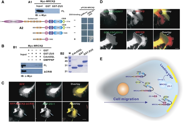
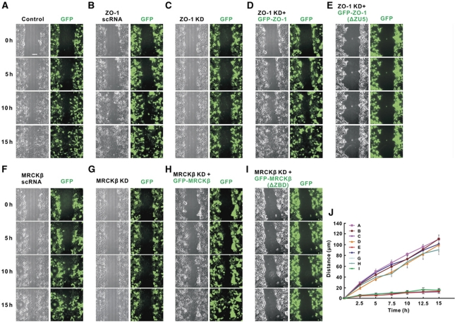
Zonula occludens (ZO)-1 is a multi-domain scaffold protein known to have critical roles in the establishment of cell-cell adhesions and the maintenance of stable tissue structures through the targeting, anchoring, and clustering of transmembrane adhesion molecules and cytoskeletal proteins. Here, we report that ZO-1 directly binds to MRCKβ, a Cdc42 effector kinase that modulates cell protrusion and migration, at the leading edge of migrating cells. Structural studies reveal that the binding of a β hairpin from GRINL1A converts ZO-1 ZU5 into a complete ZU5-fold. A similar interaction mode is likely to occur between ZO-1 ZU5 and MRCKβ. The interaction between ZO-1 and MRCKβ requires the kinase to be primed by Cdc42 due to the closed conformation of the kinase. Formation of the ZO-1/MRCKβ complex enriches the kinase at the lamellae of migrating cells. Disruption of the ZO-1/MRCKβ complex inhibits MRCKβ-mediated cell migration. These results demonstrate that ZO-1, a classical scaffold protein with accepted roles in maintaining cell-cell adhesions in stable tissues, also has an active role in cell migration during processes such as tissue development and remodelling.


Conflict of interest statement

The authors declare that they have no conflict of interest.

Figures

Figure 1
Figure 1
ZO-1 specifically binds to MRCKβ via its ZU5 domain. (A) Domain organization of ZO-1 and MRCKβ. ZO-1 contains three N-terminal PDZ domains followed by a SH3-GK tandem, and a unique C-terminal ZU5 domain. MRCKβ is composed of, in order from its N- to C-termini: a kinase domain, three coiled-coil domains, and C1-PH-CH-CRIB domains arranged in tandem. The two magenta lines underneath MRCKβ represent the seven overlapping clones identified in the Y2H screening using ZO-1 ZU5 as the bait. (B) Y2H assays showing the strong and specific interaction between an ∼0.9 kb MRCKβ fragment encoding amino-acid residues 745–1074 and the ZO-1 ZU5 domain. (C, D) Mapping of the minimal ZO-1 ZU5-binding region of MRCKβ (residues 940–1092) by a Y2H-based binding assay (C) and an in vitro GST pull-down assay (D). The corresponding region in MRCKα failed to bind to ZO-1 ZU5, showing the specificity of the interaction between ZO-1 and MRCKβ. The weak growth of the 940–1092 construct (bottom right panel of C2) presumably is due to self-activation of the fragment in the Y2H assay. (E) ZO-1 specifically interacts with MRCKβ in motile cells. ZO-1 and MRCKβ show specific co-localization at the leading edge of migrating cells (E1, lower panel). In contrast, no obvious co-localization between ZO-1 and MRCKβ could be observed when cells were cultured to near confluence (E1, upper panel). In this assay, directed cell migration was induced by scratching confluent cells with a pipette indicated by the dashed lines at the bottom panels of (E1). The leading edge of the wounded cells was identified by rhodamine-conjugated phalloidin staining of actins (Supplementary Figure S1A). Scale bar: 10 μm. Endogenous MRCKβ was co-immunoprecipitated by endogenous ZO-1 in wounded COS-7 cell lysates using an anti-ZO-1 antibody (E2). No detectable ZO-1/MRCKβ interaction was found in unwounded cells.
Figure 2
Figure 2
ZO-1 anchors MRCKβ at the leading edge of migrating cells. (A) When overexpressed, a fraction of ZO-1 and MRCKβ are co-localized at the leading edge of migrating COS-7 cells (A3). RFP and GFP served as the controls and show diffused distribution throughout the cytoplasm and nucleus (A1), and GFP alone cannot target RFP-MRCKβ to the leading edge (A2). Deletion of the ZO-1-binding domain (ZBD) from MRCKβ (A4) or removal of the ZU5 domain from ZO-1 (A5) severely impaired the leading edge localization of MRCKβ but not ZO-1. (B, C) The leading edge localization of MRCKβ was not affected by the scramble siRNA (scRNA) of ZO-1 (B1). Knockdown of ZO-1 disrupted the leading edge localization of MRCKβ (B2), which could be rescued by the RNAi-resistant WT ZO-1 (B3) but not by ZO-1 with its ZU5 domain removed (B4). Both the scRNA of MRCKβ and knockdown of MRCKβ had no effect on endogenous ZO-1 leading edge localization (B5, B6), and the RNAi-resistant WT MRCKβ was found to co-localize with ZO-1 at the leading edge (B7). The knockdown efficiencies of shRNAs were evaluated by western blot analysis (C). Scale bar: 20 μm. (D) Quantification of leading edge localizations of ZO-1 and MRCKβ in experiments is shown in (A, B). The ratio of average fluorescence intensities of membrane cortex over cytoplasm was used to measure leading edge enrichments of ZO-1 and MRCKβ in each cell. Error bars represent SEM. ‘n' represents the number of cells analysed in each experiment. ***P<0.0005 by the Student's t-test.
Figure 3
Figure 3
Solution structure of the ZO-1 ZU5 domain. (A) Stereo-view showing the backbones of 20 superimposed NMR-derived structures of ZU5_MC/AA. The two flexible loops are coloured in purple. (B) Ribbon diagram of a representative structure of ZU5_MC/AA. The secondary structures (β1–β8) are labelled. The two flexible β4/β5 and β7/β8 loops are labelled with loop I and loop II, respectively. (C) Surface representation of ZU5_MC/AA. Positively charged residues are drawn in blue, negatively charged residues in red, hydrophobic residues in yellow, and the rest in grey. The residues from the last β strand, together with the β5 and β6 strands of the domain, form a large solvent-exposed hydrophobic surface that is directly responsible for binding to its targets. (D) A stereo-view showing the hydrophobic packing of the last β strand (β8) and the extreme C-terminal Phe (Phe1748) with the β-barrel core of the ZU5 domain. The residues involved in the packing are drawn in the explicit atomic representation. The orientation of the domain is the same as that in (B). (E) Amino-acid sequence alignment of ZO-1 ZU5 from different species, showing the highly conserved nature of the domain throughout the evolution. In this alignment, the absolutely conserved amino acids are highlighted in red, and the highly conserved residues are in green. The residues forming the hydrophobic core are highlighted by orange dots, and Met1699 and Cys1700, which were substituted with Ala for the structural determination of the apo-ZO-1 ZU5 domain, are indicated by blue stars. The residues forming loops I and II are marked by two dashed boxes in purple.
Figure 4
Figure 4
Conformational requirements of ZO-1 ZU5 for binding to MRCKβ. (A) The stabilization of the closed conformation of ZO-1 ZU5 caused by the MC/AA mutation (A1), the deletion of the last Phe (A2), and the removal of the entire β8 (A3) all severely decreased the leading edge localizations of MRCKβ when each of the ZO-1 mutants was co-expressed with MRCKβ in COS-7 cells. As the control, extending ZO-1 at its C-terminal tail by adding an Ala did not have an observable impact on the leading edge localization of MRCKβ (A4). Scale bar: 20 μm. (B) GST pull-down assay showing that the MC/AA mutation of ZO-1 ZU5 disrupts its binding to MRCKβ. (C) The impairment of the MRCKβ leading edge localization induced by ZO-1 knockdown was not rescued by ZO-1 with the MC/AA mutation (C1), the deletion of Phe1748 (C2), and the deletion of the last β strand (C3), but could be rescued by the ZO-1 mutant with the C-terminal Ala extension (C4). Scale bar: 20 μm. (D) Co-IP-based assay showing the interactions between MRCKβ and the various ZO-1 mutants tested in (A). In this assay, proteins in each cell lysate were immunoprecipitated with anti-ZO-1 antibody, and co-precipitated proteins were probed with anti-MRCKβ antibody. (E) The ratio of average fluorescence intensities of membrane cortex over cytoplasm was used to measure leading edge enrichments of ZO-1 and MRCKβ in each cell. Error bars represent s.e.m. ‘n' represents the number of cells analysed in each experiment. ***P<0.0005.
Figure 5
Figure 5
Structural basis of the ZO-1 ZU5/MRCKβ complex formation. (A) Ribbon diagram representation of the ZO-1 ZU5/GRINL1A peptide complex structure. The GRINL1A peptide forms a β-hairpin structure (β9 and β10), and β9 pairs in an anti-parallel manner with two short β strands (β5 and β6) of ZO-1 ZU5. Loop I of the domain becomes structured and interacts with the target peptide. (B) Amino-acid sequence alignment of the GRINL1A β-hairpin peptide with the MRCKβ peptide fragment N-terminal to its C1 domain and the equivalent sequences from UNC5B and ankyrin ZU5. Note that the aligned MRCKβ residues corresponding to β9 and β10 of the GRINL1A peptide are highly conserved (in rat, mouse, human, fish, and chicken) and are predicted to form β strands. The residues from MRCKβ are highlighted with orange dots, which are also highly conserved in ZU5 domain from UNC5B and ankyrin, are predicted to be critical for binding to ZO-1 ZU5 based on the ZU5/GRINL1A complex structure, and the roles of these residues in ZO-1 ZU5 binding were directly tested (D). In contrast, the corresponding amino acids from MRCKα (the residues aligned to β10 in particular, highlighted by a dashed box in purple) are distinctly different. (C) A structural model of the ZO-1 ZU5/MRCKβ ZBD complex. In addition to the N-terminal β-hairpin peptide, the C1 domain of MRCKβ (shown as a blue sphere) is also required for binding to ZO-1 ZU5. (D) In vitro GST pull-down assay showing the critical roles of the residues forming the predicted β9 and β10 strands of MRCKβ in binding to ZO-1 ZU5. (E) The replacement of the MRCKα peptide fragment N-terminal to its C1 domain with the corresponding sequence in MRCKβ converted the MRCKα chimera into a ZO-1 ZU5-binding enzyme. In contrast, the WT MRCKα failed to bind to ZO-1 ZU5.
Figure 6
Figure 6
Cdc42-induced opening of the closed conformation of MRCKβ. (A) In vitro pull-down experiment showing that the full-length MRCKβ adopts a closed conformation incapable of binding to ZO-1 ZU5 (A1). Y2H-based analysis reveals that the PH-CH tandem is responsible for covering the ZO-1 ZU5-binding site of MRCKβ (A2). (B) Binding of active Cdc42 to the CRIB domain releases the closed conformation of MRCKβ, enabling the enzyme to bind to ZO-1. Deletion of the CRIB domain eliminates Cdc42-induced MRCKβ binding to ZO-1. (C) The removal of the CRIB domain also dramatically decreases the ZO-1-mediated leading edge localization capacity of MRCKβ. Scale bar: 20 μm. (D) The overexpression of a dominant negative form of Cdc42 (‘Cdc42N17') resulted in the near complete loss of the leading edge localization of the endogenous MRCKβ. However, overexpression of a constitutive active form of Cdc42 (‘Cdc42V12') did not further enhance the leading edge localization of MRCKβ. Scale bar: 20 μm. (E) A schematic model showing the Cdc42-dependent binding of MRCKβ to ZO-1. In this model, binding of Cdc42 to the CRIB domain not only activates MRCKβ, but also triggers the activated enzyme to be localized at the leading edge of migrating cells by exposing its ZO-1 ZU5-binding site. The ZO-1-anchored Cdc42/MRCKβ complex at the lamellae is likely to be critical for the directional migration of various types of cells.
Figure 7
Figure 7
The ZO-1/MRCKβ complex is essential for cell migration. (AI) Time lapse imaging analysis showing that COS-7 cells display well-characterized, wounding-induced migrations leading to the wound closure. The pSUPER alone and the empty GFP vector were used as the transfection control (A). The scRNA of ZO-1 (B) and the scRNA of MRCKβ (F) had no observable effects on the wound closure (see (J) for the quantification). The cell migrations were imaged both with phase contrast mode and with the GFP fluorescence signals. Knockdown of either ZO-1 (C) or MRCKβ (G) significantly impaired wounding-induced cell migrations. Restored expression of ZO-1 (D) or MRCKβ (H) rescued cell migration defects induced by the shRNA-mediated knockdowns of the two proteins. In contrast, neither ZO-1 with ZU5 deleted (E) nor MRCKβ with ZBD removed (I) could rescue cell migration defects. Scale bar: 100 μm. (J) Quantification of the imaging experiments are shown in (AI). In this analysis, the migration distances of the cells containing GFP signals were averaged for each condition. Error bars represent s.e.m. from n=2 experiments with 100 cells from each experiment.

Similar articles

Cited by

References

    1. Ackerman SL, Kozak LP, Przyborski SA, Rund LA, Boyer BB, Knowles BB (1997) The mouse rostral cerebellar malformation gene encodes an UNC-5-like protein. Nature 386: 838–842 - PubMed
    1. Anderson JM, Fanning AS, Lapierre L, Van Itallie CM (1995) Zonula occludens (ZO)-1 and ZO-2: membrane-associated guanylate kinase homologues (MAGuKs) of the tight junction. Biochem Soc Trans 23: 470–475 - PubMed
    1. Anderson JM, Van Itallie CM, Fanning AS (2004) Setting up a selective barrier at the apical junction complex. Curr Opin Cell Biol 16: 140–145 - PubMed
    1. Balda MS, Matter K (1998) Tight junctions. J Cell Sci 111 (Part 5): 541–547 - PubMed
    1. Bax A, Grzesiek S (1993) Methodological advaces in protein NMR. Acc Chem Res 26: 131–138

Publication types

MeSH terms