. 2011 Feb;17(2):211-5.
doi: 10.1038/nm.2284. Epub 2011 Jan 16.

The microRNA miR-34a inhibits prostate cancer stem cells and metastasis by directly repressing CD44

Affiliations

The microRNA miR-34a inhibits prostate cancer stem cells and metastasis by directly repressing CD44

Can Liu et al. Nat Med. 2011 Feb.

Abstract

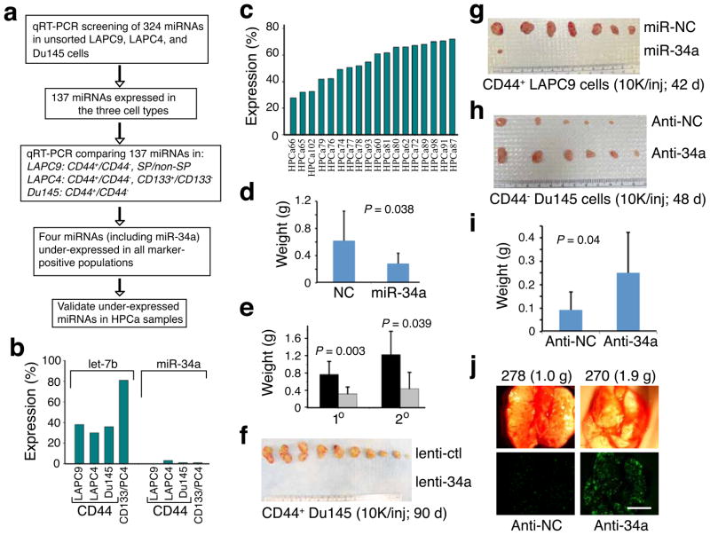
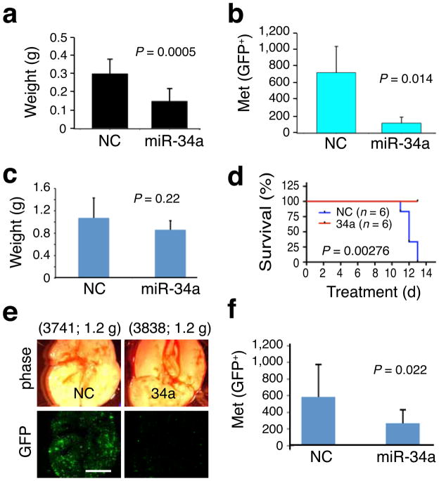
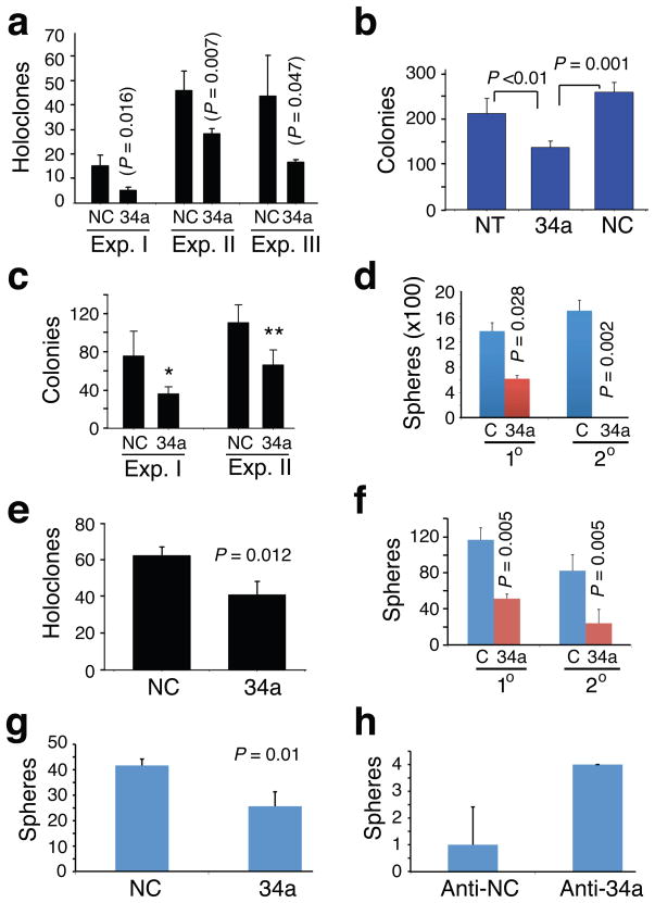
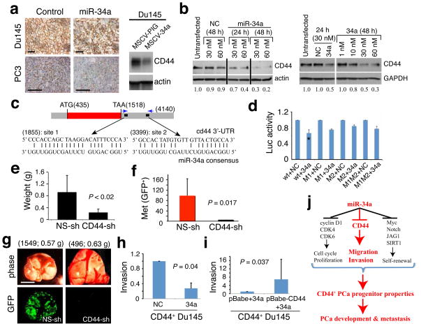
Cancer stem cells (CSCs), or tumor-initiating cells, are involved in tumor progression and metastasis. MicroRNAs (miRNAs) regulate both normal stem cells and CSCs, and dysregulation of miRNAs has been implicated in tumorigenesis. CSCs in many tumors--including cancers of the breast, pancreas, head and neck, colon, small intestine, liver, stomach, bladder and ovary--have been identified using the adhesion molecule CD44, either individually or in combination with other marker(s). Prostate CSCs with enhanced clonogenic and tumor-initiating and metastatic capacities are enriched in the CD44(+) cell population, but whether miRNAs regulate CD44(+) prostate cancer cells and prostate cancer metastasis remains unclear. Here we show, through expression analysis, that miR-34a, a p53 target, was underexpressed in CD44(+) prostate cancer cells purified from xenograft and primary tumors. Enforced expression of miR-34a in bulk or purified CD44(+) prostate cancer cells inhibited clonogenic expansion, tumor regeneration, and metastasis. In contrast, expression of miR-34a antagomirs in CD44(-) prostate cancer cells promoted tumor development and metastasis. Systemically delivered miR-34a inhibited prostate cancer metastasis and extended survival of tumor-bearing mice. We identified and validated CD44 as a direct and functional target of miR-34a and found that CD44 knockdown phenocopied miR-34a overexpression in inhibiting prostate cancer regeneration and metastasis. Our study shows that miR-34a is a key negative regulator of CD44(+) prostate cancer cells and establishes a strong rationale for developing miR-34a as a novel therapeutic agent against prostate CSCs.


Conflict of interest statement

COMPETING INTERESTS STATEMENT

K.K, J.F.W, A.G.B, and D.B are employees of Mirna Therapeutics, Inc., which develops miRNA-based therapeutics. Other authors declare no competing financial interests.

Figures

Figure 1
Figure 1. Underexpression and tumor-inhibitory effects of miR-34a
a. Experimental scheme. b,c. Lower miR-34a levels in CD44+ xenograft (b; PC4, LAPC4) or primary tumor (HPCa; c) cells. Results are expressed as the mean % of marker-positive over marker-negative cells. d,e. miR-34a inhibited LAPC9 (d) and HPCa58 (e; black, lenti-ctl; grey, lenti-34a; n = 10 and n = 7 for 1° and 2° experiments) tumor growth (mean ± S.D). f. CD44+ Du145 cells infected with lenti-ctl or lenti-34a were injected (10,000 cells each) s.c in NOD-SCID mice. Tumor incidence was 10/10 and mean weight was 0.6 g for lenti-ctl group whereas the incidence for lenti-34a group was 0/10. g. Purified CD44+ LAPC9 cells were transfected with miR-NC or miR-34a and s.c injected. Tumor incidence was 7/8 and mean weight was 0.5 g for miR-NC group whereas incidence was 1/8 and tumor weight was 0.03 g for miR-34a group (P = 0.016, incidence). h. Purified CD44 Du145 cells were transfected with anti-NC or anti-34a and s.c injected. Tumor incidence was 5/8 and mean weight was 0.05 g for anti-NC group whereas incidence was 6/8 and tumor weight was 0.2 g for anti-34a group (P = 0.038, weight). i. Bulk LAPC9 cells were transfected with anti-NC or anti-34a oligos and implanted (100,000 cells) in the DP. Mice were terminated at d 46. Tumor incidences were 5/8 and 7/8 for anti-NC and anti-34a groups, respectively. j. Representative microphotographs (animal number and tumor weight indicated on top; scale bar, 100 μm) showing increased lung metastasis by anti-34a (also see Supplementary Fig. 7c–d).
Figure 2
Figure 2. Therapeutic effects of miR-34a
a. Tail vein-injected miR-34a inhibited orthotopic PC3 tumor growth (n = 9 each). bd. Tail vein-injected miR-34a oligos inhibited metastasis (GFP+ foci in the endpoint lungs; mean ± S.D, n = 6/group) of orthotopic LAPC9-GFP tumors (b) without significantly affecting tumor growth (c) and extended animal survival (d; Kaplan-Meier analysis and Log-Rank test). e,f. The fourth set of therapeutic experiment in LAPC9 cells. Shown are representative lung images (e; animal number and tumor weight indicated on top; scale bar, 100 μm) and quantification of lung metastases (f; mean ± S.D, n = 10/group).
Figure 3
Figure 3. miR-34a inhibits clonal and clonogenic properties of PCa cells
a. Holoclone assays in Du145 cells. Cells transfected with miR-NC (NC) or miR-34a (34a) oligos were used in three experiments (Exp. I, 100 cells/well scored on d 9; Exp. II, 100 cells/well scored on d 13; Exp. III, 500 cells/well scored on d 7). b. Clonogenic assays in Du145 cells. Cells (3,000/well) were plated in MG and colonies counted on day 13. NT, non-transfected. c. MG clonogenic assays in LAPC4 cells. Two experiments were performed (Exp. I, 1,250 cells/well scored on d 5, *P = 0.005; Exp. II, 25,000 cells/well scored on d 5, **P = 0.015). d. Sphere assays in LAPC4 cells. LAPC4 cells infected with lenti-ctl (C) or lenti-34a were plated (10,000 cells/well) for both 1° and 2° assays and spheres scored on d 15. e. Holoclone assays in PPC-1 cells. Cells transfected with miR-NC or miR-34a oligos were plated (500 cells/well) in triplicate and holoclones quantified on d 5. f. Sphere assays in HPCa101 (Gleason 9) cells. Purified HPCa101 cells infected with lenti-ctl (C) or lenti-34a were plated (20,000 cells/well) for both 1° and 2° and spheres scored 3 weeks later. g,h. Sphere assays in purified CD44+ HPCa116 (Gleason 7) cells transfected with NC or miR-34a oligos (g) or CD44 HPCa116 cells transfected with anti-NC or anti-34a oligos (h). Spheres were scored on d 15.
Figure 4
Figure 4. CD44 as a direct and functional target of miR-34a
a. Representative CD44 IHC images in Du145 tumors from cells infected with MSCV-PIG (control) or MSCV-34a vectors (Western blot on the right) and PC3 tumors harvested from animals treated with miR-NC or miR-34a oligos. Scale bars, 10 μm. b. miR-34a downregulates CD44 in Du145 (left) and PPC-1 (right) cells. Relative levels of CD44 indicated at the bottom. c. Schematic of two putative miR-34a binding sites in the CD44 3’-UTR. d. Luciferase experiments in Du145 cells (*P <0.01). e. CD44 knockdown inhibits LAPC4 tumor regeneration (see Supplementary Fig. 12). f,g. CD44 knockdown inhibits PC3 cell metastasis evidenced by both quantification (f) and images (g; scale bar, 100 μm). h,i. Invasion assays. miR-34a oligos inhibit Matrigel invasion of CD44+ Du145 cells (h), which was partially overcome by overexpression of a human CD44 cDNA lacking the miR-34a binding sites at the 3’-UTR (i). Invasion was expressed as values relative to the corresponding controls. j. A schematic summary. The part highlighted in red refers to the novel findings made in the present study.

Comment in

  • Stemming a tumor with a little miR.
    Wicha MS. Wicha MS. Nat Med. 2011 Feb;17(2):162-4. doi: 10.1038/nm0211-162. Nat Med. 2011. PMID: 21297608 No abstract available.
  • Microrna: Micromanaging CD44.
    McCarthy N. McCarthy N. Nat Rev Cancer. 2011 Mar;11(3):156. doi: 10.1038/nrc3024. Epub 2011 Feb 10. Nat Rev Cancer. 2011. PMID: 21451552 No abstract available.

References

    1. Visvader JE, Lindeman GJ. Cancer stem cells in solid tumours: accumulating evidence and unresolved questions. Nat Rev Cancer. 2008;8:755–768. - PubMed
    1. Croce CM, Calin GA. miRNAs, cancer, and stem cell division. Cell. 2005;122:6–7. - PubMed
    1. Melton C, Judson RL, Blelloch R. Opposing microRNA families regulate self-renewal in mouse embryonic stem cells. Nature. 2010;463:621–626. - PMC - PubMed
    1. Yu F, et al. let-7 regulates self-renewal and tumorigenicity of breast cancer cells. Cell. 2007;131:1109–1123. - PubMed
    1. Shimono Y, et al. Downregulation of miRNA-200c links breast cancer stem cells with normal stem cells. Cell. 2009;138:592–603. - PMC - PubMed

Publication types

MeSH terms