. 2011 Feb;17(2):216-22.
doi: 10.1038/nm.2290. Epub 2011 Jan 16.

The Ngal reporter mouse detects the response of the kidney to injury in real time

Affiliations

The Ngal reporter mouse detects the response of the kidney to injury in real time

Neal Paragas et al. Nat Med. 2011 Feb.

Abstract

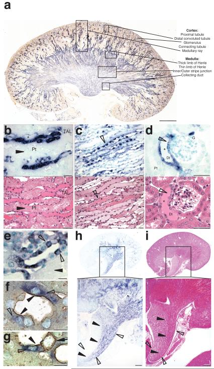
Many proteins have been proposed to act as surrogate markers of organ damage, yet for many candidates the essential biomarker characteristics that link the protein to the injured organ have not yet been described. We generated an Ngal reporter mouse by inserting a double-fusion reporter gene encoding luciferase-2 and mCherry (Luc2-mC) into the Ngal (Lcn2) locus. The Ngal-Luc2-mC reporter accurately recapitulated the endogenous message and illuminated injuries in vivo in real time. In the kidney, Ngal-Luc2-mC imaging showed a sensitive, rapid, dose-dependent, reversible, and organ- and cell-specific relationship with tubular stress, which correlated with the level of urinary Ngal (uNgal). Unexpectedly, specific cells of the distal nephron were the source of uNgal. Cells isolated from Ngal-Luc2-mC mice also revealed both the onset and the resolution of the injury, and the actions of NF-κB inhibitors and antibiotics during infection. Thus, imaging of Ngal-Luc2-mC mice and cells identified injurious and reparative agents that affect kidney damage.


Figures

Figure 1
Figure 1
Ngal-Luc2/mC visualized kidney damage in real time in vivo. (a) Heterozygous Ngal-Luc2/mC female mice were subjected to left kidney ischemia for 15 (top panel) or 30min (bottom panel) and visualized in a Bioimager (30s). Ngal-Luc2 was nearly specific to the left kidney. (b) Ngal-Luc2/mC radiated from the medulla 12h after injury, but not from the contralateral, uninjured kidney. (c) Photon emission (Panel A) was plotted by subtracting background radiance of the contralateral kidney, and normalizing the data for luminescence at 0h. The average radiance (ps−1cm−2sr−1) within a constant region of interest (ROI) was converted to fold change and displayed on the Y-axis. (d) Immunoblot detection of uNgal from the ischemic mouse (15min) shown in Panel A. Recombinant mouse non-glycosylated Ngal was used as a standard. (e) Heterozygous Ngal-Luc2/mC albino female mice were subjected to bilateral ischemia for 15min and NGAL-Luc2 was examined 12h later. (f) Ngal-Luc2 activity rose significantly by 6h after 15min of ischemia (compared to time=0: t=3h, P=0.085; t=6h, P=0.0009; t=12h, P=0.008; t=24h, P=0.04; t=48h, P=0.12; n=4), while a significant elevation of sCr lagged until 12h (compared to time=0: t=3h, P=0.35; t=6h, P=0.18; t=12h, P=0.028; t=24h, P=0.055; t=48h, P=0.15; n=10). uCr fell within a narrow interval (baseline vs: 0-3h, 0.22 fold, P=0.027; 3-6h, 0.41 fold, P=0.15), then returned to steady state (baseline vs: 6-12h, 1.08 fold, P=0.31; 12-24h, 1.27 fold, P=0.19; 24-48h, a 1.22 fold change p=0.15). Median±s.e. (g) uNgal protein from the mouse in Panel E paralleled the expression of kidney Ngal-Luc2.
Figure 2
Figure 2
Ngal-Luc2/mC reported kidney cellular damage in vivo, which was induced by cisplatin and lipid A. (a) Ngal-Luc2/mC expression in both kidneys of a mouse 168h after cisplatin (20 mg/kg) exposure. (b) Treated kidneys revealed prominent Ngal-Luc2/mC expression in the medulla and abundant uNgal. (c) Ngal-Luc2/mC kidney cells responded to cisplatin (10 μM). (d) Ngal-Luc2/mC fluorescence was elicited by lipid A in a dose-dependent manner in the kidney, liver, spleen, lung and trachea. Low level expression of Ngal-Luc2/mC is also seen in the skin of the feet, consistent with expression of TLR4. (e) 5, 15 and 30 mg/Kg of lipid A led to a 17, 21 and 34 fold increase in total Ngal-reporter expression after 24h (n=3). sCr is shown for each dose. (f) In situ hybridization demonstrated Ngal mRNA in TAL and CD in the outer stripe of the inner medulla. (g) H&E staining revealed cast formation 24h after a 5 mg/kg lipid A challenge. High magnification images of the delineated regions (f and g) showed that Ngal RNA was localized to presumptive intercalated cells (open arrowhead) of CDs that contained casts and cellular debris (asterisks).
Figure 3
Figure 3
Damaged nephron is the source of kidney Ngal. (a) In situ hybridization shows Ngal mRNA expression in TAL and CD, weaker expression in distal convoluted tubules and absent expression in proximal tubules and thin limbs of Henle. (b) Ngal mRNA was expressed in the outer stripe of the outer medulla and cortical TAL of Henle (in medullary rays) containing casts (asterisks), in CD (open arrowheads) containing casts (asterisks, c), and the macula densa (open arrowhead) which had undergone epithelial flattening and cast formation (asterisks, d), but not in the necrotic pars recta of proximal tubules (closed arrowhead, b and e). Top panels show in situ hybridization and the bottom panels show H&E staining in paraffin sections. (e) High magnification image revealed that Ngal mRNA was specifically expressed by intercalated cells (open arrowheads). (f) Anti v-ATPase IHC identified Ngal expressing (open arrowhead) CD cells as α-type intercalated cells (α-IC) in which the v-ATPase locates to the apical surface (closed arrowhead). (g) Outer medullary CD expressed Ngal mRNA (open arrowhead) in α-IC cells, while adjacent β-type intercalated cells (closed arrow) failed to express NGAL. (h, i) A polar (segmental) renal artery was subjected to 30min ischemia, and after 24h, Ngal mRNA expression was located by in situ hybridization. Ngal mRNA (h) was expressed in the ischemic zone (i) where histological damage was evident (closed arrowheads). Closed arrowheads indicate the boundary of damaged and non-damaged tubules in the papilla, and open arrowheads depict the width of the papilla.
Figure 4
Figure 4
Volume depletion failed to activate Ngal-reporter expression. (a, b) Volume depletion induced mild hypernatremia (serum sodium increased on average 8 mmol/L±2.5 s.e. (n=3, * P< 0.05; ** P< 0.02), b) mild azotemia with a rise in sCr to 0.3±0.2 s.e. (n=3), and approximately a 21.7±4% weight loss. (c) NGAL-Luc2 was not upregulated in any organ (the kidney is circled) following simple volume depletion. Male mice tonically express Ngal in testis (circled), which serves as an internal positive control.
Figure 5
Figure 5
(a) Uropathogenic E. coli (CFT073; 104 CFU/ml)) and lipid A stimulate Ngal-Luc2 expression in primary kidney cells (105 cells/well) isolated from reporter mice. When these cells were pre-treated (1h) or post-treated (1 and 6h) with gentamicin (100 μg/ml), there was a significant reduction in Ngal-Luc2 expression compared to the uninhibited growth (n=3, two independent experiments), pretreatment being more effective. (b) Ngal-Luc2 activity was induced by lipid A (4 μg/ml), but pretreatment with either a known NF-κB inhibitor, MG132 (0.5-5 μM), or novel NF-κB inhibitors (5 μM) reduced Ngal-Luc2 activity by 15-80%. (*P<0.05, **P<0.01, ***P<0.001; Student’s t-Test two-tailed unequal variance; n=3 independent experiments). Bars represent fold change from DMSO control ± s.e. (c) Primary cells isolated from the inner medulla and papilla of Ngal-Luc2/mC kidneys (n=6) demonstrated enrichment of distal tubular markers Aqp2 and Umod and de-enrichment of Aqp1, a proximal tubular marker. Bars represent the ratio of gene expression (medullary vs cortical) ± s.e. (d) Ngal-Luc2 expression was markedly increased by lipid A (4 μg/ml, 24h) in inner medullary and papillary primary cells (Luc2 increased by 17.2×10³±6.14×10² photon/mg protein), but not in the cortical primary cells (increase of 2.11×10³ ±0.61×10³ photon/mg protein). Bars represent total photon/mg protein and inset is the net change in photon/mg protein from the control ± s.e.

References

    1. Bosch JP, et al. Renal functional reserve in humans. Effect of protein intake on glomerular filtration rate. The American journal of medicine. 1983;75:943–950. - PubMed
    1. Lassnigg A, et al. Minimal changes of serum creatinine predict prognosis in patients after cardiothoracic surgery: a prospective cohort study. Journal of the American Society of Nephrology : JASN. 2004;15:1597–1605. - PubMed
    1. Christensen EI, Verroust PJ. Interstitial fibrosis: tubular hypothesis versus glomerular hypothesis. Kidney international. 2008;74:1233–1236. - PubMed
    1. Supavekin S, et al. Differential gene expression following early renal ischemia/reperfusion. Kidney international. 2003;63:1714–1724. - PubMed
    1. Mori K, et al. Endocytic delivery of lipocalin-siderophore-iron complex rescues the kidney from ischemia-reperfusion injury. J Clin Invest. 2005;115:610–621. - PMC - PubMed

Publication types

MeSH terms