. 2011 Feb;29(2):158-63.
doi: 10.1038/nbt.1742. Epub 2011 Jan 16.

Efficient mucosal vaccination mediated by the neonatal Fc receptor

Affiliations

Efficient mucosal vaccination mediated by the neonatal Fc receptor

Lilin Ye et al. Nat Biotechnol. 2011 Feb.

Abstract

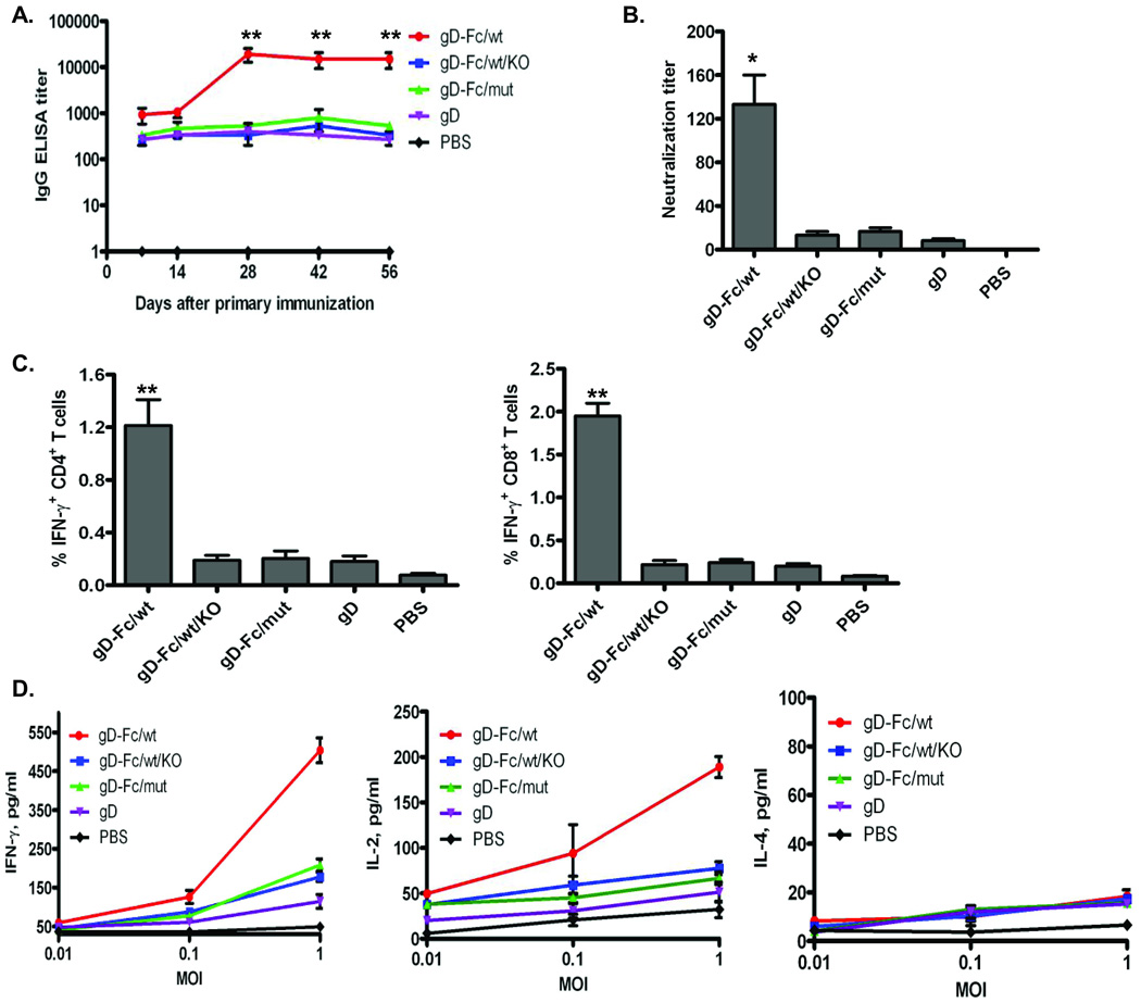
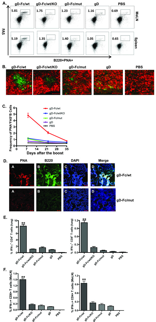
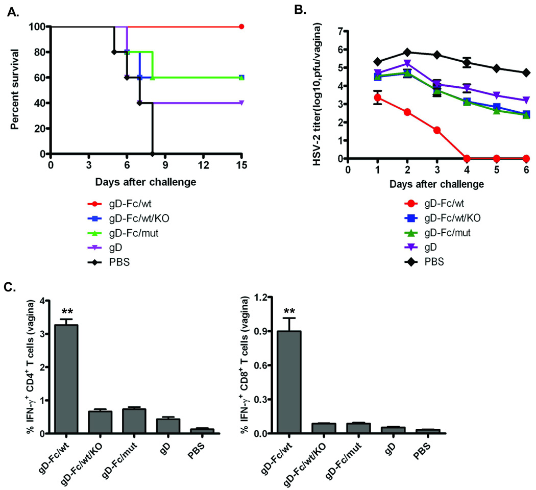
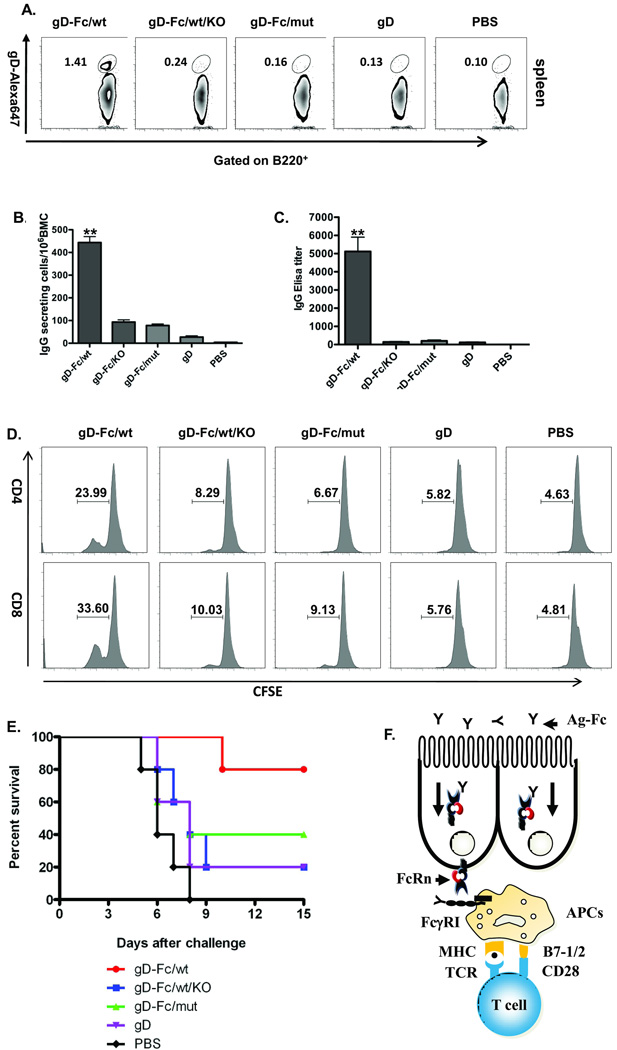
Almost all infectious diseases are initiated at mucosal surfaces, yet intramuscular or subcutaneous vaccination usually provides only minimal protection at sites of infection owing to suboptimal activation of the mucosal immune system. The neonatal Fc receptor (FcRn) mediates the transport of IgG across polarized epithelial cells lining mucosal surfaces. We mimicked this process by fusing a model antigen, herpes simplex virus type-2 (HSV-2) glycoprotein gD, to an IgG Fc fragment. Intranasal immunization, together with the adjuvant CpG, completely protected wild-type, but not FcRn knockout, mice after intravaginal challenge with virulent HSV-2 186. This immunization strategy induced efficient mucosal and systemic antibody, B- and T-cell immune responses, with stable protection for at least 6 months after vaccination in most of the immunized animals. The FcRn-IgG transcellular transport pathway may provide a general delivery route for subunit vaccines against many mucosal pathogens.


Figures

Fig. 1
Fig. 1. FcRn-targeted mucosal vaccination induces enhanced gD–specific antibody and T cell responses
The 20 µg gD-Fc/wt, gD-Fc/mut, gD, or PBS in combination with 20 µg CpG were i.n. administered into wild-type (WT) or FcRn knockout (KO) mice. (A). Measurement of anti-HSV-2 gD-specific IgG antibody titers in serum before and after the boost immunization. HSV-2 gD-specific IgG antibody at indicated days was measured in serum by ELISA. Immunization conditions are displayed at the right. The curves represent mean values for each group (±S.E.M.). Values marked with asterisk in this and subsequent figures: *P < 0.05; **P < 0.01. (B). Test of neutralizing activity in the immunized sera. Sera were heat-inactivated, diluted 10-fold, then in twofold steps in MEM with 2% FBS. HSV-2 (50 PFU) was added and incubated at 37°C for 1 hr. Finally, the mixture were removed and washed, overlaid with 0.8% methylcellulose in 2% FBS containing DMEM and further incubated for 72 hr at 37°C. The titers were expressed as the reciprocal of the twofold serial dilution preventing the appearance of the cytopathic effects (CPE) over control sera. Each assay was done in triplicate. (C). The percentage of IFN-γ producing T cells in the spleen 4 days after the boost. Spleen cells from the immunized mice were stimulated for 10 hr with purified gD or medium control. Lymphocytes were gated by forward and side scatter and T cells labeled with anti-CD3 and identified by their respective surface markers CD4 and CD8 and intracellular IFN-γ staining. Immunization conditions are displayed on the bottom. Numbers represent the percentage of IFN-γ+ CD3+ CD4+ (left panel) or IFN-γ+ CD3+ CD8+ (right panel) T cells. Isotype controls included FITC-mouse-IgG1 with baseline response. (D) Cytokine secretions from the stimulated spleen T cells. Spleen cells were collected on day 4 after the boost. Cells were stimulated in vitro specifically with different multiplicity of infection (MOI) of inactivated HSV-2 virus as indicated for 24 hr. Cytokines IFN-γ, IL-2, and IL-4 in the culture supernatant were detected by ELISA. Data are representative of three experiments with three immunized mice pooled in each group.
Fig. 2
Fig. 2. Local immune responses induced by FcRn-targeted mucosal immunization
(A). Accumulation of activated B cells in germinal centers (GCs) in the mediastinal lymph nodes (MeLNs) and spleen. Representative flow cytometric analyses of GC B cells among CD19+B220+ B cells in the MeLNs and spleen 10 days after the boost. B220+PNAhigh cells are B cells that exhibit the phenotypic attributes of GC B cells. The GC staining in spleen was used as a positive control. Numbers are the percentage of activated GC B cells (PNA+FAS+) among gated B cells and are representative of three independent experiments. (B). GC formation and presence of activated B cells following immunizations as indicated. Frozen MeLN sections at day 10 from the immunized mice were co-stained with biotin–PNA (developed with avidin–FITC) and Alexa647 labeled anti-IgD. Scale bar represents 50 µm. (C). Quantitative analysis of GCs following immunization. The dynamics of the frequency of germinal center B cells (FAS+PNA+, gated on CD19+B220+ cells) were plotted on day 10, 22 and 35 after the boost. Data indicate the mean and S.E.M., n=5 mice. (D). The formation of inducible bronchus-associated lymphoid tissue (iBALT). Frozen serial sections of the lung were stained with biotin-PNA (GC, red) and anti-B220 (B cells, green), followed by Alexa 488-conjugated IgG of corresponding species and Alexa 555-Avidin. The nucleus is stained with DAPI (blue). A germinal center-like structure is shown in the merged panel by the white color. The data are representative of sections from at least three independent mice. Images were originally obtained at 10× magnification. Scale bars represent 100 µm. (E) + (F). Presence of HSV-2 gD-specific T lymphocytes in the lung (E) and MeLNs (F). Lung or MeLN cells from mice 4 days after the boost were collected. Lymphocytes were gated based on their forward scatter (FSC) vs. side scatter (SSC) profile. Intracellular staining for IFN-γ was performed after surface staining of CD4 and CD8 molecules. The profiles shown are representative of five mice from three separate experiments. Numbers indicate percentages of IFN-γ-producing T lymphocytes from gated CD4+ and CD8+ T cells.
Fig. 3
Fig. 3. FcRn-targeted mucosal immunization provides protective immunity to intravaginal (ivag) challenge with virulent HSV-2 186
(A) Mean survival following genital HSV-2 challenge. Four weeks after the immunization, groups of five mice were challenged intravaginally with 1×10⁴ pfu of HSV-2 strain 186. Percentage of mice from protection on the indicated days is calculated as the number of mice surviving divided by the number of mice in each group and represented two similar experiments. (B) Mean of viral titers following HSV-2 challenge. Virus titers were measured from vaginal washes by taking swabs on the indicated days after HSV-2 inoculation based on a plaque assay on Vero cell monolayers. (C). Increased presence of HSV-specific T lymphocytes in the vaginal epithelium after challenge. Lymphocytes were harvested from collagenase-digested vaginal tissues 4 days intravaginal inoculation of virus. Intracellular staining for IFN-γ expression on CD4+ and CD8+ T cells was analyzed after gating on viable CD3+ lymphocytes. The numbers in each column show the percentage of IFN-γ-positive T lymphocytes from the gated CD4+ or CD8+ T cells. Data shown are of a representative from three experiments using 3 mice per experiment.
Fig. 4
Fig. 4. Increased memory immune response in FcRn-targeted mucosal immunization
(A). Induction of gD specific memory B cells in the spleen. The frequency of gD-specific memory B cells was assessed 6 months after the boost. Memory B cells, defined as B220+ gD-surface+, were analyzed 6 months after the boost by FACS. Purified gD proteins were labeled with Alexa Fluro647. Spleen cells (2 × 10⁶) were incubated with the 1 µg Alexa Fluro647-labeled gD proteins and B220 antibody. Numbers in the quadrants are the percentage of gD-specific memory B lymphocytes. (B). Long-lived HSV gD-specific antibody-secreting cells in the bone marrow. Bone marrow cells removed 6 months after the boost were placed on gD-coated plates and quantified by ELISPOT analysis of IgG-secreting plasma cells. Data were pooled from two separate experiments with five mice in each experiment. The graphs were plotted based on the average ELISPOT for replicate wells. Values marked with asterisk are significantly greater (P<0.01) from the gD-Fc/wt protein-immunized mice than those of other groups as indicated. (C). Durability of HSV-2 gD-specific serum IgG response. In two separate experiments, HSV-2 gD-specific IgG was quantified by ELISA in serum by endpoint titer from five mice at 6 months after the boost. HSV-specific IgG antibody was not detected in PBS-immunized mice. (D). Long-lived gD specific T cell memory to FcRn-targeted mucosal vaccination. Spleen cells were isolated from the immunized mice six months after the boost, stained with CFSE, and stimulated in vitro with 20 µg/ml of purified gD for 4 days. Data are expressed in CFSE histograms of fluorescence intensity versus the number of fluorescing cells, indicating the percentage of the cell population positive for CD4 and CD8 antigen. Numbers in the quadrants are the percentage of CD4+ and CD8+ proliferating T cells. Representative flow cytometry profiles of two similar experiments with three mice per group are shown. Immunization conditions are displayed on the top. (E). Mean survival following genital HSV-2 challenge six months following the boost. The immunized mice were challenged intravaginally with 1×10⁴ pfu of HSV-2. Percentage of mice protected on the indicated days is calculated as the number of mice surviving divided by the number of mice in each group (n=5). (F). Proposed model of FcRn-mediated mucosal vaccine delivery. The Fc-fused antigens are transported by FcRn and targeted to the mucosal antigen presenting cells (APCs), such as dendritic cells. Antigen is taken up by pinocytosis or FcγRI-mediated endocytosis in APCs, then processed and presented to T cells.

Comment in

  • A mucosal gateway for vaccines.
    McGhee JR. McGhee JR. Nat Biotechnol. 2011 Feb;29(2):136-8. doi: 10.1038/nbt.1766. Nat Biotechnol. 2011. PMID: 21301439 No abstract available.

References

    1. Neutra MR, Kozlowski PA. Mucosal vaccines: the promise and the challenge. Nat. Rev. Immunol. 2006;6:148–158. - PubMed
    1. Holmgren J, Czerkinsky C. Mucosal immunity and vaccines. Nat. Med. 2005;11(4 Suppl):S45–S53. - PubMed
    1. McGhee JR, et al. The mucosal immune system: from fundamental concepts to vaccine development. Vaccine. 1992;10:75–88. - PubMed
    1. Gallichan WS, Rosenthal KL. Long-term immunity and protection against herpes simplex virus type 2 in the murine female genital tract after mucosal but not systemic immunization. J. Infect. Dis. 1988;177:1155–1161. - PubMed
    1. Neutra MR, Mantis NJ, Kraehenbuhl J-P. Collaboration of epithelial cells with organized mucosal lymphoid tissues. Nat. Immunol. 2001;2:1004–1009. - PubMed

Publication types

MeSH terms