. 2011 Feb;18(2):185-90.
doi: 10.1038/nsmb.1981. Epub 2011 Jan 16.

A processed noncoding RNA regulates an altruistic bacterial antiviral system

Affiliations

A processed noncoding RNA regulates an altruistic bacterial antiviral system

Tim R Blower et al. Nat Struct Mol Biol. 2011 Feb.

Abstract

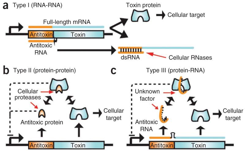
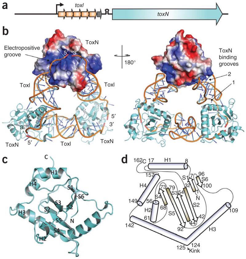
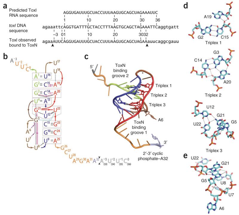
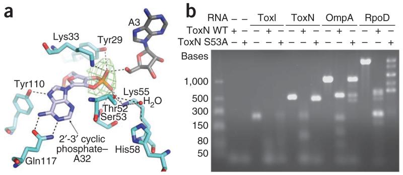
The ≥ 10³⁰ bacteriophages on Earth relentlessly drive adaptive coevolution, forcing the generation of protective mechanisms in their bacterial hosts. One such bacterial phage-resistance system, ToxIN, consists of a protein toxin (ToxN) that is inhibited in vivo by a specific RNA antitoxin (ToxI); however, the mechanisms for this toxicity and inhibition have not been defined. Here we present the crystal structure of the ToxN-ToxI complex from Pectobacterium atrosepticum, determined to 2.75-Å resolution. ToxI is a 36-nucleotide noncoding RNA pseudoknot, and three ToxI monomers bind to three ToxN monomers to generate a trimeric ToxN-ToxI complex. Assembly of this complex is mediated entirely through extensive RNA-protein interactions. Furthermore, a 2'-3' cyclic phosphate at the 3' end of ToxI, and catalytic residues, identify ToxN as an endoRNase that processes ToxI from a repetitive precursor but is regulated by its own catalytic product.


Figures

Figure 1
Figure 1
Overview of TA systems. In general, the antitoxic gene (orange) precedes the toxin gene (cyan), as part of a bicistronic operon. (a) In type I TA systems, the short antisense antitoxic RNA forms a duplex with a short region of the full-length mRNA. This duplex prevents translation of the toxin gene and promotes degradation by a cellular RNase such as RNase III. (b) In type II TA systems, the protein antitoxin forms a complex with the protein toxin. Either as a complex or by itself the antitoxic protein often negatively regulates transcription of the operon. Cellular proteases such as Lon and Clp degrade the antitoxin, releasing the toxin. (c) In our proposed type III TA system, a proteinaceous toxin interacts with an antitoxic RNA. A transcriptional terminator (black stem-loop) between the antitoxin and toxin genes regulates relative levels of ToxI and ToxN transcript. As a complex, the toxin and antitoxin negatively regulate transcription. Cellular factor(s) that have yet to be identified trigger toxin activity by degrading the antitoxic RNA, decreasing the level of transcription from the locus or releasing the toxin from the RNA antitoxin. dsRNA, double-stranded RNA.
Figure 2
Figure 2
ToxIN complex structure. (a) Schematic of the toxIN locus, which is transcribed from a single promoter (black arrow). The toxN gene is downstream of a transcriptional terminator (black stem-loop) and toxI, which encodes 5.5 36-nt repeats. A whole repeat is shown as an orange arrow and the half repeat as a gray arrow. (b) An overview of the ToxIN complex structure. Two ToxN protomers are shown as cartoons (colored cyan) and the third as a surface representation, with the positively charged surface in blue and the negatively charged surface in red. The phosphate sugar backbones of the ToxI molecules are shown as orange cartoons and the bases as single sticks that are colored in a gradient from cyan to blue. Right, 180° rotation about the vertical axis. (c) Cartoon representation of monomeric ToxN, with α-helices labeled H1 to H4 and β-strands labeled S1 to S6. (d) Schematic representation of ToxN topology, with α-helices shown as cylinders and β-strands as arrows. Numbers indicate the amino acid residues at the limits of the secondary structures. ToxN monomers in c and d are orientated as in the surface representation ToxN in a (left).
Figure 3
Figure 3
ToxI pseudoknot structure. (a) Sections of the toxI DNA and the predicted corresponding RNA repeat are shown with the ToxI RNA repeat that is seen in the crystal structure. Capitals indicate the 36-nt repeats. Arrows indicate the cleavage sites of a single active ToxI RNA from the longer ToxI transcript. (b) Overview of hydrogen bonding in the ToxI pseudoknot. Nucleotides −3 to 32 correspond to one 36-nt RNA oligomer in the crystal structure. Nucleotides 1 to 36 correspond to a single consensus toxI 36-nt repeat. Black arrows between nucleotides 32 and 33 indicate the putative ToxN cutting site in ToxI. The black-outlined, open letters for nucleotides 33–36 represent the 5′-most 4 nt of a second 36-nt ToxI oligomer from the crystal structure. Three interacting sections are shown in green (nucleotides 1–4), blue (nucleotides 9–16) and red (nucleotides 19–25), separated by brown loops (nucleotides 5–8 and 17–18). Duplex and triplex base pairs are highlighted by gray boxes. The single-stranded RNA tail nucleotides are shown in orange, except the termini, with A3 in gray and A32 in violet. Base-base hydrogen bonds are shown as black lines. Ribose 2′OH-base hydrogen bonds are shown as magenta lines, ribose 2′OH-phosphate hydrogen bonds as violet lines and a phosphate-base hydrogen bond as a light blue line; arrows point from the first partner. Black dashed lines depict selected stacking interactions. (c) ToxI pseudoknot structure, showing the locations of base triplexes. The main chain backbone and bases are colored as in b. (d) Detail of each of the three ToxI base triplexes, drawn as cyan sticks with oxygen in red, nitrogen in blue and phosphorus in orange. Hydrogen bonding interactions within 2.6 Å to 3.3 Å are indicated by dashed lines. (e) The network of hydrogen bonds extending from ToxI G5 supports the formation of a loop structure and facilitates the presentation of A6 for interaction with ToxN. Hydrogen bonding interactions within 2.5 Å to 3.5 Å are indicated by black dashed lines. The color scheme is the same as in d.
Figure 4
Figure 4
ToxN has an endoRNase active site. (a) ToxN active site interactions with ToxI. A simulated annealing omit map, at 3.75σ level, was calculated using our model, omitting the proposed 2′-3′ cyclic phosphate group, and is shown in green. Hydrogen bonding interactions within 2.6 Å to 3.5 Å are indicated by black dashed lines, and a water molecule is shown as a red sphere. Coloring is as in Figure 3. (b) In vitro ToxN RNase assay. The ability of the purified wild-type (WT) ToxN protein and the ToxN S53A mutant to digest in vitro–transcribed RNAs was assessed. RNA substrates were selected to allow investigation of autoregulation (full-length ToxI RNA (5.5 36-nt repeats) and ToxN RNA) and to examine the effect on transcripts of highly active genes (OmpA and RpoD RNA). Protein was mixed with RNA at a 4:1 molar ratio and incubated. The products were analyzed by agarose gel electrophoresis.
Figure 5
Figure 5
Identification of ToxN–ToxI residues that are vital for toxicity and interaction. (a) The N-terminal section of ToxN H3 forms an extensive hydrogen bonding network with ToxI groove 1 (Fig. 3c). (b) View of groove 1 after a rotation of roughly 90° about the vertical axis relative to a. ToxI A6 is held in a hydrophobic pocket on the ToxN surface. Coloring in a and b is the same as in Figure 3. (c) In vivo Abi activity of ToxN mutants. The efficiencies of plating (EOP) of phages ΦM1 and ΦS61 were assessed upon strains of P. atrosepticum 1043 carrying either wild-type (WT) toxIN or mutant toxIN plasmids. (d) In vivo toxicity of ToxN mutants and antitoxic activity of ToxI mutants to interact. Strains of E. coli DH5α carrying two independently inducible ToxN and ToxI plasmids were tested for their viability in the presence of WT ToxN or a ToxN mutant protein either with or without WT ToxI or a ToxI mutant RNA. In c and d, error bars indicate s.d.
Figure 6
Figure 6
Structural comparisons in the ToxN family. (a) Cartoon representation of ToxN in orange. (b) Cartoon representation of Kid (PDB 1M1F) in green. (c) Structural overlay of ToxN in orange and Kid in green. Structures in ac are shown in the same orientation.

References

    1. Wommack KE, Colwell RR. Virioplankton: viruses in aquatic ecosystems. Microbiol. Mol. Biol. Rev. 2000;64:69–114. - PMC - PubMed
    1. Lima-Mendez G, Toussaint A, Leplae R. Analysis of the phage sequence space: the benefit of structured information. Virology. 2007;365:241–249. - PubMed
    1. Labrie SJ, Samson JE, Moineau S. Bacteriophage resistance mechanisms. Nat. Rev. Microbiol. 2010;8:317–327. - PubMed
    1. Chopin MC, Chopin A, Bidnenko E. Phage abortive infection in lactococci: variations on a theme. Curr. Opin. Microbiol. 2005;8:473–479. - PubMed
    1. Shub DA. Bacterial viruses. Bacterial altruism? Curr. Biol. 1994;4:555–556. - PubMed

Publication types

MeSH terms

LinkOut - more resources