. 2011 Feb;18(2):198-204.
doi: 10.1038/nsmb.1980. Epub 2011 Jan 16.

Structural basis of substrate discrimination and integrin binding by autotaxin

Affiliations

Structural basis of substrate discrimination and integrin binding by autotaxin

Jens Hausmann et al. Nat Struct Mol Biol. 2011 Feb.

Abstract

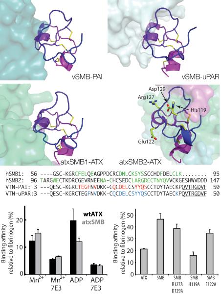
Autotaxin (ATX, also known as ectonucleotide pyrophosphatase/phosphodiesterase-2, ENPP2) is a secreted lysophospholipase D that generates the lipid mediator lysophosphatidic acid (LPA), a mitogen and chemoattractant for many cell types. ATX-LPA signaling is involved in various pathologies including tumor progression and inflammation. However, the molecular basis of substrate recognition and catalysis by ATX and the mechanism by which it interacts with target cells are unclear. Here, we present the crystal structure of ATX, alone and in complex with a small-molecule inhibitor. We have identified a hydrophobic lipid-binding pocket and mapped key residues for catalysis and selection between nucleotide and phospholipid substrates. We have shown that ATX interacts with cell-surface integrins through its N-terminal somatomedin B-like domains, using an atypical mechanism. Our results define determinants of substrate discrimination by the ENPP family, suggest how ATX promotes localized LPA signaling and suggest new approaches for targeting ATX with small-molecule therapeutic agents.


Figures

Figure 1
Figure 1
The structure of ATX. (a) A schematic view of the domain structure of ATX; (b) Cartoon representation with the SMB domains coloured in pink and magenta, the PDE domain in green, the NUC domain in blue, the lasso loop wrapping around the NUC domain in orange (the lasso loop interacts with the rest of the proteins through 18 hydrogen bonds, 8 salt bridges, and a disulphide bond between residues 566 and 666) and the short loop connecting the SMB domains to the PDE domain in cyan; the phosphate and the zinc ions bound to the active site as well as cystine bridges are shown in stick-and-sphere representation, with the Sγ atoms in yellow, phosphorous in orange, zinc in pink; (c) The same cartoon as above in a different orientation, showing the active site with the bound phosphate and zinc ions and the lasso loop wrapping around the NUC domain to the left; the colored boxes relate to panels (d,e,f) showing the interface between the PDE and NUC domains; (d) The essential Cys-bridge that connects the PDE and NUC domains. (e) The essential glycan chain; (f) the C-terminal helix highlighted in lemon-green, with the important Lys-852 and its two salt-bridge partners, Glu-830 and Asp-836. All structural figures were created with Pymol (http://www.pymol.org)
Figure 2
Figure 2
ATX domains and closest structural homologues. (a) The two SMB domains of ATX/ENPP2, the ENPP SMB1 domain (PDB:2YS0, unpublished), and two structures of the vitronectin SMB domain (PDB:1OC0, 3BT138) are shown side by side, colored blue to magenta from N to C terminus with cystine bridges shown as sticks, with the Sγ atom atoms in yellow. The ATX SMB domains share 38% sequence identity with the SMB domain of vitronectin, and 56% with that of ENPP1, displaying in both cases an rmsd of about 1.3 Å over 40 superposed Cαs; (b) The ATX and XaNPP phosphodiesterase (PDB:2GSN24) domains shown in matched orientations; the short loop (residues 274–280) that is more extended in XaNPP (residues 154–178) is shown thicker and in ruby and the bound zinc ions are shown as grey spheres; the sequence identity between the two is 32%, with 1.5 Å rmsd over 335 superposed Cαs; (c) Side by side comparison of the ATX nuclease domain with the A.sp nuclease (PDB:1ZM830); the sequence identity is 19%, with 2.1 Å rmsd over 335 superposed Cαs. The EF-hand-like calcium (brown sphere) binding motif is at the bottom left and is absent in the bacterial nuclease; the A.sp ion binding sites are indicated with magenta spheres and are absent in the ATX domain.
Figure 3
Figure 3
The ATX catalytic site and substrate binding pocket. (a) The active site of ATX and (b) of XaNPP (PDB: 2GSO24). Side chains atoms are shown as sticks, with the Ca atoms as spheres; all oxygen and nitrogen atoms are in red and blue respectively; Zn+2 ions are grey spheres; phosphate atoms are coloured orange; and vanadium atoms magenta; (c) A surface representation of the ATX binding site with the HA155 inhibitor buried in the binding pocket and shown as sticks with carbon atoms colored grey and the covalently bound boron pink; the ATX surface is green with the short loop that is more extended in XaNPP and ENPP1 (residues 274–280) colored ruby; (d) The surface of XaNPP coloured in aquamarine, superposed on the ATX complex with HA155; the extended XaNPP- and ENPP1-specific loop (residues 154–178) in ruby, highlighting the absence of the hydrophobic pocket in XaNPP; (e) The ATX substrate binding site shown as in (c) but with AMP superposed from the XaNPP complex structure; (f) The XaNPP surface with bound AMP (PDB: 2GSU24) shown as sticks; (g) The ATX surface colored by electrostatic potential (−70 to- 70 kT) to highlight the hydrophobic nature of the binding pocket, is shown together with a stick model of a docked LPC molecule; (h) A view similar to (g) colored as in (c),(e) and with the second SMB domain in magenta, highlighting the close interaction of the SMB domain with the lipid binding groove. The narrow tunnel is visible as a white hole in the middle.
Figure 4
Figure 4
Biochemical analysis of human ATX and mutants. (a) A simplified drawing of the ATX binding pocket. The residues used for mutagenesis studies are indicated; (b) Modulation of LPC and pNP-TMP hydrolysis activity by ATX mutants, indicating the importance of specific residues for selectivity in LPC and nucleotide hydrolysis; (c) The A218V mutants prefers shorter chain lengths; the activity for WT hATX and A218V mutant was normalized to 100% for LPC(14:0); (d) LPA(18:1) inhibition of nucleotide hydrolysis for wild-type ATX and ΔSMB-ATX. All error bars represent the s.e.m.
Figure 5
Figure 5
The SMB domains and integrin-mediated interaction with activated platelets. (a) The structure of the vitronectin SMB domain (vSMB) with plasminogen activator inhibitor 1 (PAI-1, PDB:1OC037). The SMB domain is shown as a cartoon, colored from purple through blue from the N- to the C-terminus, and the Cys-bridges are shown as yellow sticks. PAI-1 is shown as a surface colored aquamarine. The vSMB interaction surface with PAI-1 is 656 Å2, making five hydrogen bonds and five salt bridges; (b) vSMB in complex with the urokinase-type plasminogen activator receptor (uPAR complex, PDB:3BT138) in light cyan. The vSMB interaction surface with uPAR is 620 Å2, making seven hydrogen bonds and five salt bridges; (c) The first SMB domain of ATX (atxSMB1) highlighting the interaction with the rest of ATX, shown as a surface colored as in Fig.1. The atxSMB1 interaction surface with the PDE is 620 Å2, making nine hydrogen bonds and a salt bridge; (d) The second SMB domain (atxSMB2) with the rest of ATX. The atxSMB2 interaction surface with the PDE is 571 Å2, but forming only three hydrogen bonds; (e) An alignment of vitronectin and ATX SMB sequences; residues involved in interaction with binding partners are in color; the RGD motifs are underlined; (f) Binding of wild-type ATX and atxSMB to activated platelets is similar in magnitude, depends on Mn+2 and ADP, and can be lowered by interference with an integrin β3 binding antibody. (g) Binding of the atxSMB domains to platelets is significantly attenuated by mutation of His-119 to alanine and less affected by mutation of the RDB motif. All error bars represent the s.e.m.

Comment in

References

    1. Stracke ML, et al. Identification, purification, and partial sequence analysis of autotaxin, a novel motility-stimulating protein. J Biol Chem. 1992;267:2524–2529. - PubMed
    1. Tokumura A, et al. Identification of human plasma lysophospholipase D, a lysophosphatidic acid-producing enzyme, as autotaxin, a multifunctional phosphodiesterase. J Biol Chem. 2002;277:39436–39442. - PubMed
    1. Umezu-Goto M, et al. Autotaxin has lysophospholipase D activity leading to tumor cell growth and motility by lysophosphatidic acid production. J Cell Biol. 2002;158:227–233. - PMC - PubMed
    1. Noguchi K, Herr D, Mutoh T, Chun J. Lysophosphatidic acid (LPA) and its receptors. Curr Opin Pharmacol. 2009;9:15–23. - PubMed
    1. van Meeteren LA, Moolenaar WH. Regulation and biological activities of the autotaxin-LPA axis. Prog Lipid Res. 2007;46:145–160. - PubMed

Publication types

MeSH terms