

doi: 10.1080/08998280.2009.11928558.
Chest wall chondrosarcoma
Affiliations
- PMID: 21240303
- PMCID: PMC2760173
- DOI: 10.1080/08998280.2009.11928558
Item in Clipboard
Chest wall chondrosarcoma
Proc (Bayl Univ Med Cent).
2009 Oct.
No abstract available
Figures
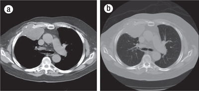
CT images through the chest at the level of the carina in both (a) soft tissue and (b) bone windows demonstrate a lytic mass involving the right anterior third rib, centered at the costochondral junction. A large, lobulated soft tissue component elevates the pectoralis musculature. A few scattered internal calcifications are suggestive of chondroid matrix.
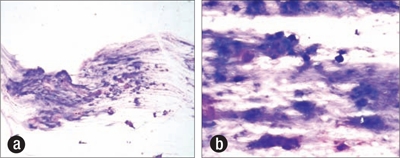
Scrape preparation at time of ultrasound-guided biopsy demonstrating a myxoid chondroid matrix, increased cellularity, and chondrocytes. Diff-Quik stain, (a) ×100, (b) ×400.
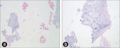
Paraffin cell block prepared from ultrasound-guided biopsy showing focal increased cellularity, chondroid matrix, and chondrocytes. Hematoxylin and eosin stain, (a) ×40, (b) ×100.
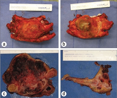
Gross images of the resected tumor, showing the lesion (a) anteriorly, (b) posteriorly, (c) in cross-section, and (d) invading and destroying the third rib.
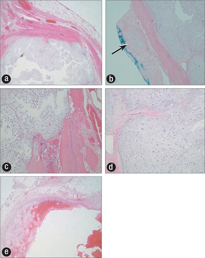
(a) Subpleural tumor nodule (×100). (b) Tumor at anterior resection margin (arrow) (×40). (c, d) Tumor expanding marrow cavity with markedly increased cellularity, nuclear atypia, and occasional multinucleated cells (×400). (e) Tumor expanding blood vessel.
References
-
- Mazanet R, Antman KH. Sarcomas of soft tissue and bone. Cancer. 1991;68(3):463–473. - PubMed
-
- Cakir O, Topal U, Bayram AS, Tolunay S. Sarcomas: rare primary malignant tumors of the thorax. Diagn Interv Radiol. 2005;11(1):23–27. - PubMed
-
- Murphey MD, Walker EA, Wilson AJ, Kransdorf MJ, Temple HT, Gannon FH. From the archives of the AFIP: imaging of primary chondrosarcoma: radiologic-pathologic correlation. Radiographics. 2003;23(5):1245–1278. - PubMed
-
- Weatherby RP, Dahlin DC, Ivins JC. Postradiation sarcoma of bone: review of 78 Mayo Clinic cases. Mayo Clin Proc. 1981;56(5):294–306. - PubMed
-
- Sauter ER, Keller SM, Curran WJ, Russo J, Langer CJ. Radiation-induced chest-wall chondrosarcoma following surgical resection and radiotherapy for non-small-cell lung cancer. J Natl Cancer Inst. 1993;85(2):162–163. - PubMed
LinkOut - more resources
Full Text Sources