. 2011 Apr;10(2):327-37.
doi: 10.1111/j.1474-9726.2011.00673.x. Epub 2011 Feb 25.

E47 retroviral rescue of intrinsic B-cell defects in senescent mice

Affiliations

E47 retroviral rescue of intrinsic B-cell defects in senescent mice

Ana M Landin et al. Aging Cell. 2011 Apr.

Abstract

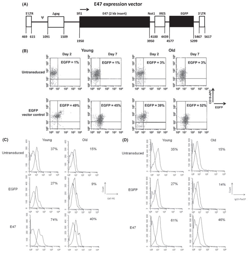
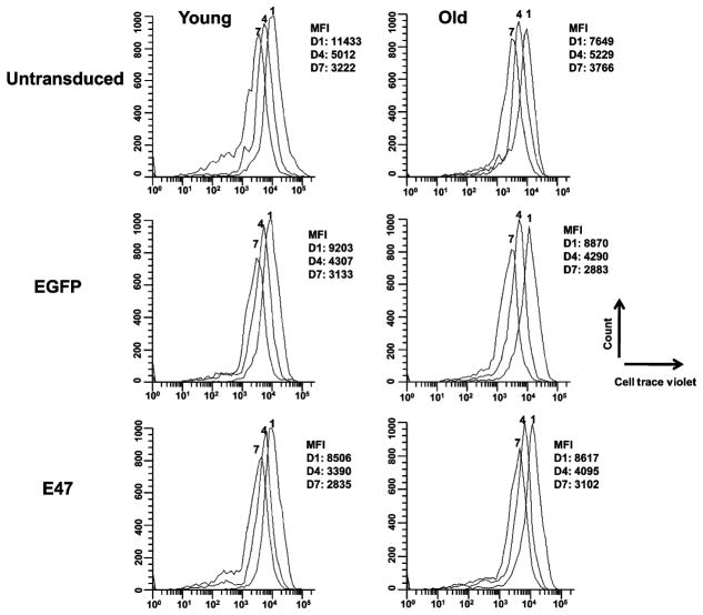
In aging, immune responses are dramatically impaired, specifically the ability to produce protective antibodies. We previously showed that with age there is a B-cell intrinsic decrease in class switch recombination (CSR) because of a decrease in activation-induced cytidine deaminase (AID). One mechanism we have demonstrated for decreased AID includes increased mRNA degradation of the transcription factor E47, critical for AID transcription. Here, we show by means of a retroviral construct containing the DsRED reporter and the 3'UTR of E47 that the 3'UTR lowers mRNA expression, and particularly in B cells from old mice. This is the first demonstration that the E47 3'UTR directly regulates its degradation. The AID mRNA was not differentially regulated by degradation in aging. Therefore, we have here further established critical components for decreased AID with age. The major aim of this study was to establish conditions for the rescue of the intrinsic defect of aged B cells with retroviral addition of the coding region of E47 in splenic B cells to restore their ability to produce optimal AID and class switch to IgG. In this study, we show that young and old primary B cells overexpressing a stable E47 mRNA up-regulate E47, AID, and CSR and improve B-cell immune responses in senescent murine B cells. Our results provide a proof of principle for the rescue of intrinsic B-cell defects and the humoral immune response in senescence.


Figures

Fig. 1
Fig. 1
Construction of DsRED vectors. (A) DsRED-E47 3′UTR or –ku80 3′UTR retroviral vector. A retroviral vector pRevTRE- was used to prepare the E47 3′UTR construct by inserting the 638-bp fragment containing the cDNA for mouse E47 3′UTR or the ku80 3′UTR construct by inserting the 235-bp fragment containing the cDNA for mouse ku80 3′UTR see Materials and Methods. The → denotes the direction of transcription. The Psi (Ψ) is the retroviral packaging signal, the promoter (⇨) is a minimal CMV promoter directly upstream from the red fluorescent protein (DsRED), denoted by (■). B. The 3′-UTR of E47 induces mRNA degradation of DsRED. BJAB cells were left untransduced, transduced with the DsRED vector control, or with DsRED-ku80 3′UTR, or DsRED-E47 3′UTR vectors. BJAB cells were then assessed for DsRED RNA and DNA by qPCR. Results are expressed as DsRED qPCR values, normalized to GAPDH first and also to DsRED DNA, as in Materials and Methods, to take into account potential differences in transduction efficiency. Results are referred to DsRED qPCR values taken as 100. Vertical bars are means ± SE from three independent experiments. The difference between DsRED vector control and DsRED-ku80 3′UTR is not statistically significant (P = 0.07). The difference between DsRED vector control and DsRED-E47 3′UTR is statistically significant (P = 0.0008).
Fig. 2
Fig. 2
Activation-induced cytidine deaminase (AID) mRNA stability in young and old splenic B cells is similar. Splenic B cells from young and old mice were stimulated with lipopolysaccharide (LPS) + IL-4 for 5 days. Cells were then treated with Actinomycin D for 0, 10, 45, and 90 min, and harvested for RNA extraction and qPCR. Results are expressed as AID RNA expression normalized to GAPDH ± SE from three pairs of young (□) and old (◆) mice. Both young and old untreated samples are expressed as 100% at time 0, but the actual value of the old is 23 ± 5%when compared to the young. The difference between young and old mice is not significant at any Actinomycin D time point, as determined by the two-tailed Student’s t test.
Fig. 3
Fig. 3
E47 retroviral vector and E47 rescue in aged splenic B cells. (A) A bicistronic retroviral vector pMXs-IG was used to prepare our construct pMXs-E47-IG (E47 vector) by inserting the 2-kb fragment containing the cDNA for mouse E47 without its 5′ and 3′UTR into the Sfi I and Not I sites. The → denotes the direction of transcription. The promoter is within the 5′ long terminal repeat (LTR), Psi (Ψ) is the packaging signal, Δgag is a truncated gag sequence, E47 and the enhanced green fluorescent protein (EGFP) are denoted by (■). (B) Transduction efficiency in young and old B cells is comparable. Splenic B cells were activated with LPS and IL-4 for 24 h and then left untransduced (top panels) or transduced with the EGFP vector control (bottom panels). Days indicate days after transduction. The y axis shows Flow cytometric expression for CD19-APC and the x axis shows EGFP expression. The % EGFP+ for both the CD19+ and CD19 populations is given (in the CD19+ quadrant). Data are representative of cells from five young and six old mice, and averages are given in the text. (C) Retroviral transduction with E47 elevates E47 in young and old B cells. Splenic B cells for (C) and (D) were treated as in (B) but with the addition of the E47-vector-transduced group. This is a representative figure from five young and six old mice showing the E47 intracellular staining at day 2 post-transduction (peak). Isotype controls were used to set the gate for the E47 staining and subtracted from the E47 percentage before young and old fold difference comparison (see Results). Cell counts (y axis) and E47 expression (x axis) are shown. The%E47 is from all cells derived from the CD19+ cultured cells. Untransduced and EGFP-transduced young or old B cells are not statistically different, as determined by the two-tailed Student’s t test, n = 5. The difference between young and old untransduced is significant at P = 0.01. The difference between young vector control and young E47 vector transduced is significant at P = 0.001. The difference between old vector control and old E47 vector transduced is significant at P = 0.01. The difference between young untransduced and old E47 transduced is not significant showing the rescue of the depleted old phenotype for E47. (D). E47 transduction elevates IgG in young and old B cells. This is a representative figure of IgG1 membrane staining at day 7 from five young and six old mice. Isotype controls were used to set the gate for the IgG1 staining, and subtracted from the IgG1 percentage before young and old fold difference comparison (see Results). Cell count (y axis) and IgG1 (x axis) expression are shown. Untransduced and EGFP-transduced young or old B cells are not significantly different as determined by the two-tailed Student’s t test. The difference between young and old untransduced is significant at P = 0.02. The difference between young vector control and young E47 vector transduced is significant at P = 0.01. The difference between old vector control and old E47 vector transduced is significant at P = 0.03. The difference between young vector control and old E47 transduced is not significant supporting a rescue in class switch recombination (CSR) and IgG expression.
Fig. 4
Fig. 4
(A) Transduction of both young and old B cells with E47 significantly increases E47 mRNA levels. Splenic B cells from young and old mice were treated as in Fig. 3C. At day 2 after transduction, cells were harvested and qPCR was performed. Results are E47 mRNA expression normalized to GAPDH ± SE from five young and six old mice, untransduced (white bars), EGFP transduced (striped bars) or E47 transduced (black bars). Results are normalized to young untransduced taken as 100%. Untransduced and EGFP-transduced B cells (either for young or old) are not statistically different, as determined by the two-tailed Student’s t test. The difference between young and old untransduced is significant at P = 0.0001. The difference between young untransduced and young E47 transduced is significant at P = 0.036. The difference between old untransduced and old E47 transduced is significant at P = 0.005. The difference between young untransduced and old E47 transduced is not significant, consistent with the flow data (Fig. 3C) B. Exogenous E47 mRNA stability is increased in old E47-vector-transduced-splenic B cells. Splenic B cells from young and old mice were stimulated with LPS + IL-4 and either left untransduced or transduced with the E47-vector. Two days post-transduction, cells were treated with Actinomycin D for 0, 30 and 90 min, and harvested for RNA extraction and qPCR. Here, the endogenous E47 is represented by (◆) and the exogenous E47 is represented by (□). The exogenous E47 levels were determined by subtracting the E47 mRNA levels of the untransduced samples from the E47-vector-transduced samples. Results are expressed as E47 mRNA normalized to GAPDH ± SE from three pairs of young and old mice. Both young and old untreated samples are expressed as 100% at time 0, but the actual % expressed relative to the young untransduced values were: for the untransduced young 95 ± 2, the E47-vector-transduced-young 235 ± 26, the untransduced old 42 ± 5, and the E47-vector-transduced-old 130 ± 7. The difference of stability between endogenous and exogenous E47 levels in the young is not significant at any Actinomycin D time point, as determined by the two-tailed Student’s t test. Difference in stability between endogenous and exogenous E47 levels in the old is significant at all Actinomycin D time points, as determined by the two-tailed Student’s t test at 0 min P = 0.0004, 30 min P = 0.01, and 90 min P = 0.04.
Fig. 5
Fig. 5
Transduction of both young and old B cells with E47 increases AID mRNA levels. Splenic B cells from young and old mice were treated as in Fig. 3C. At day 7 post-transduction, cells were harvested and qPCR was performed. Results are expressed as AID mRNA qPCR normalized to GAPDH±SE from five young and six old mice, untransduced (white bars), EGFP transduced (striped bars) or E47 transduced (black bars). Young untransduced are taken as 100%. The difference between young and old untransduced is significant at P = 0.02. The difference between young untransduced and young E47-vector transduced is significant at P = 0.005. The difference between old untransduced and old E47-vector transduced is significant at P = 0.01. The difference between young untransduced and old E47 transduced is not significant, again showing rescue, here of AID, in the old B cells with E47 transduction.
Fig. 6
Fig. 6
Transduction of young and old splenic B cells does not change the cell proliferation rates. Splenic B cells were activated with lipopolysaccharide and IL-4 for 24 h and then left untransduced or transduced with enhanced green fluorescent protein vector control or E47-vector. The cells were loaded with CellTrace Violet 24 h post-transduction and assayed after 1, 4, and 7 days by flow cytometry. This is a representative figure of stimulated B cells from three young and three old mice.
Fig. 7
Fig. 7
Production of IgG1 measured by ELISA was up-regulated in both young and old B cells after transduction with E47. Splenic B cells from young and old mice were treated as in Fig. 3C. At day 7 post-transduction, cell supernatants were collected and assayed by ELISA to evaluate IgG1 production. Vertical bars are means ± SE from five young and six old mice, EGFP transduced (striped bars) or E47 transduced (black bars). The EGFP vector control is represented here as the control, because the untransduced and EGFP controls are not statistically different and only the vector control supernatants were assayed here. The difference between young and old EGFP transduced is significant at P = 0.008. The difference between young EGFP transduced and young E47 transduced is significant at P = 0.007, as determined by the two-tailed Student’s t test. The difference between old EGFP transduced and old E47 transduced is significant at P = 0.0014. The difference between young EGFP transduced and old E47 transduced is not significant P = 0.08, again showing the rescue of the old phenotype.

Similar articles

Cited by

References

    1. Asanuma H, Hirokawa K, Uchiyama M, Suzuki Y, Aizawa C, Kurata T, Sata T, Tamura S. Immune responses and protection in different strains of aged mice immunized intranasally with an adjuvant-combined influenza vaccine. Vaccine. 2001;19:3981–3989. - PubMed
    1. Cancro MP, Hao Y, Scholz JL, Riley RL, Frasca D, Dunn-Walters DK, Blomberg BB. B cells and aging: molecules and mechanisms. Trends Immunol. 2009;30:313–318. - PMC - PubMed
    1. Diaz M, Storb U. A novel cytidine deaminase AIDs in the delivery of error-prone polymerases to immunoglobulin genes. DNA Repair (Amst) 2003;2:623–627. - PubMed
    1. Dorshkind K, Montecino-Rodriguez E, Signer RA. The ageing immune system: is it ever too old to become young again? Nat Rev Immunol. 2009;9:57–62. - PubMed
    1. Ephrussi A, Church GM, Tonegawa S, Gilbert W. B lineage – specific interactions of an immunoglobulin enhancer with cellular factors in vivo. Science. 1985;227:134–140. - PubMed

Publication types

MeSH terms

Substances