. 2011 Jan 17:7:7.
doi: 10.1186/1744-8069-7-7.

The blockade of the transient receptor potential vanilloid type 1 and fatty acid amide hydrolase decreases symptoms and central sequelae in the medial prefrontal cortex of neuropathic rats

Affiliations

The blockade of the transient receptor potential vanilloid type 1 and fatty acid amide hydrolase decreases symptoms and central sequelae in the medial prefrontal cortex of neuropathic rats

Vito de Novellis et al. Mol Pain. .

Abstract

Background: Neuropathic pain is a chronic disease resulting from dysfunction within the "pain matrix". The basolateral amygdala (BLA) can modulate cortical functions and interactions between this structure and the medial prefrontal cortex (mPFC) are important for integrating emotionally salient information. In this study, we have investigated the involvement of the transient receptor potential vanilloid type 1 (TRPV1) and the catabolic enzyme fatty acid amide hydrolase (FAAH) in the morphofunctional changes occurring in the pre-limbic/infra-limbic (PL/IL) cortex in neuropathic rats.

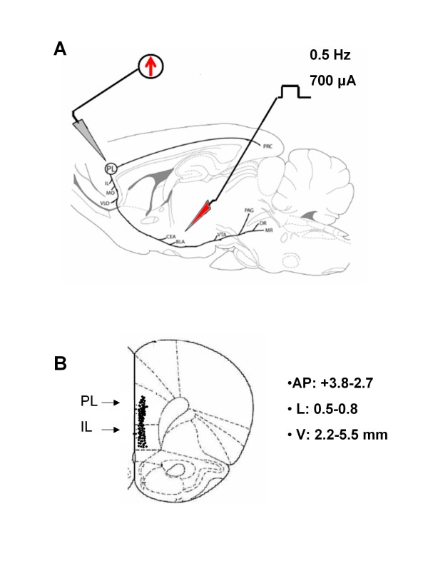
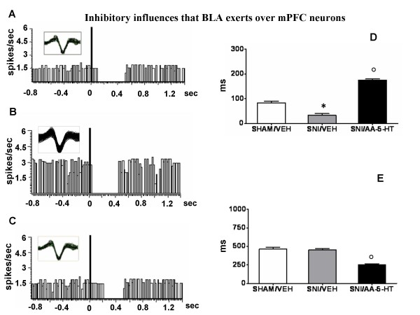
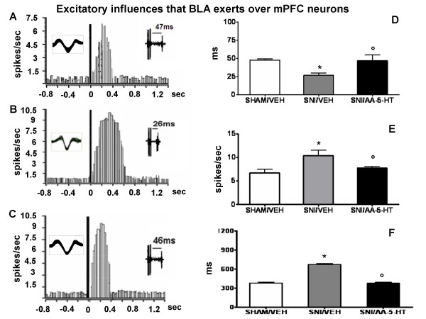
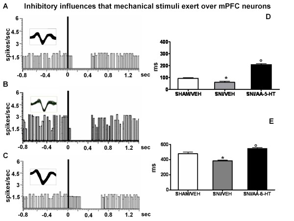
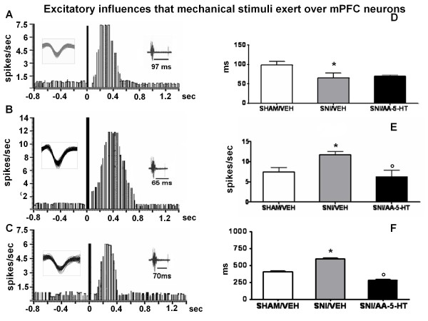
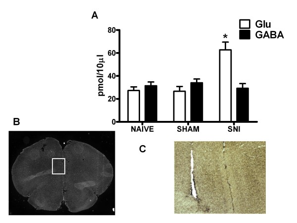
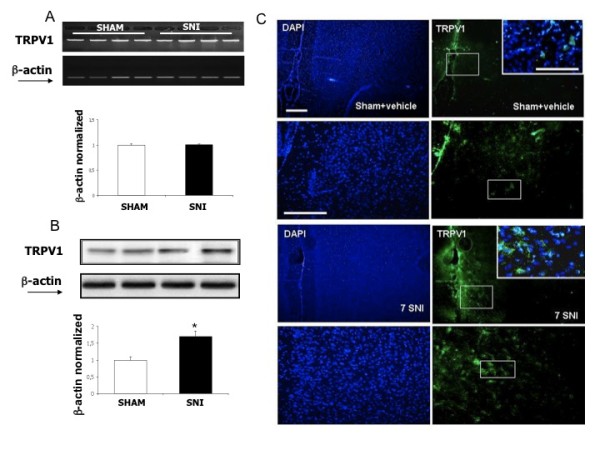
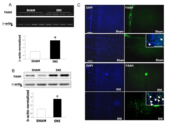
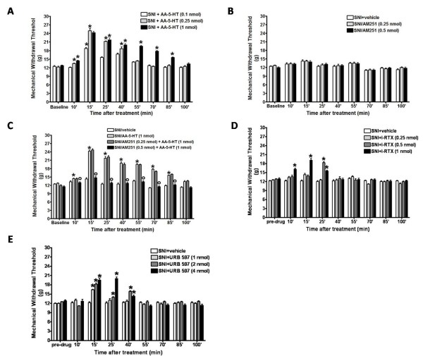
Results: The effect of N-arachidonoyl-serotonin (AA-5-HT), a hybrid FAAH inhibitor and TPRV1 channel antagonist, was tested on nociceptive behaviour associated with neuropathic pain as well as on some phenotypic changes occurring on PL/IL cortex pyramidal neurons. Those neurons were identified as belonging to the BLA-mPFC pathway by electrical stimulation of the BLA followed by hind-paw pressoceptive stimulus application. Changes in their spontaneous and evoked activity were studied in sham or spared nerve injury (SNI) rats before or after repeated treatment with AA-5-HT. Consistently with the SNI-induced changes in PL/IL cortex neurons which underwent profound phenotypic reorganization, suggesting a profound imbalance between excitatory and inhibitory responses in the mPFC neurons, we found an increase in extracellular glutamate levels, as well as the up-regulation of FAAH and TRPV1 in the PL/IL cortex of SNI rats. Daily treatment with AA-5-HT restored cortical neuronal activity, normalizing the electrophysiological changes associated with the peripheral injury of the sciatic nerve. Finally, a single acute intra-PL/IL cortex microinjection of AA-5-HT transiently decreased allodynia more effectively than URB597 or I-RTX, a selective FAAH inhibitor or a TRPV1 blocker, respectively.

Conclusion: These data suggest a possible involvement of endovanilloids in the cortical plastic changes associated with peripheral nerve injury and indicate that therapies able to normalize endovanilloid transmission may prove useful in ameliorating the symptoms and central sequelae associated with neuropathic pain.


Figures

Figure 1
Figure 1
Representative schematic illustration showing the location of the recording electrode in the PL/IL cortex and of the stimulation electrode in the BLA (A). Number values refer to stimulation parameters. Abbreviations: CEA, central nucleus of amygdala; BLA, basolateral nucleus of amygdala; VTA, ventral tegmental area; PAG, periaqueductal gray; DR, dorsal raphe nucleus; PL, prelimbic cortex; IL, infralimbic cortex; MO, medial orbital cortex; PRC, perirhinal cortex. Reprinted from Vertes [99]. Schematic illustration of the PL/IL cortex recording sites (B). Filled circles represent mPFC cell recorded sites. Number values indicate distance to bregma.
Figure 2
Figure 2
BLA stimulation evokes inhibitory responses in the PL/IL cortex of BLA → PL/IL (-) neurons. The figure shows different parameters of BLA-evoked inhibition, including the onset and duration of the inhibition, in sham and SNI rats treated for 7 days with vehicle (veh) or AA-5-HT (5 mg/kg, i.p.). "A", "B" and "C" show representative ratemater records of a BLA → PL/IL (-) neurons of sham/veh, SNI/veh or SNI/AA-5-HT rat, respectively. "D" and "E" show the onset and duration (mean ± SEM) of the inhibition, respectively, in different groups of rats. * indicates statistically significant difference versus sham/veh and º versus SNI/veh. P < 0.05 was considered as value of significance and n = 10 was used for each group.
Figure 3
Figure 3
BLA stimulation evokes excitatory responses in the PL/IL cortex of BLA → PL/IL (+) neurons. The figure shows different parameters of BLA-evoked excitation, including the onset, the frequency and the duration of excitation, in sham and SNI rats treated for 7 days with vehicle (veh) or AA-5-HT (5 mg/kg, i.p.). "A", "B" and "C" show a representative ratemater record of a BLA → PL/IL (+) neurons of sham/veh, SNI/veh and SNI/AA-5-HT rat. "D","E" and F show the onset, onset, the frequency and the duration (mean ± SEM) of the excitation, respectively, in the different groups of rats. * indicates statistically significant difference versus sham/veh and º versus SNI/veh. P < 0.05 was considered as value of significance and n = 10 was used for each group.
Figure 4
Figure 4
Mechanical nociceptive stimulation evokes inhibitory responses in the PL/IL cortex of BLA → PL/IL (-) neurons. The figure shows different parameters of mechanical nociceptive stimulation-evoked inhibition, including the onset and duration of the inhibition, in sham and SNI rats treated for 7 days with vehicle (veh) or AA-5-HT (5 mg/kg, i.p.). "A", "B" and "C" show a representative ratemater record of a mechanical stimulus → PL/IL (-) neurons of sham/veh, SNI/veh and SNI/AA-5-HT rat, respectively. "D" and "E" show the onset and duration (mean ± SEM) of the inhibition, respectively, in the different groups of rats. * indicates statistically significant difference versus sham/veh and º versus SNI/veh. P < 0.05 was considered as value of significance and n = 10 was used for each group.
Figure 5
Figure 5
Mechanical stimulation evokes excitatory responses in the PL/IL cortex neurons of BLA → PL/IL (+) neurons. The figure shows different parameters of mechanical stimulus-evoked excitation, including the onset, the frequency and the duration of excitation, in sham and SNI rats treated for 7 days with vehicle (veh) or AA-5-HT (5 mg/kg, i.p.). "A", "B" and "C" show a representative ratemater record of a mechanical stimulus → PL/IL (+) neurons of sham/veh, SNI/veh and SNI/AA-5-HT rat. "D","E" and F show the onset, the frequency and the duration (mean ± SEM) of the excitation, respectively, in the different groups of rats. * indicates statistically significant difference versus sham/veh and º versus SNI/veh. P < 0.05 was considered as value of significance and n = 10 was used for each group.
Figure 6
Figure 6
"A" shows the release of glutamate and GABA in naïve, sham or SNI rats 7 days after injury. The values of extracellular GABA and glutamate in the mPFC were expressed as pmol in 10 μl of perfusate. * indicate significant difference vs sham rats. Each point represents the mean ± S.E.M of 7-8 animals per group. P values < 0.05 were considered statistically significant. "B" shows a panoramic picture of the pre-frontal cortex, the square indicates the pre/infra-limbic area. "C" shows a high magnification of the microdialysis probe location for aminoacid collection within the the pre/infra-limbic cortex. Coronal brain slices containing the sites of implantation of the microdialysis probes were obtained after the experiment and processed for histological analysis.
Figure 7
Figure 7
TRPV1 mRNA and protein levels in the pre/infra-limbic cortex of sham and SNI rats. "A" shows the unchanged TRPV1 mRNA levels normalized vs β-actin in neuropathic vs sham rats. "B" shows the enhanced TRPV1 protein levels normalized vs β-actin in neuropathic vs sham rats. "C" shows the increased TRPV1 staining in the layer II-III of the rat pre/infra-limbic cortex. Data are represented as a mean ± SEM n = 3 rats per group. P < 0.05 was considered statistically significant. ANOVA, post hoc Tukey. Scale bars = 100 μm.
Figure 8
Figure 8
FAAH mRNA and protein levels in the pre/infra-limbic cortex of sham and SNI rats. "A" shows the enhanced FAAH mRNA levels normalized vs β-actin in neuropathic vs sham rats. "B" shows the enhanced FAAH protein levels normalized vs β-actin in neuropathic vs sham rats. "C" shows the increased FAAH staining in the layer II-III of the rat pre/infra-limbic cortex. Data are represented as a mean ± SEM n = 3 rats per group. P < 0.05 was considered statistically significant. ANOVA, post hoc Tukey. Scale bars = 100 μm.
Figure 9
Figure 9
Effects of a single injection of vehicle, AA-5-HT (0.1-0.25-1 nmol) (A), AM251 (0.25-0.5 nmol) (B), AM251+AA-5-HT (C), I-RTX (0.25-0.5-1 nmol) (D) or URB 597 (1-2-4 nmol) (E) on mechanical withdrawal threshold (mean ± S.E.M.) in spared nerve injury (SNI) rats. Each point represents the mean ± S.E.M. of 8-10 rats per group. *p < 0.05 vs SNI/veh.

Similar articles

Cited by

References

    1. Gusnard DA, Akbudak E, Shulman GL, Raichle ME. Medial prefrontal cortex and self-referential mental activity: relation to a default mode of brain function. Proc Natl Acad Sci USA. 2001;98:4259–4264. doi: 10.1073/pnas.071043098. - DOI - PMC - PubMed
    1. Phelps EA, Delgado MR, Nearing KI, LeDoux JE. Extinction learning in humans: role of the amygdala and vmPFC. Neuron. 2004;43:897–905. doi: 10.1016/j.neuron.2004.08.042. - DOI - PubMed
    1. Lorenz J, Cross DJ, Minoshima S, Morrow TJ, Paulson PE, Casey KL. A unique representation of heat allodynia in the human brain. Neuron. 2002;35:383–393. doi: 10.1016/S0896-6273(02)00767-5. - DOI - PubMed
    1. Porro CA, Baraldi P, Pagnoni G, Serafini M, Facchin P, Maieron M, Nichelli P. Does anticipation of pain affect cortical nociceptive systems? J Neurosci. 2002;22:3206–3214. - PMC - PubMed
    1. Rainville P, Duncan GH, Price DD, Carrier B, Bushnell MC. Pain affect encoded in human anterior cingulate but not somatosensory cortex. Science. 1997;277:968–971. doi: 10.1126/science.277.5328.968. - DOI - PubMed

MeSH terms

LinkOut - more resources