. 2011 Jan 17;13(1):R5.
doi: 10.1186/bcr2809.

Proliferation-associated POU4F2/Brn-3b transcription factor expression is regulated by oestrogen through ERα and growth factors via MAPK pathway

Affiliations

Proliferation-associated POU4F2/Brn-3b transcription factor expression is regulated by oestrogen through ERα and growth factors via MAPK pathway

Samir Ounzain et al. Breast Cancer Res. .

Abstract

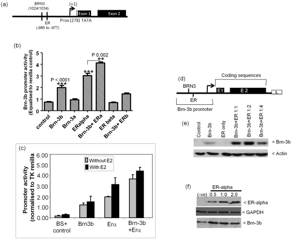
Introduction: In cancer cells, elevated transcription factor-related Brn-3a regulator isolated from brain cDNA (Brn-3b) transcription factor enhances proliferation in vitro and increases tumour growth in vivo whilst conferring drug resistance and migratory potential, whereas reducing Brn-3b slows growth both in vitro and in vivo. Brn-3b regulates distinct groups of key target genes that control cell growth and behaviour. Brn-3b is elevated in >65% of breast cancer biopsies, but mechanisms controlling its expression in these cells are not known.

Methods: Bioinformatics analysis was used to identify the regulatory promoter region and map transcription start site as well as transcription factor binding sites. Polymerase chain reaction (PCR) cloning was used to generate promoter constructs for reporter assays. Chromatin immunoprecipitation and site-directed mutagenesis were used to confirm the transcription start site and autoregulation. MCF-7 and Cos-7 breast cancer cells were used. Cells grown in culture were transfected with Brn-3b promoter and treated with growth factors or estradiol to test for effects on promoter activity. Quantitative reverse transcriptase PCR assays and immunoblotting were used to confirm changes in gene and protein expression.

Results: We cloned the Brn-3b promoter, mapped the transcription start site and showed stimulation by estradiol and growth factors, nerve growth factor and epidermal growth factor, which are implicated in breast cancer initiation and/or progression. The effects of growth factors are mediated through the mitogen-activated protein kinase pathway, whereas hormone effects act via oestrogen receptor α (ERα). Brn-3b also autoregulates its expression and cooperates with ERα to further enhance levels.

Conclusions: Key regulators of growth in cancer cells, for example, oestrogens and growth factors, can stimulate Brn-3b expression, and autoregulation also contributes to increasing Brn-3b in breast cancers. Since increasing Brn-3b profoundly enhances growth in these cells, understanding how Brn-3b is increased in breast cancers will help to identify strategies for reducing its expression and thus its effects on target genes, thereby reversing its effects in breast cancer cells.


Figures

Figure 1
Figure 1
Identification and cloning of transcription start site in transcription factor-related Brn-3a regulator isolated from brain cDNA (Brn-3b) promoter. (a) Homology plots (VISTA Genome Browser) showing regions of similarity in Brn-3b gene and 5' upstream sequences between human (top) and dog, horse and mouse (bottom) genome sequences. Regions of homology are indicated by peaks and grey shading, and positions of Brn-3b exons and intronic sequences are shown. (b) Schematic showing cloned BstX1 (B)/Stu1 (S)/Xho1 (X) (BSX) construct containing putative Brn-3b promoter and regulatory sequences or the BSX exon-intron-exon (BSXEIE) expression construct containing the promoter, regulatory and coding sequences, which were used for subsequent studies. Promoter and regulatory sequences are shown in grey-striped area. 5' noncoding sequences at the beginning of exon 1 are indicated by the black bar. The white stripe at the start of exon 2 represents unique sequences that are present in Brn-3b(s) transcripts, but not in Brn-3b(l) transcripts. The positions of restriction enzyme sites BstX1 (B), Stu1 (S) and Xho1 (X) used for cloning are also shown. (c) Luciferase activity of Brn-3b reporter constructs following transfection into MCF-7 cells is compared with baseline luciferase activity of the empty reporter vector. Values, shown as relative luciferase units (RLU),, were equalised with Renilla internal control (d) Schematic showing positions of putative start sites identified by in silico analysis. Initiator element and proximal TATA sequences are shown relative to ATG in exon 1, and putative intronic TATA sequences are indicated. Half-arrows show relative positions of primers used for polymerase chain reaction (PCR) assay following chromatin immunoprecipitation (ChIP) assay with α-TATA box binding protein (α-TBP) antibody (Ab) to analyse TBP binding to the different sites. (e) PCR products obtained when ChIP DNA (obtained with α-TBP Ab or secondary control Ab) was used for amplification with primers that flanked the upstream initiator elements (-1,048 bp) (A), -278TATA (B) or the intronic TA sequences (C). Primers for sequences within exon 2 (> 1 kb from ATG) were used for amplification in the negative control (D). Positive controls represent PCR products derived by using primers that amplified the known start site of the glyceraldehyde 3-phosphate dehydrogenase (GAPDH) gene using α-TBP or control ChIP DNA (E). Input represents amplification using one-tenth of DNA isolated before performing the ChIP assay. Right column (labeled 2nd Ab) shows the products obtained following amplification of control ChIP Ab (α-rabbit secondary Ab) using the indicated primers.
Figure 2
Figure 2
Analysing the transcription start sites of Brn-3b promoter. (a) Analysis of Brn-3b promoter (BSX) activity following the mutation of key sites to identify the transcriptional start site. Wild-type (WT) promoter in row 1 (right) is represented as 100%, and all mutations are expressed as percentages of the WT promoter. Schematic shows positions of mutations at key sites (indicated by x, left), for example, upstream initiator alone (row 2, right) or in combination with other sites (rows 7 to 9, right). The proximal -278TATA mutation is shown alone (row 3, right) or in combination (rows 7 and 9, right). The effects of mutation of different intronic TA alone (rows 4 to 6, right) or in combination (rows 8 to 10, right) are also shown. Values are equalised to internal control (Renilla luciferase), and the results represent data from three independent experiments expressed as means + SD. * is used to show statistical significance between different mutant promoter constructs and wild-type constructs. (b) Western blot analysis of Brn-3b protein expression in cellular extracts prepared from MCF7 cells transfected with BSXE1E expression constructs in which Brn-3b promoter drives expression of the Brn-3b gene. WT promoter activity is shown in left column. Middle column (Δ-278TATA shows the reduction in Brn-3b protein from expression constructs containing a mutation within the proximal -278TATA promoter of an otherwise, intact construct. Right column shows untransfected control cells with no reporter and therefore represents levels of endogenous Brn-3b protein in these cells.
Figure 3
Figure 3
Analysing the effect of growth factors on Brn-3b promoter activity. (a) Brn-3b promoter (BSX) activity was measured following transfection of the reporter construct into MCF-7 cells and treatment with different growth factors. Values were equalised with internal control and Renilla luciferase activity and expressed as a percentage of promoter only (set at 100%). The data shown represent means ± SD from at least three independent experiments. NGF, nerve growth factor; EGF, epidermal growth factor; IGF, insulin-like growth factor; TGF, transforming growth factor; cAMP, cyclic adenosine monophosphate. (b) Schematic showing the position of growth factor response element (EGRF) and serum response element (SRE) sites in the Brn-3b promoter. The location of two Sma1 restriction enzyme sites, which were used to generate the deletion (Sma1/Sma1) construct, BS-SS, is shown in relation to the DNA binding sites. The resultant truncated promoter is represented schematically below. (c) Luciferase activity of intact (BS) promoter (control) or Sma1/Sma1 deletion promoter (BS-SS) is shown following transfection into MCF-7 cells. Grey bars represent WT promoter activity either alone or following treatment with NGF or EGF, whereas stippled bars show the activity of the Sma1/Sma1 deletion construct with or without growth factors. Values represent means ± SE of three independent experiments.
Figure 4
Figure 4
Brn-3b promoter is activated via the mitogen-activated protein kinase/extracellular signal-regulated kinase (MAPK/ERK) pathway in breast cancer cells. (a) Brn-3b promoter activity is shown following treatment of transfected cells with inhibitors of different signalling pathways (as indicated), in absence or presence of NGF or EGF. Values have been adjusted using internal Renilla luciferase control and represent means ± SD of at least three independent experiments. (b) Increase in Brn-3b promoter activity following treatment of transfected cells with the protein kinase C analogue phorbol 12,13-dibutyrate in the absence or presence of the ERK inhibitor, PD98059. Values represent relative luciferase activity after adjusting for Renilla internal control. (c) Brn-3b promoter activity is shown following cotransfection with either dominant-negative mitogen-activated protein kinase kinase (dnMEK) or WT MEK compared with activity in the presence of the ERK inhibitor PD98059. Values represent relative luciferase activity after adjusting for Renilla internal control.
Figure 5
Figure 5
Brn-3b promoter activity is strongly stimulated by 17β-estradiol via activation of oestrogen receptor α (ERα). (a) Brn-3b promoter activity following treatment of transfected MCF-7 cells with different concentration of 17β-estradiol. Values have been adjusted with internal control, Renilla luciferase and represent means ± SD of three independent experiments ***indicate statistical significance of p < 0.0001 compared with control. (b) Effect of different oestrogen receptors, ERα or ERβ on Brn-3b (BSX) promoter activity are shown after cotransfection into sensitised MCF-7 cells (grown in stripped serum medium for 48 hours). Promoter activity is adjusted to internal Renilla control and expressed as percentage of levels seen with empty vector only (set at 100%). Values represent data from three independent experiments, expressed as means ± SD. (c) Activation of Brn-3b promoter by ERα can be blocked by the receptor antagonist tamoxifen. Promoter activity is shown after cotransfection of Brn-3b promoter with ERα into sensitised MCF-7 cells (grown in oestrogen-depleted medium for 48 hours) in the presence of 1 μM 4 hydroxy tamoxifen (TAM) compared with untreated control. Promoter activity is adjusted to internal Renilla control and expressed as percentages of levels seen with untreated controls (set at 100%). (d) Brn-3b promoter activity in ER-negative Cos-7 cells treated with estradiol and either transfected ERα or control vector. Data shows luciferase activity in cells transfection of Brn-3b promoter and treatment with either vehicle (-E2) or 10 nM estradiol (+E2) in the absence or presence of ERα.
Figure 6
Figure 6
Cooperation between Brn-3b and ER can stimulate promoter activity. (a) Schematic diagram showing position of Brn-3 consensus binding sites and oestrogen response element (ERE) site relative to the proximal -278TATA, (now designated +1). (b) Promoter activity following cotransfection of the BSX reporter construct with Brn-3b, Brn-3a, ERα or ERβ alone or in different combinations into MCF-7 cells. Values have been equalised with internal Renilla control [renilla reporter gene driven by minimal tyrosine kinase (TK) promoter] and represent means ± SD of three independent experiments. (c) Brn-3b promoter activity when coexpressed with Brn-3b and ERα, alone or together, in cells grown in oestrogen-depleted medium and either untreated or treated with estradiol, after transfection. Grey bars show inducibility in the absence of estradiol, and black bars demonstrate the effects of adding estradiol after transfection with the constructs described. (d) Schematic showing the modified expression construct, BSXE1E, in which the Brn-3b promoter drives expression of the Brn-3b gene upstream of luciferase reporter (L). (e) Immunoblot showing changes in Brn-3b protein produced from the BSXE1E expression construct when cotransfected with Brn-3b or ERα alone or in combination (at different ratios of Brn-3b:ER, 1:1 to 1:4). Actin immunoblot was used to indicate variation in protein loading. To demonstrate that Brn-3b protein levels was dependent on the ratio of Brn-3b to ER, shorter exposure times and therefore smaller increases in Brn-3b expression are not evident when ER only is expressed. (f) Immunoblot to show increases in ER protein following transfection with expression constructs (top immunoblot) and a corresponding changes in endogenous Brn-3b protein that were dependent on concentrations of ERα (middle immunoblot). GAPDH immunoblot is included to show variation in protein loading.
Figure 7
Figure 7
Mutation of Brn-3 site reduces the inducibility of promoter by ERα and also abolishes cooperativity. (a) Testing effects of ERα with or without Brn-3b on activity of deletion promoter, BS-SS, in which putative binding site for Brn-3b and ERα are lost. Reporter gene activity was adjusted using internal control, Renilla luciferase. Values are shown relative to BS promoter activity in the presence of control empty vector, LTR, set at 100%. Effects of ERα or Brn-3b, alone or together on the intact BS promoter or deletion (BS-SS) promoter are compared (b) Reporter gene activity following cotransfection of Brn-3b or ER (alone or together) with WT or mutant Brn-3b reporter construct in which Brn-3b site is mutated. Values have been adjusted for internal Renilla luciferase control and are expressed as percentages of activity in empty vector transfected cells (set at 100%) for the respective promoters. Results represent means ± SD from three independent experiments. (c) Similar reporter assays were used to show changes in activity of Brn-3b promoter containing mutation of ERE alone (ERE mutant) or double-mutant lacking both sites (Brn-3/ERE mutant) when coexpressed with Brn-3b +/- ERα. The data represent means ± SD of three independent experiments. (d) Representative PCR product obtained using ChIP DNA (immunoprecipitated with Brn-3b antibody) and primers to amplify and promoter region containing the putative Brn-3b binding site. Input column represents PCR amplification using one-tenth of isolated DNA before ChIP. Middle column (-ve Ab) shows product following ChIP assay with negative control α-rabbit Ab only). Right column (Brn-3b Ab) shows product resulting from ChIP assay using Brn-3b Ab.

References

    1. Budhram-Mahadeo VS, Latchman DS. Targeting Brn-3b in breast cancer therapy. Expert Opin Ther Targets. 2006;10:15–25. doi: 10.1517/14728222.10.1.15. - DOI - PubMed
    1. Irshad S, Pedley RB, Anderson J, Latchman DS, Budhram-Mahadeo V. The Brn-3b transcription factor regulates the growth, behavior, and invasiveness of human neuroblastoma cells in vitro and in vivo. J Biol Chem. 2004;279:21617–21627. doi: 10.1074/jbc.M312506200. - DOI - PubMed
    1. Dennis JH, Budhram-Mahadeo V, Latchman DS. The Brn-3b POU family transcription factor regulates the cellular growth, proliferation, and anchorage dependence of MCF7 human breast cancer cells. Oncogene. 2001;20:4961–4971. doi: 10.1038/sj.onc.1204491. - DOI - PubMed
    1. Samady L, Dennis J, Budhram-Mahadeo V, Latchman DS. Activation of CDK4 Gene Expression in Human Breast Cancer Cells by the Brn-3b POU Family Transcription Factor. Cancer Biol Ther. 2004;3:317–323. doi: 10.4161/cbt.3.3.698. - DOI - PubMed
    1. Budhram-Mahadeo VS, Irshad S, Bowen S, Lee SA, Samady L, Tonini GP, Latchman DS. Proliferation-associated Brn-3b transcription factor can activate cyclin D1 expression in neuroblastoma and breast cancer cells. Oncogene. 2008;27:145–154. doi: 10.1038/sj.onc.1210621. - DOI - PubMed

Publication types

MeSH terms

Substances

LinkOut - more resources