

A combined approach for comparative exoproteome analysis of Corynebacterium pseudotuberculosis
- PMID: 21241507
- PMCID: PMC3025830
- DOI: 10.1186/1471-2180-11-12
A combined approach for comparative exoproteome analysis of Corynebacterium pseudotuberculosis
Abstract
Background: Bacterial exported proteins represent key components of the host-pathogen interplay. Hence, we sought to implement a combined approach for characterizing the entire exoproteome of the pathogenic bacterium Corynebacterium pseudotuberculosis, the etiological agent of caseous lymphadenitis (CLA) in sheep and goats.
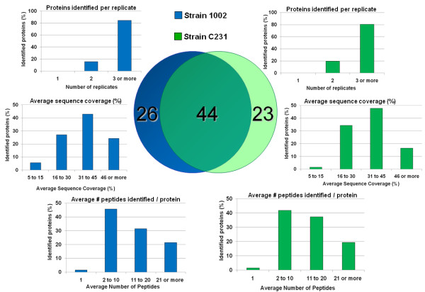
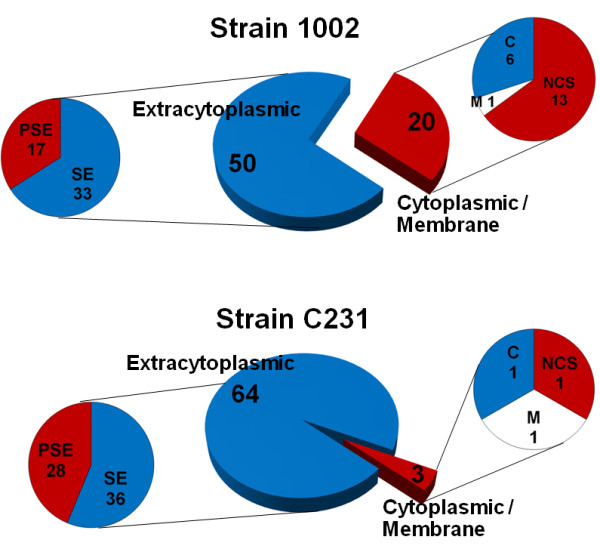
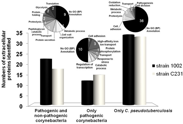
Results: An optimized protocol of three-phase partitioning (TPP) was used to obtain the C. pseudotuberculosis exoproteins, and a newly introduced method of data-independent MS acquisition (LC-MSE) was employed for protein identification and label-free quantification. Additionally, the recently developed tool SurfG+ was used for in silico prediction of sub-cellular localization of the identified proteins. In total, 93 different extracellular proteins of C. pseudotuberculosis were identified with high confidence by this strategy; 44 proteins were commonly identified in two different strains, isolated from distinct hosts, then composing a core C. pseudotuberculosis exoproteome. Analysis with the SurfG+ tool showed that more than 75% (70/93) of the identified proteins could be predicted as containing signals for active exportation. Moreover, evidence could be found for probable non-classical export of most of the remaining proteins.
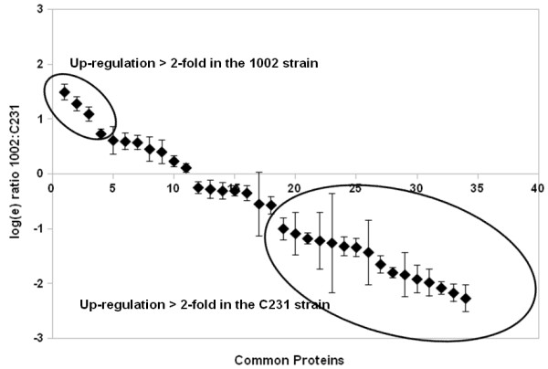
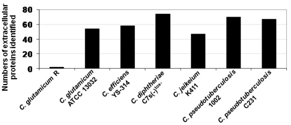
Conclusions: Comparative analyses of the exoproteomes of two C. pseudotuberculosis strains, in addition to comparison with other experimentally determined corynebacterial exoproteomes, were helpful to gain novel insights into the contribution of the exported proteins in the virulence of this bacterium. The results presented here compose the most comprehensive coverage of the exoproteome of a corynebacterial species so far.
Figures
Similar articles
-
Identification of 11 new exoproteins in Corynebacterium pseudotuberculosis by comparative analysis of the exoproteome.Microb Pathog. 2013 Aug-Sep;61-62:37-42. doi: 10.1016/j.micpath.2013.05.004. Epub 2013 May 16. Microb Pathog. 2013. PMID: 23684727
-
Differential Exoproteome analysis of two corynebacterium pseudotuberculosis biovar ovis strains isolated from goat (1002) and sheep (C231).Curr Microbiol. 2013 Oct;67(4):460-5. doi: 10.1007/s00284-013-0388-4. Epub 2013 May 23. Curr Microbiol. 2013. PMID: 23699973
-
The Corynebacterium pseudotuberculosis in silico predicted pan-exoproteome.BMC Genomics. 2012;13 Suppl 5(Suppl 5):S6. doi: 10.1186/1471-2164-13-S5-S6. Epub 2012 Oct 19. BMC Genomics. 2012. PMID: 23095951 Free PMC article.
-
A description of genes of Corynebacterium pseudotuberculosis useful in diagnostics and vaccine applications.Genet Mol Res. 2008 Mar 18;7(1):252-60. doi: 10.4238/vol7-1gmr438. Genet Mol Res. 2008. PMID: 18551390 Review.
-
Vaccines for caseous lymphadenitis: up-to-date and forward-looking strategies.Appl Microbiol Biotechnol. 2021 Mar;105(6):2287-2296. doi: 10.1007/s00253-021-11191-4. Epub 2021 Mar 2. Appl Microbiol Biotechnol. 2021. PMID: 33651132 Free PMC article. Review.
Cited by
-
Multi-Omics of Corynebacterium Pseudotuberculosis 12CS0282 and an In Silico Reverse Vaccinology Approach Reveal Novel Vaccine and Drug Targets.Proteomes. 2022 Nov 23;10(4):39. doi: 10.3390/proteomes10040039. Proteomes. 2022. PMID: 36548458 Free PMC article.
-
Identification of a novel zinc metalloprotease through a global analysis of Clostridium difficile extracellular proteins.PLoS One. 2013 Nov 26;8(11):e81306. doi: 10.1371/journal.pone.0081306. eCollection 2013. PLoS One. 2013. PMID: 24303041 Free PMC article.
-
Comparative Proteomic Analyses Between Biofilm-Forming and Non-biofilm-Forming Strains of Corynebacterium pseudotuberculosis Isolated From Goats.Front Vet Sci. 2021 Feb 16;8:614011. doi: 10.3389/fvets.2021.614011. eCollection 2021. Front Vet Sci. 2021. PMID: 33665217 Free PMC article.
-
A journey through the Corynebacterium pseudotuberculosis proteome promotes insights into its functional genome.PeerJ. 2021 Dec 23;9:e12456. doi: 10.7717/peerj.12456. eCollection 2021. PeerJ. 2021. PMID: 35036114 Free PMC article.
-
Quantitative Proteomic Analysis Reveals Changes in the Benchmark Corynebacterium pseudotuberculosis Biovar Equi Exoproteome after Passage in a Murine Host.Front Cell Infect Microbiol. 2017 Jul 25;7:325. doi: 10.3389/fcimb.2017.00325. eCollection 2017. Front Cell Infect Microbiol. 2017. PMID: 28791255 Free PMC article.
References
Publication types
MeSH terms
Substances
LinkOut - more resources
Full Text Sources
Other Literature Sources
