. 2011 Jan 18;18(1):6.
doi: 10.1186/1423-0127-18-6.

Hedgehog overexpression leads to the formation of prostate cancer stem cells with metastatic property irrespective of androgen receptor expression in the mouse model

Affiliations

Hedgehog overexpression leads to the formation of prostate cancer stem cells with metastatic property irrespective of androgen receptor expression in the mouse model

Han-Hsin Chang et al. J Biomed Sci. .

Abstract

Background: Hedgehog signalling has been implicated in prostate tumorigenesis in human subjects and mouse models, but its effects on transforming normal basal/stem cells toward malignant cancer stem cells remain poorly understood.

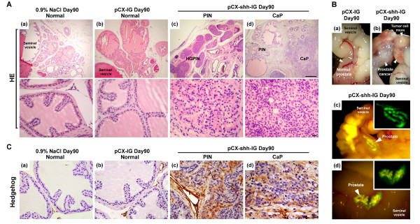
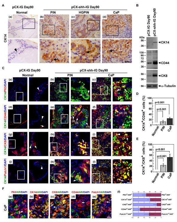
Methods: We produced pCX-shh-IG mice that overexpress Hedgehog protein persistently in adult prostates, allowing for elucidation of the mechanism during prostate cancer initiation and progression. Various markers were used to characterize and confirm the transformation of normal prostate basal/stem cells into malignant cancer stem cells under the influence of Hedgehog overexpression.

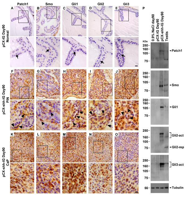
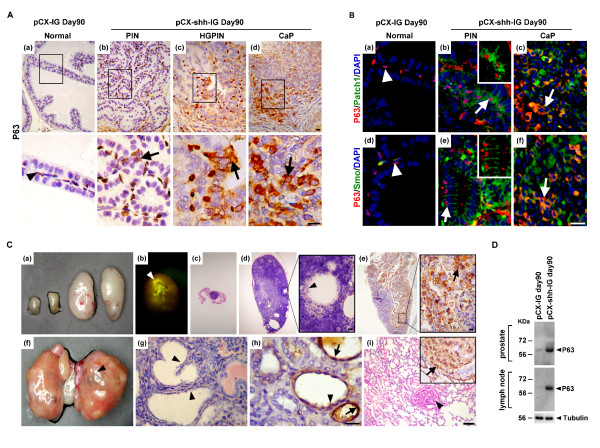
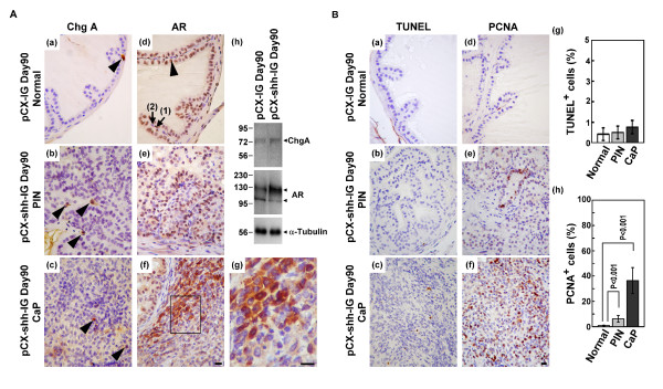
Results: The pCX-shh-IG mice developed prostatic intraepithelial neoplasia (PIN) that led to invasive and metastatic prostate cancers within 90 days. The prostate cancer was initiated through activation of P63+ basal/stem cells along with simultaneous activation of Hedgehog signalling members, suggesting that P63+/Patch1+ and P63+/Smo+ cells may serve as cancer-initiating cells and progress into malignant prostate cancer stem cells (PCSCs). In the hyperplastic lesions and tumors, the progeny of PCSCs differentiated into cells of basal-intermediate and intermediate-luminal characteristics, whereas rare ChgA+ neuroendocrine differentiation was seen. Furthermore, in the metastatic loci within lymph nodes, kidneys, and lungs, the P63+ PCSCs formed prostate-like glandular structures, characteristic of the primitive structures during early prostate development. Besides, androgen receptor (AR) expression was detected heterogeneously during tumor progression. The existence of P63+/AR-, CK14+/AR- and CD44+/AR- progeny indicates direct procurement of AR- malignant cancer trait.

Conclusions: These data support a cancer stem cell scenario in which Hedgehog signalling plays important roles in transforming normal prostate basal/stem cells into PCSCs and in the progression of PCSCs into metastatic tumor cells.


Figures

Figure 1
Figure 1
Persistent Hedgehog overexpression induces mouse prostate tumorigenesis. (A) Histopathological analysis with hematoxylin-eosin stain showing characteristics of progressive tumor formation through stages of PIN and CaP in a pCX-shh-IG-injected prostate at day 90 as compared to sham (0.9%NaCl) and vehicle (pCX-IG) injection controls. The lower pictures are higher magnifications corresponding to the tissue sections shown in the upper pictures. (B) Tumor formation induced by Hedgehog overexpression (arrowheads in (b) indicate tumor mass), as compared to vehicle control (arrowhead in (a)). The prostate gland displayed GFP signals at day 90 after pCX-shh-IG injection (arrowhead-indicated in (c) and (d), also magnified in the inlets). (C) The pCX-shh-IG-injected prostate tissue sections were stained strongly for Hedgehog protein in PIN and CaP, in contrast to those of the sham and vehicle controls. Scale bars: 50 μm in upper (d) of panel A; 10 μm in lower (d) of panel A and in (d) of panel C. CaP: prostate cancer; HE: hematoxylin-eosin stain; PIN: prostatic intraepithelial neoplasia; HGPIN: high grade prostatic intraepithelial neoplasia.
Figure 2
Figure 2
Overexpression of Hedgehog signaling members, including Patch1, Smo, Gli1, Gli2, and Gli3, in pCX-shh-IG-injected prostates at day 90 after injection. The lower pictures are magnifications of the boxed areas shown in the upper pictures. Hedgehog signalling members were expressed in the PIN (F-J) and CaP (K-O) of the pCX-shh-IG-injected prostates, in contrast to the absence of evident signal in the pCX-IG-injected vehicle controls (A-E; basal/stem cells indicated by arrows). Note that some cells positive for Hedgehog signalling (indicated by arrowheads in F-J) appeared to be basal/stem cells in morphology. Western blot analysis (P) confirmed that Hedgehog signalling members were highly expressed in the pCX-shh-IG- injected prostates, as compared to those sham injections with 0.9% saline or vehicle controls with pCX-IG vector. Tubulin detections served as loading controls. Scale bars: 10 μm in lower E, lower J and lower O. CaP: prostate cancer; PIN: prostatic intraepithelial neoplasia; Gli2-act: active form of Gli2; Gli2-rep: repressed form of Gli2; Gli3-act: active form of Gli3.
Figure 3
Figure 3
P63+ basal cells are involved in prostate tumorigenesis and progressed into metastatic cancer cells with Hedgehog overexpression. The boxed areas in (A) and (C) are magnified respectively and inlets in (B) correspond to arrow-indicated areas. (A) Increased P63+ basal/stem cell density and altered cellular morphology, including bigger cell size, disoriented polarity, and displaced localization were found along with tumor initiation and progression in the pCX-shh-IG-injected prostate (arrow-indicated in (b), (c) and (d)), in contrast to the normal P63+ cells in the pCX-IG-injected prostate (indicated by arrowhead in (a)). (B) Patch1 and Smo proteins were located within P63+ basal cells in the PIN and CaP of pCX-shh-IG-injected prostate. The P63+/Patch1+ and P63+/Smo+ cells are arrow-indicated respectively in (b) and (e), as compared to those in the pCX-IG-injected prostate (arrowhead in (a) and (d)). Patch1 or Smo was co-expressed with P63+ in some cancer cells of the advanced prostate cancer (arrow-indicated respectively in (c) and (f)). (C) P63+ cancer cells recapitulated prostate-like glandular structure formation in the metastatic loci (arrowhead-indicated in (d), (f), (g), (h) and (i)). Lymph node metastasis is shown by two enlarged specimens on the right of (a) and in (b), (d), (e), as compared to the normal small lymph node in (c) and the two other small specimens on the left side of (a). Note that GFP signals can be detected in the lymph node in (b). Kidney metastasis is shown in (f-h) and lung metastasis in (i). Arrows in (e), (h), and (i) indicate P63+ metastatic cancer cells. (D) Western blot analysis confirmed increased P63+ cells in the prostates and the lymph nodes of the pCX-shh-IG-injected mice. All scale bars: 10 μm. CaP: prostate cancer; PIN: prostatic intraepithelial neoplasia; HGPIN: high grade prostatic intraepithelial neoplasia.
Figure 4
Figure 4
Characterization of mouse prostate cancer under the influence of Hedgehog overexpression. (A) The ChgA+ neuroendocrine cells were rarely detected in the pCX-IG-injected vehicle control prostate (arrowhead-indicated in (a)) and in PIN and CaP lesions of the pCX-shh-IG-injected prostates (arrowhead-indicated in (b) and (c)). All luminal cells (arrowhead-indicated in (d)) and some basal cells (arrow (1)-indicated in (d)) in the normal prostate expressed AR in the nucleus, in contrast to the nucleus or cytosolic AR localization in the PIN and CaP lesions ((e), (f) and (g)). Some AR- basal cells (arrow (2)-indicated in (d)) and AR- progeny of PCSCs were found in the CaP ((f) and the magnified boxed area of (f) as shown in (g)). Western blot analysis confirmed the status of ChgA and AR expression in the vehicle control and pCX-shh-IG-injected prostate ((h)). (B) The PIN and CaP lesions ((b), (c) and (g)) showed similar apoptosis status as compared to the normal prostate ((a) and (g)). PCNA+ proliferative cells were increased in the PIN and CaP lesions ((e), (f) and (h)) as compared to normal prostate ((d) and (h)). All scale bars represent 10 μm in length. CaP: prostate cancer; PIN: prostatic intraepithelial neoplasia; HGPIN: high grade prostatic intraepithelial neoplasia.
Figure 5
Figure 5
Differentiation status and AR expression profile of PCSCs under the influence of Hedgehog overexpression. The boxed areas in the pictures are further magnified and shown in the corresponding lower or right pictures. (A) Increased CK14+ cells along with tumorigenic progression in the pCX-shh-IG-injected prostate ((b), (c) and (d)), as compared to the normal prostate ((a)). (B) Western blot analysis indicated up-regulation of CK14, CD44 and CK8 in the pCX-shh-IG-injected prostates as compared to the normal pCX-IG-injected prostates. (C) Characterization of PCSCs by double-immunofluorescence staining showing differentiation toward CK14+ progeny ((a), (b) and (c)), CK14+ cells toward CD44+ progeny ((d), (e) and (f)), and CK14+ cells toward CK8+ progeny ((g), (h) and (i)). CK14+ differentiation involved Hedgehog signalling activation, as indicated by co-localized Patch1 expression ((j), (k) and (l)). The basal-intermediate (CK14+/CD44+) and intermediate-luminal (CK14+/CK8+) populations were increased in PIN and CaP of pCX-shh-IG-injected prostates as compared to those of the pCX-IG-injected vehicle controls (D and E). (F) Some PCSCs were AR- as indicated by arrows in (a), (b), (c), (d), and (e), even though they were P63+, CK14+, CK8+, CD44+, or Patch1+. The relative proportions of AR+ and AR- cells among P63+, CK14+, CK8+, CD44+, and Patch1+ cell populations were shown respectively in (f). All scale bars represent 10 μm in length. CaP: prostate cancer; PIN: prostatic intraepithelial neoplasia; HGPIN: high grade prostatic intraepithelial neoplasia.

Similar articles

Cited by

References

    1. Wang Y, Hayward S, Cao M, Thayer K, Cunha G. Cell differentiation lineage in the prostate. Differentiation. 2001;68:270–279. doi: 10.1046/j.1432-0436.2001.680414.x. - DOI - PubMed
    1. Uzgare AR, Xu Y, Isaacs JT. In vitro culturing and characteristics of transit amplifying epithelial cells from human prostate tissue. J Cell Biochem. 2004;91:196–205. doi: 10.1002/jcb.10764. - DOI - PubMed
    1. Tokar EJ, Ancrile BB, Cunha GR, Webber MM. Stem/progenitor and intermediate cell types and the origin of human prostate cancer. Differentiation. 2005;73:463–473. doi: 10.1111/j.1432-0436.2005.00047.x. - DOI - PubMed
    1. Liu AY, True LD, LaTray L, Nelson PS, Ellis WJ, Vessella RL, Lange PH, Hood L, van den Engh G. Cell-cell interaction in prostate gene regulation and cytodifferentiation. Proc Natl Acad Sci USA. 1997;94:10705–10710. doi: 10.1073/pnas.94.20.10705. - DOI - PMC - PubMed
    1. Schalken JA, van Leenders G. Cellular and molecular biology of the prostate: stem cell biology. Urology. 2003;62:11–20. doi: 10.1016/S0090-4295(03)00758-1. - DOI - PubMed

Publication types

MeSH terms