. 2011 Jan 18:8:22.
doi: 10.1186/1743-422X-8-22.

Oncolysis of malignant human melanoma tumors by Coxsackieviruses A13, A15 and A18

Affiliations

Oncolysis of malignant human melanoma tumors by Coxsackieviruses A13, A15 and A18

Gough G Au et al. Virol J. .

Abstract

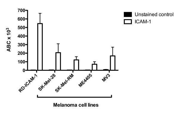
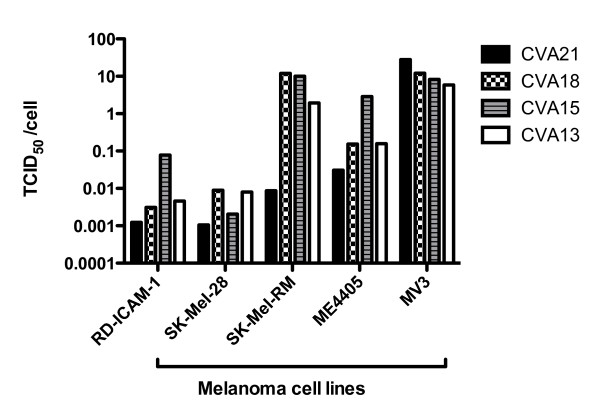
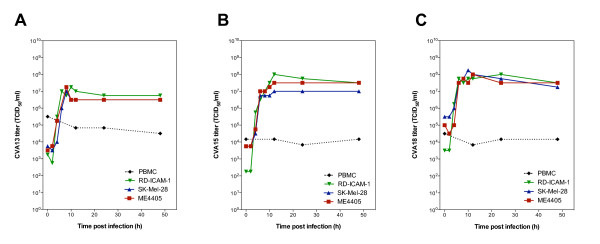
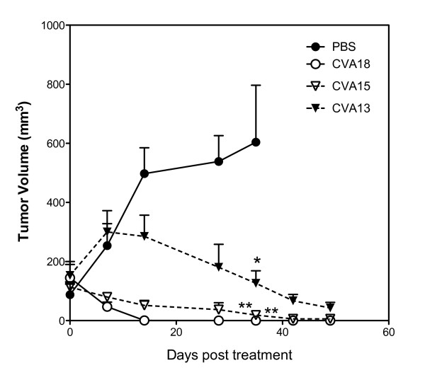
Many RNA viruses are displaying great promise in the field of oncolytic virotherapy. Previously, we reported that the picornavirus Coxsackievirus A21 (CVA21) possessed potent oncolytic activity against cultured malignant melanoma cells and melanoma xenografts in mice. In the present study, we demonstrate that three additional Group A Coxsackieviruses; Coxsackievirus A13 (CVA13), Coxsackievirus A15 (CVA15) and Coxsackievirus A18 (CVA18), also have similar oncolytic activity against malignant melanoma. Each of the viruses grew quickly to high titers in cancer cells expressing ICAM-1 and intratumoral injection of preformed subcutaneous SK-Mel-28 xenografts in mice with CVA13, CVA15 and CVA18 resulted in significant tumor volume reduction.As preexisting immunity could potentially hinder oncolytic virotherapy, sera from stage IV melanoma patients and normal controls were tested for levels of protective antibody against the panel of oncolytic Coxsackieviruses. Serum neutralization assays revealed that 3 of 21 subjects possessed low levels of anti-CVA21 antibodies, while protective antibodies for CVA13, CVA15 and CVA18 were not detected in any sample. Serum from individuals who were seropositive for CVA21 failed to exhibit cross-neutralization of CVA13, CVA15 and CVA18. From these studies it can be concluded that the administration of CVA13, CVA15 or CVA18 could be employed as a potential multivalent oncolytic therapy against malignant melanoma.


Figures

Figure 1
Figure 1
ICAM-1 expression on SK-Mel-28, ME4405 and RD-ICAM-1 cells. Quantitative flow cytometric analysis of virus-entry receptor ICAM-1 expression on melanoma cell lines SK-Mel-28, SK-Mel-RM, ME4405, MV3; and ICAM-1 transfected RD cell line (RD-ICAM-1). The level of ICAM-1 antibodies bound per cell (ABC) are denoted by the white bars whereas the unstained control is shown in black.
Figure 2
Figure 2
Destruction of melanoma cells by CVA13, CVA15, CVA18 and CVA21. Cultures of SK-Mel-28, SK-Mel-RM, ME4405, MV3 and RD-ICAM-1 monolayers were infected with 10-fold serial dilutions of virus ranging from 1:10 to 1:107. After incubation for 48 h, the plates were fixed and stained with a crystal violet/methanol and TCID50/cell required to induce monolayer destruction was calculated. SK-Mel-28 and RD-ICAM-1 were the most sensitive lines to the panel of CVAs requiring only low concentrations of virus to achieve cell lysis.
Figure 3
Figure 3
Growth curves of CVA13, CVA15 and CVA18 in SK-Mel-28, ME4405 and RD-ICAM-1 cells compared to PBMCs. SK-Mel-28, ME4405, RD-ICAM-1 and PBMCs were challenged with CVA13 (A), CVA15 (B) or CVA18 (C) for 1 h, and after the removal of unbound virus, cells and media were collected at the indicated time points for assessment of virus progeny by endpoint titration.
Figure 4
Figure 4
SK-Mel-28 tumors are responsive to intratumoral CVA13, CVA15 and CVA18 therapy in SCID mice. Severe combined immunodeficient (SCID) mice bearing preformed s.c. tumors (approximately 150 mm3) growing on the flanks after injection of 1 × 10⁶ SK-Mel-28 cells, received an intratumoral injection with a single dose of CVA13, CVA15, CVA18 (105 TCID50) or PBS (n = 5 for each group). The average relative tumor sizes were measured externally with calipers and are expressed as the means of five treated mice ± S.E. * and ** indicate statistical significance of tumor volumes compared to the control, P < 0.05 and P < 0.01 respectively (One-way ANOVA).

References

    1. Au GG, Lindberg AM, Barry RD, Shafren DR. Oncolysis of vascular malignant human melanoma tumors by Coxsackievirus A21. Int J Oncol. 2005;26:1471–1476. - PubMed
    1. Shafren DR, Au GG, Nguyen T, Newcombe NG, Haley ES, Beagley L, Johansson ES, Hersey P, Barry RD. Systemic therapy of malignant human melanoma tumors by a common cold-producing enterovirus, coxsackievirus a21. Clin Cancer Res. 2004;10:53–60. doi: 10.1158/1078-0432.CCR-0690-3. - DOI - PubMed
    1. Shokrollahi K, Whitaker I. Malignant melanoma: perspectives, diagnostics and treatment. Ann Plast Surg. 2010;64:132–133. doi: 10.1097/SAP.0b013e3181cf9f32. - DOI - PubMed
    1. Johnson JP. Cell adhesion molecules in the development and progression of malignant melanoma. Cancer Metastasis Rev. 1999;18:345–357. doi: 10.1023/A:1006304806799. - DOI - PubMed
    1. Johnson JP, Stade BG, Holzmann B, Schwable W, Riethmuller G. De novo expression of intercellular-adhesion molecule 1 in melanoma correlates with increased risk of metastasis. Proc Natl Acad Sci USA. 1989;86:641–644. doi: 10.1073/pnas.86.2.641. - DOI - PMC - PubMed

Publication types