. 2011 Apr 1;81(7):910-6.
doi: 10.1016/j.bcp.2011.01.005. Epub 2011 Jan 15.

ERK1/2 deactivation enhances cytoplasmic Nur77 expression level and improves the apoptotic effect of fenretinide in human liver cancer cells

Affiliations

ERK1/2 deactivation enhances cytoplasmic Nur77 expression level and improves the apoptotic effect of fenretinide in human liver cancer cells

Hui Yang et al. Biochem Pharmacol. .

Abstract

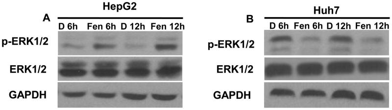
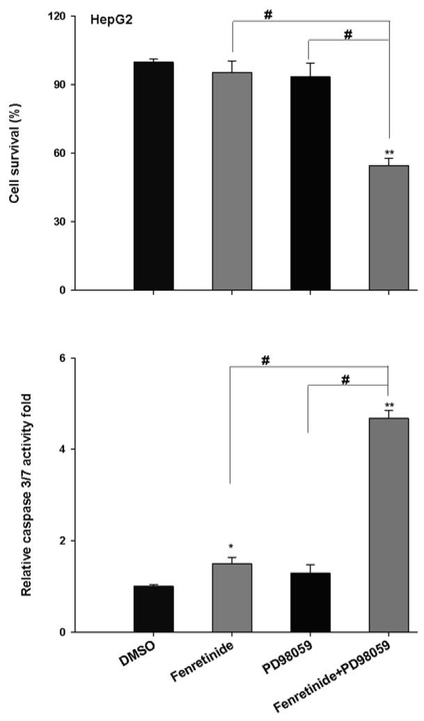
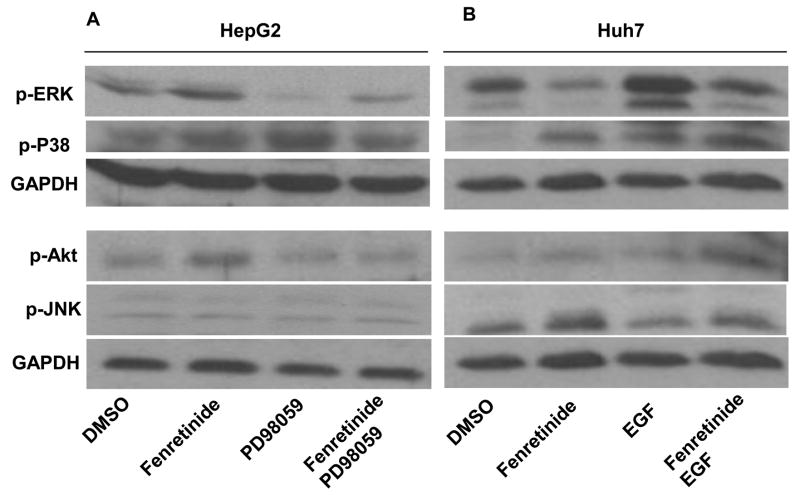
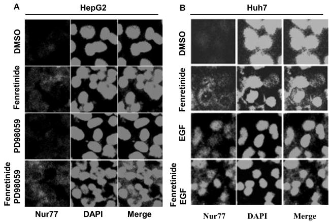
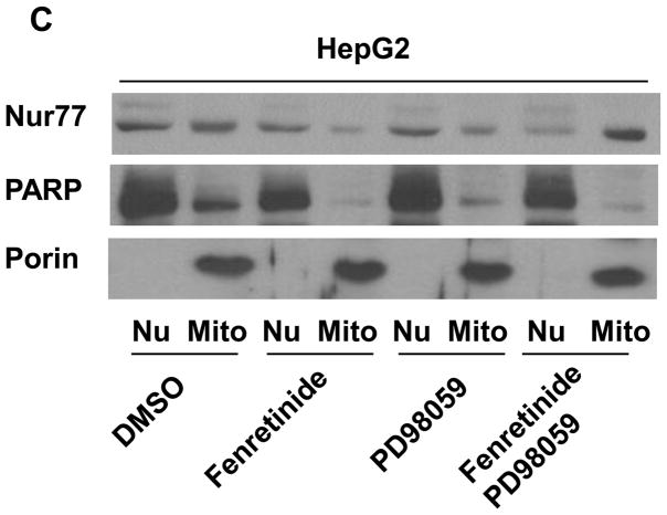
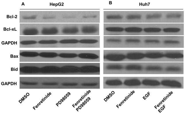
Fenretinide, a synthetic retinoid, is a promising anticancer agent based on many in vitro, animal, and chemoprevention clinical trial studies. However, cells such as HepG2 human liver cancer cells are resistant to the apoptotic effect of fenretinide. Previously, we have shown that fenretinide-induced apoptosis is Nur77 dependent, and the sensitivity of the cancer cells to fenretinide-induced apoptosis is positively associated with cytoplasmic enrichment of Nur77. The goal of current study was to identify means to modulate nuclear export of Nur77 in order to improve the efficacy of fenretinide. Fenretinide treatment deactivated ERK1/2 in Huh7 cells, but activated ERK1/2 in HepG2 cells, which was positively associated with the sensitivity of cells to the apoptotic effect of fenretinide. Neither fenretinide nor ERK1/2 inhibitor PD98059 alone could affect the survival of HepG2 cells, but the combination of both induced cell death and increased caspase 3/7 activity. In fenretinide sensitive Huh7 cells, activation of ERK1/2 by epidermal growth factor (EGF) prevented fenretinide-induced cell death and caspase 3/7 induction. In addition, modulation of ERK1/2 changed the intracellular localization of Nur77. Fenretinide/PD98059-induced cell death of HepG2 cell was positively associated with induction and cytoplasmic location as well as mitochondria enrichment of Nur77. The effect was specific for ERK1/2 because other mitogen activated protein kinases such as P38, Akt, and JNK did not have correlated changes in their phosphorylation levels. Taken together, the current study demonstrates that ERK1/2-modulated Nur77 intracellular location dictates the efficacy of fenretinide-induced apoptosis.


Figures

Figure 1
Figure 1
Differential effect of fenretinide on ERK1/2 activation in Huh7 and HepG2 cells. Huh7 and HepG2 cells were treated with fenretinide (10 μM) for 6 and 12 hrs. Phosphorylation of ERK1/2 was analyzed by Western blotting using antibody specific for phosphorylated ERK1/2. Fenretinide increased phosphorylated ERK1/2 in HepG2 cells (A), but decreased phosphorylated ERK1/2 in Huh7 cells (B).
Figure 2
Figure 2
Inhibition of ERK1/2 sensitizes HepG2 cells to the apoptotic effect of fenretinide. HepG2 cells were seeded onto a 96-well plate and treated with fenretinide (10 μM) or PD98059 (20 uM). For the combination treatment, HepG2 cells were exposed to PD98059 for 2 hrs and then co-exposed to fenretinide and PD98059 for additional 24 hrs. Caspase 3/7 activity and cell survival were determined by Caspase-Glo® 3/7 and CellTiter-Glo® Luminescent Cell Viability Assay (Promega), respectively as described in the Materials and Methods. Data were expressed as mean ± SD from three independent experiments, * *p<0.01, vs. DMSO; #p <0.05.
Figure 3
Figure 3
Activation of ERK1/2 by EGF protects Huh7 cells from fenretinide-induced apoptosis. Cells were seeded onto a 96-well plate and treated with fenretinide (10 μM) or EGF (0.2 μg/ml) for 24 hrs. For the combination treatment, Huh7 cells were treated with EGF for 2 hrs followed by EGF and fenretinide combination treatment for additional 24 hrs. Caspase 3/7 activity and cell survival were determined by Caspase-Glo® 3/7 and CellTiter-Glo® Luminescent Cell Viability Assay (Promega), respectively as described in the Materials and Methods. Data were expressed as mean ± SD from three independent experiments, **p<0.01, vs. DMSO; #p<0.05.
Figure 4
Figure 4
Fenretinide differentially regulates ERK1/2 activation in HepG2 (A) and Huh7 (B) cells. HCC cells were treated as described in figure legends 2 and 3. Proteins were extracted for western blot using specific antibodies as described in the Materials and Methods. GAPDH level was used for loading control. Representative data are shown from three independent experiments.
Figure 5
Figure 5
Modulation of ERK1/2 activation changes the intracellular localization of Nur77. HepG2 (A) and Huh7 (B) cells were treated as described in figure legends 2 and 3, respectively for 24 hrs. Immunofluorescence staining was performed using anti-Nur77 antibody and nuclear counterstaining with DAPI and viewed by confocal microscopy. Representative images of three independent experiments are shown. (C) Nuclear (Nu) and Mitochondria (Mit) enriched fractions were isolated from treated HepG2 cells. Proteins were fractionated followed by western blot using antibodies specific to Nur77, PARP, and Porin.
Figure 5
Figure 5
Modulation of ERK1/2 activation changes the intracellular localization of Nur77. HepG2 (A) and Huh7 (B) cells were treated as described in figure legends 2 and 3, respectively for 24 hrs. Immunofluorescence staining was performed using anti-Nur77 antibody and nuclear counterstaining with DAPI and viewed by confocal microscopy. Representative images of three independent experiments are shown. (C) Nuclear (Nu) and Mitochondria (Mit) enriched fractions were isolated from treated HepG2 cells. Proteins were fractionated followed by western blot using antibodies specific to Nur77, PARP, and Porin.
Figure 6
Figure 6
The expression levels of Bcl-2, Bcl-xL, Bax, and Bid are not associated with the apoptotic effect induced by fenretinide, PD98059, or EGF treatments. HepG2 (A) and Huh7 (B) cells were treated as described in figure legends 2 and 3, respectively. Proteins were extracted for western blot using specific antibodies as described in the Materials and Methods. GAPDH level was used for loading control. Representative data are shown from three independent experiments.
Figure 7
Figure 7
Modulation ERK1/2 activity does not affect fenretinide-generated ROS in HCC cells. HepG2 (A) and HuH7 (B) cells were treated as described in figure legends 2 and 3 for 6 hrs followed by staining using MitoSOX™ Red mitochondrial superoxide indicator. Cells were analyzed by flow cytometry. The results were generated from three independent experiments. *p<0.05 vs. DMSO.

Similar articles

Cited by

References

    1. Moon RC, Mehta RG. Chemoprevention of experimental carcinogenesis in animals. Prev Med. 1989;18:576–91. - PubMed
    1. Hail N, Jr, Kim HJ, Lotan R. Mechanisms of fenretinide-induced apoptosis. Apoptosis. 2006;11:1677–94. - PubMed
    1. Wu JM, DiPietrantonio AM, Hsieh TC. Mechanism of fenretinide (4-HPR)-induced cell death. Apoptosis. 2001;6:377–88. - PubMed
    1. Sogno I, Vene R, Ferrari N, De Censi A, Imperatori A, Noonan DM, et al. Angioprevention with fenretinide: targeting angiogenesis in prevention and therapeutic strategies. Crit Rev Oncol Hematol. 2010;75:2–14. - PubMed
    1. Preitner F, Mody N, Graham TE, Peroni OD, Kahn BB. Long-term Fenretinide treatment prevents high-fat diet-induced obesity, insulin resistance, and hepatic steatosis. Am J Physiol Endocrinol Metab. 2009;297:E1420–9. - PMC - PubMed

Publication types

MeSH terms

Substances