Comparative Study
. 2011 Mar 16:1379:11-22.
doi: 10.1016/j.brainres.2011.01.029. Epub 2011 Jan 15.

Hippocampal responsiveness to 17β-estradiol and equol after long-term ovariectomy: implication for a therapeutic window of opportunity

Affiliations
Comparative Study

Hippocampal responsiveness to 17β-estradiol and equol after long-term ovariectomy: implication for a therapeutic window of opportunity

Ryan T Hamilton et al. Brain Res. .

Abstract

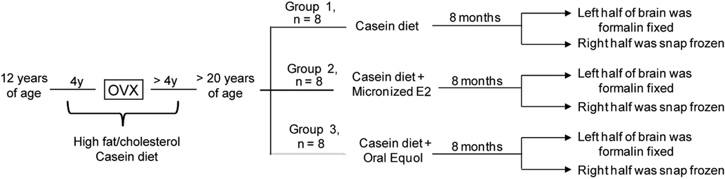
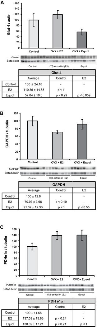
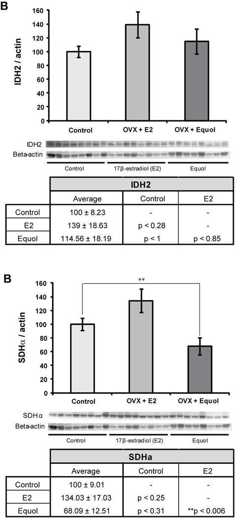
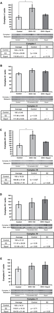
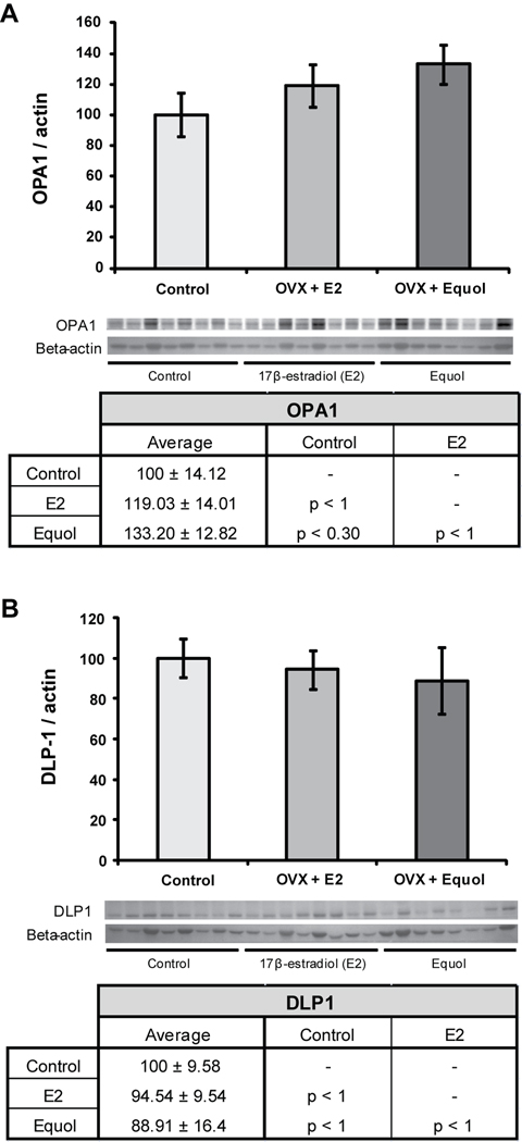
A 'critical window of opportunity' has been proposed for the efficacy of ovarian hormone intervention in peri- and post-menopausal women. We sought to address this hypothesis using a long-term ovariectomized non-human primate (NHP) model, the cynomolgus macaque (Macaca fascicularis). In these studies, we assessed the ability of 17β-estradiol and equol to regulate markers of hippocampal bioenergetic capacity. Results indicated that 17β-estradiol treatment significantly increased expression of mitochondrial respiratory chain proteins complex-I and -III in the hippocampus when compared to non-hormone-treated animals. Expression of the TCA cycle protein succinate dehydrogenase α was decreased in animals treated with equol compared to those treated with 17β-estradiol. There were no significant effects of either 17β-estradiol or equol treatment on glycolytic protein expression in the hippocampus, nor were there significant effects of treatment on expression levels of antioxidant enzymes. Similarly, 17β-estradiol and equol treatment had no effect on mitochondrial fission and fusion protein expression. In summary, findings indicate that while 17β-estradiol induced a significant increase in several proteins, the overall profile of bioenergetic system proteins was neutral to slightly positively responsive. The profile of responses with the ERβ-preferring molecule equol was consistent with overall nonresponsiveness. Collectively, the data indicate that long-term ovariectomy is associated with a decline in response to estrogens and estrogen-like compounds. By extension, the data are consistent with a primary tenet of the critical window hypothesis, i.e., that the brains of post-menopausal women ultimately lose their ability to respond positively to estrogenic stimulation.


Figures

Figure 1
Figure 1. Experimental paradigm and diets
A detailed description of the experimental protocol, showing the two hormone treatment conditions and the control condition.
Figure 2
Figure 2. 17β-estradiol and equol modulation of glucose uptake and glycolysis
Western blot analysis of Glut-4 (A), GAPDH (B), and PDHe1α (C) expression was performed on NHP hippocampal samples from both of the hormone treatment paradigms as well as controls. Expression levels for each sample were normalized to beta-actin levels (Glut-4) and beta-tubulin levels (GAPDH and PDHe1α). Expression levels were then normalized to the control animals (controls were set to 100%). Statistically significant differences were calculated using a two-tailed, one-way analysis of variance (ANOVA) followed by a Bonferroni post-hoc correction. Hormone treatment did not significant affect expression levels of these three proteins in the NHP hippocampus. Bars represent % control ± S.E.M., n = 8 for each condition.
Figure 3
Figure 3. 17β-estradiol and equol modulation of TCA cycle
Western blot analysis of IDH2 (A) and SDHα (B) expression was performed on NHP hippocampal samples from both of the hormone treatment paradigms as well as controls. Expression levels for each sample were normalized to beta-actin levels. Expression levels were then normalized to the control animals (controls were set to 100%). Statistically significant differences were calculated using a two-tailed, one-way analysis of variance (ANOVA) followed by a Bonferroni post-hoc correction. Hormone treatment significantly affected expression of SDHα in the NHP hippocampus. Bars represent % control ± S.E.M., n = 8 for each condition, **p<0.01.
Figure 4
Figure 4. 17β-estradiol and equol modulation of oxidative phosphorylation
Western blot analysis of complex-I (A), complex-II (B), complex-III (C), complex-IV-II (D) and complex-Vα (E) expression was performed on hippocampal samples from long-term ovariectomized NHP in both the hormone treatment conditions as well as controls. Expression levels for each sample were normalized to beta-actin levels. Statistically significant differences were determined by a two-tailed one-way analysis of variance (ANOVA) assuming unequal variances, followed by a Bonferroni post-hoc correction. 17β-estradiol therapy induced an increase in expression levels of complex-I (A) and complex-III (C) when compared to the control condition. No other significant changes from control were seen. Bars represent % control ± S.E.M., n = 8 for each condition, *p<0.05.
Figure 5
Figure 5. 17β-estradiol and equol modulation of antioxidant defense
Western blot analysis of MnSOD (A) and PRDX-V (B) expression was performed on hippocampal samples from long-term ovariectomized NHP. Expression levels for each sample were normalized to beta-actin levels. Statistically significant differences were determined by a two-tailed one-way analysis of variance (ANOVA) assuming unequal variances, followed by a Bonferroni post-hoc correction. Neither hormone treatment paradigm induced significant changes from control. Bars represent % control ± S.E.M., n = 8 for each condition.
Figure 6
Figure 6. 17β-estradiol and equol modulation of mitochondrial fission and fusion
Western blot analysis of OPA1 (A) and DLP-1 (B) expression was performed on hippocampal samples from long-term ovariectomized NHP. Expression levels for each sample were normalized to beta-actin levels. Statistically significant differences were determined by a two-tailed one-way analysis of variance (ANOVA) assuming unequal variances, followed by a Bonferroni post-hoc correction. Neither hormone treatment paradigm induced significant changes from control. Bars represent % control ± S.E.M., n = 8 for each condition

Similar articles

Cited by

References

    1. Adams MM, Shah RA, Janssen WG, Morrison JH. Different modes of hippocampal plasticity in response to estrogen in young and aged female rats. Proc. Natl. Acad. Sci. U. S. A. 2001;98:8071–8076. - PMC - PubMed
    1. Adams MM, Fink SE, Shah RA, Janssen WG, Hayashi S, Milner TA, McEwen BS, Morrison JH. Estrogen and aging affect the subcellular distribution of estrogen receptor-alpha in the hippocampus of female rats. J Neurosci. 2002;22(9):3608–3614. - PMC - PubMed
    1. Alzheimer's Association. Alzheimer's disease facts and figures. 2010 Mar - PubMed
    1. Appt SE, Clarkson TB, Register TC, Chen H. Dietary equol does not reproduce effects of dietary soy on cardiovascular disease risk variables; observations from postmenopausal monkeys. The North American Menopause Society; 16th Annual Meeting; September 28–October 1, 2005; San Diego. 2005.
    1. Barha CK, Galea LA. Motherhood alters the cellular response to estrogens in the hippocampus later in life. Neurobiol Aging. 2009 - PubMed

Publication types