. 2011 Mar 15;190(1):62-72.
doi: 10.1016/j.cbi.2011.01.009. Epub 2011 Jan 15.

Differentiation of human embryonic stem cells along a hepatic lineage

Affiliations

Differentiation of human embryonic stem cells along a hepatic lineage

Stephanie M Zamule et al. Chem Biol Interact. .

Abstract

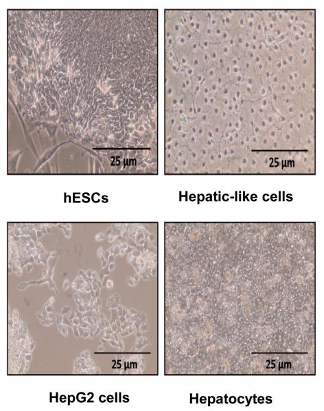
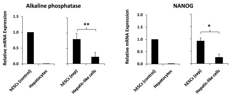
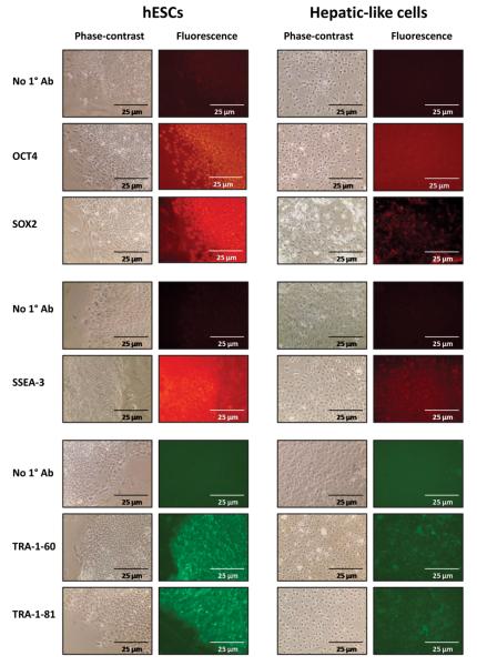
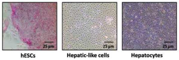
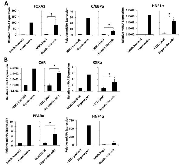
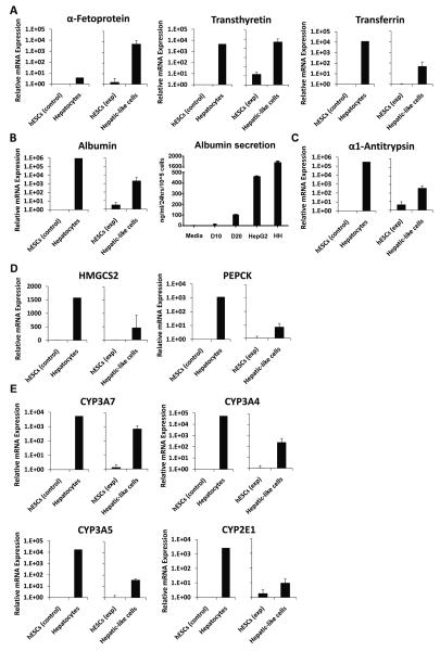
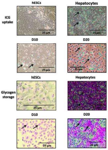
The limited availability of hepatic tissue suitable for the treatment of liver disease and drug discovery research advances the generation of hepatic-like cells from alternative sources as a valuable approach. In this investigation we exploited a unique hepatic differentiation approach to generate hepatocyte-like cells from human embryonic stem cells (hESCs). hESCs were cultured for 10-20 days on collagen substrate in highly defined and serum free hepatocyte media. The resulting cell populations exhibited hepatic cell-like morphology and were characterized with a variety of biological endpoint analyses. Real-time PCR analysis demonstrated that mRNA expression of the 'stemness' marker genes NANOG and alkaline phosphatase in the differentiated cells was significantly reduced, findings that were functionally validated using alkaline phosphatase activity detection measures. Immunofluorescence studies revealed attenuated levels of the 'stemness' markers OCT4, SOX2, SSEA-3, TRA-1-60, and TRA-1-81 in the hepatic-like cell population. The hepatic character of the cells was evaluated additionally by real-time PCR analyses that demonstrated increased mRNA expression of the hepatic transcription factors FOXA1, C/EBPα, and HNF1α, the nuclear receptors CAR, RXRα, PPARα, and HNF4α, the liver-generated plasma proteins α-fetoprotein, transthyretin, transferrin, and albumin, the protease inhibitor α-1-antitrypsin, metabolic enzymes HMGCS2, PEPCK, and biotransformation enzymes CYP3A7, CYP3A4, CYP3A5, and CYP2E1. Indocyanine green uptake albumin secretion and glycogen storage capacity further confirmed acquisition of hepatic function. These studies define an expeditious methodology that facilitates the differentiation of hESCs along a hepatic lineage and provide a framework for their subsequent use in pharmacological and toxicological research applications requiring a renewable supply of human hepatocytes.


Figures

Figure 1
Figure 1. HESCs subjected to hepatic differentiation exhibit hepatic cell-like morphology
Images depict phase-contrast micrographs of an undifferentiated hESC colony (hESCs) and hESC-derived hepatic-like cells generated by culturing passage-matched hESCs for 10 days on type I rat-tail collagen in hepatocyte media (Hepatic-like cells), in parallel to the undifferentiated hESC negative controls. HepG2 hepatoma cells (HepG2 cells) and cultures of primary human hepatocytes (Hepatocytes) are included as positive controls.
Figure 2
Figure 2. ‘Stemness’ marker mRNA expression is significantly decreased in hepatic-like cells
Passage-matched HESCs were cultured in parallel on either hFF feeder layers in hESC media (hESCs exp) or on collagen in hepatocyte media (Hepatic-like cells). After 10 days in culture cells were harvested and RNA was isolated, converted to cDNA, subjected to real-time PCR, and the data analyzed using the ΔΔCT method to determine fold expression levels of NANOG and alkaline phosphatase relative to low-passage hESCs. Control data (left graph) depict means of expression levels of replicate determinations of a typical low-passage culture of hESCs (hESCs control), in contrast to mean expression levels of replicate determinations performed using a pooled sample of six adult primary human hepatocyte donors (Hepatocytes). In these control experiments, the replicate values were reproducible within a ≤ 5% error range. Experimental data (right graph) depict expression levels of hESCs (hESCs exp) and passage-matched hESCs subject to our hepatic differentiation protocol (Hepatic-like cells). Experimental data are expressed as mean +/− standard deviations of at least two independent trials using hESCs from different passages. * p < 0.05; ** p < 0.01.
Figure 3
Figure 3. ‘Stemness’ marker protein expression is reduced in hepatic-like cells
Passage-matched hESCs were cultured in parallel on either hFF feeder layers in hESC media (hESCs) or on collagen in hepatocyte media (Hepatic-like cells). At 10 days in culture cells were probed for OCT4, SOX2, SSEA-3, TRA-1-60, and TRA-1-81 protein expression by immunofluorescence using antibodies specified in the Methods. Cells were examined microscopically and phase-contrast and fluorescence images were captured.
Figure 4
Figure 4. Hepatic-like cells exhibit decreased ‘stemness’ function
Passage-matched hESCs were cultured in parallel on either hFF feeder layers in hESC media (hESCs) or on collagen in hepatocyte media (Hepatic-like cells). At 10 days in culture cells were stained for alkaline phosphatase activity (pink) and phase-contrast images were captured. Primary adult human hepatocytes (Hepatocytes) stained in parallel serve as a negative control.
Figure 5
Figure 5. Hepatic transcription factor mRNA expression is increased in hepatic-like cells
Passage-matched hESCs were cultured in parallel on either hFF feeder layers in hESC media (hESCs exp) or on collagen in hepatocyte media (Hepatic-like cells). After 10 days in culture, cells were harvested and RNA was isolated, converted to cDNA, subjected to real-time PCR, and the data analyzed using the ΔΔCT method [32] to determine fold expression levels relative to low-passage hESCs (hESCs control) of: (A), the hepatic transcription factors FOXA1, C/EBPα, and HNF1α; and (B), the hepatic nuclear receptors CAR, RXRα, PPARα, and HNF4α. Control data (left graph for each endpoint) depict means of expression levels of replicate determinations of a typical low-passage culture of hESCs (hESCs control), in contrast to mean expression levels of replicate determinations performed using a pooled sample of six adult primary human hepatocyte donors (Hepatocytes). In these control experiments, the replicate values were reproducible within a ≤ 5% error range. Experimental data (right graph) depict expression levels of hESCs (hESCs exp) and passage-matched hESCs subject to our hepatic differentiation protocol (Hepatic-like cells). Experimental data are expressed as mean +/− standard deviations of at least three independent trials using hESCs from different passages. * p < 0.05.
Figure 6
Figure 6. Hepatic marker mRNA expression is augmented in hepatic-like cells
Passage-matched hESCs were cultured in parallel on either hFF feeder layers in hESC media (hESCs exp) or on collagen in hepatocyte media (Hepatic-like cells). After either 10 or 20 days in culture, cells were harvested and RNA was isolated, converted to cDNA, subjected to real-time PCR, and the data analyzed using the ΔΔCT method [32] to determine fold expression levels relative to low-passage hESCs (hESCs control) of: (A) the plasma proteins α-fetoprotein, transthyretin, and transferrin; (B) albumin transcript expression and Elisa-based protein secretion analysis; (C) the protease inhibitor α-1-antitrypsin; (D), the metabolic enzymes HMGCS2 and PEPCK; and (E), the biotransformation enzymes CYP3A7, CYP3A4, CYP3A5, and CYP2E1. Control data (left graph) depict means of expression levels of replicate determinations of a typical low-passage culture of hESCs (hESCs control), in contrast to mean expression levels of replicate determinations performed using a pooled sample of six adult primary human hepatocyte donors (Hepatocytes). In these control experiments, the replicate values were reproducible within a ≤ 5% error range. Experimental data (right graph) depict expression levels of hESCs (hESCs exp) and passage-matched hESCs subject to our hepatic differentiation protocol (Hepatic-like cells). Experimental data are expressed as mean +/− standard deviations of at least three independent trials using hESCs from different passages.
Figure 7
Figure 7. Hepatic-like cells exhibit the hepatic-specific functions of ICG uptake and glycogen storage
Passage-matched hESCs were cultured in parallel on either hFF feeder layers in hESC media (hESCs) or on collagen in hepatocyte media (Hepatic-like cells) for either 10 or 20 days. Cells were evaluated for ICG uptake (cyan-green) by incubation with 100 μg/ml ICG and for glycogen storage capacity (pink) using periodic acid-Schiff (PAS) staining, both as described in Materials and Methods. Cells were examined microscopically and phase-contrast images were captured. Cultures of adult primary human hepatocytes (Hepatocytes) assessed in parallel were included as positive controls.

Similar articles

Cited by

References

    1. Spear BT, Jin L, Ramasamy S, Dobierzewska A. Transcriptional control in the mammalian liver: liver development, perinatal repression, and zonal gene regulation. Cell Mol Life Sci. 2006;63:2922–38. - PMC - PubMed
    1. Cao S, Esquivel CO, Keeffe EB. New approaches to supporting the failing liver. Annu Rev Med. 1998;49:85–94. - PubMed
    1. Malhi H, Gupta S. Hepatocyte transplantation: new horizons and challenges. J Hepatobiliary Pancreat Surg. 2001;8:40–50. - PubMed
    1. Schwartz RE, Linehan JL, Painschab MS, Hu WS, Verfaillie CM, Kaufman DS. Defined conditions for development of functional hepatic cells from human embryonic stem cells. Stem Cells Dev. 2005;14:643–55. - PubMed
    1. Lemaigre F, Zaret KS. Liver development update: new embryo models, cell lineage control, and morphogenesis. Curr Opin Genet Dev. 2004;14:582–90. - PubMed

Publication types

MeSH terms