. 2011 Apr;228(2):222-31.
doi: 10.1016/j.expneurol.2011.01.006. Epub 2011 Jan 15.

A brain slice culture model of viral encephalitis reveals an innate CNS cytokine response profile and the therapeutic potential of caspase inhibition

Affiliations

A brain slice culture model of viral encephalitis reveals an innate CNS cytokine response profile and the therapeutic potential of caspase inhibition

Kalen R Dionne et al. Exp Neurol. 2011 Apr.

Abstract

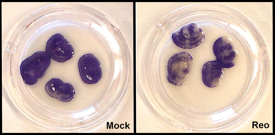
Viral encephalitis is a significant cause of human morbidity and mortality in large part due to suboptimal diagnosis and treatment. Murine reovirus infection serves as a classic experimental model of viral encephalitis. Infection of neonatal mice with T3 reoviruses results in lethal encephalitis associated with neuronal infection, apoptosis, and CNS tissue injury. We have developed an ex vivo brain slice culture (BSC) system that recapitulates the basic pathological features and kinetics of viral replication seen in vivo. We utilize the BSC model to identify an innate, brain-tissue specific inflammatory cytokine response to reoviral infection, which is characterized by the release of IL6, CXCL10, RANTES, and murine IL8 analog (KC). Additionally, we demonstrate the potential utility of this system as a pharmaceutical screening platform by inhibiting reovirus-induced apoptosis and CNS tissue injury with the pan-caspase inhibitor, Q-VD-OPh. Cultured brain slices not only serve to model events occurring during viral encephalitis, but can also be utilized to investigate aspects of pathogenesis and therapy that are not experimentally accessible in vivo.


Figures

Figure 1
Figure 1. BSCs support viral replication
(A) Anatomical organization of a freshly cut brain slice containing thalamic, hippocampal, and cortical tissue. Red inset delineates the frontoparietal cortical area pictured in Figure 1B. (B) At 5dpi, BSCs were removed from media and fixed in 4% paraformaldehyde before being cryosectioned. Resliced sections were stained with anti-reovirus monoclonal σ3 primary antibodies and anti-mouse Cy3-conjugated secondary antibodies. High levels of reovirus antigen were found within cells of the cortex, localized to discrete foci. 100X magnification. (C) 5dpi BSCs were fixed and cryosectioned prior to incubation with primary antibodies: monoclonal reovirus σ3 antibody and mouse anti-neuronal nucleus antibody (NeuN) or anti-glial fibrillary acidic protein (GFAP). Sections were then incubated with secondary antibodies: Alexa Fluor 488-conjugated goat anti-mouse IgG and/or Cy3-conjugated goat anti-rabbit IgG. Reovirus antigen is located specifically within the cytoplasm of neurons, as represented by white arrows. 630X magnification. (D) RNA was purified from BSCs at stated times post-infection (N≥2). Quantification of L1 gene of reovirus relative to β-actin transcript revealed exponential viral growth in the first week in culture, yielding significant differences (p≤0.014) between early (dpi≤1) and late (dpi≥3) timepoints. t=0 was defined as a reference value equal to 1. (E) BSCs (N≥3) were harvested at given time points and processed for absolute viral titer determination by plaque assay on L929 mouse fibroblasts. Titers of all later time points were significantly higher (p≤0.0071) than the titer at the time of inoculation (t=0).
Figure 2
Figure 2. Viral infection induces tissue injury in BSCs
(A) At 7dpi, slices were incubated with 3µM propidium iodide for 1h prior to fluorescence determination by plate reader (N=3). Fluorescence was significantly greater (p=0.004) in reovirus-infected samples, suggesting viral-induced tissue injury. (B) Media was taken at specified time points and LDH was detected via colorimetric assay (N≥3). In mock samples, tissue injury was evident in the days following slicing, but decreased progressively. However, tissue injury was sustained in reovirus-infected BSCs, suggesting injury induced by the virus. (C) At 11dpi, BSC media was replaced with fresh media containing 0.5 mg/mL MTT and photographs were taken 45 minutes later. Live tissue cleaved the substrate to form purple formazan crystals, whereas dead tissue remained pale/white. Cortical and subcortical tissue death is evident in reovirus-infected BSCs.
Figure 3
Figure 3. Viral infection induces apoptosis in BSCs
(A) BSCs (N≥3) were harvested into lysis buffer at indicated times before Caspase 3 activity was determined by fluorogenic activity assay. At all time points Caspase 3 activity was increased in reovirus-infected BSCs when compared to mock samples, with significant differences (p≤0.029) occurring at 7, 9, and 11dpi. (B) BSCs were harvested into lysis buffer at indicated times and ran on SDS-PAGE gel. Cleaved Caspase 3 (cC3), an apoptotic marker, was markedly increased in reovirus-infected BSCs. (C) BSCs were harvested into lysis buffer at specified times and ran on SDS-PAGE gel. PARP cleavage, a downstream result of Caspase 3 activation, was detected in reovirus-infected samples to a significant degree.
Figure 4
Figure 4. Inflammatory cytokines are released into the media of virus-infected BSCs
The four cytokines identified to be released in response to reovirus infection by ELISArray were quantified on individual ELISA plates. At 9dpi, media from experimentally similar wells was pooled into a single aliquot for independent experiments (N≥3) and serially diluted for quantification of cytokines based on standard curves (R2 >0.97). The release of KC (murine IL8 analog), RANTES, CXCL10, and IL6 was significantly greater (p≤0.037) in reovirus-infected BSCs.
Figure 5
Figure 5. Q-VD-OPh treatment limits tissue injury and apoptosis, but not viral replication, in virus-infected BSCs
Approximately 12h after being infected, negative control peptide or Q-VD-OPh was applied dropwise to the top of each slice at a concentration of 500 µg/mL to yield a final media concentration of 36.4 µg/mL. In a similar manner, additional treatments were made at each subsequent media change. (A) BSCs were harvested into lysis buffer at 7dpi and ran on SDS-PAGE gel. Cleaved Caspase 3 (cC3), an apoptotic marker, was detected in negative control treated samples but not in Q-VD-OPh treated samples. Western blot is representative of two independent experiments. (B) BSC media from mock (no virus/no peptide treatment), infected/negative control peptide treatment, and infected/O-VD-OPh treated samples (N=5) was harvested at 7dpi for LDH assay determination of tissue injury. With mock samples defined as a 0% injury reference and infected/negative control peptide defined as a 100% injury reference, infected/Q-Vd-OPh-treated BSCs had a 35% reduction (p=0.008) in terms of virus-induced injury. (C) At 11dpi, BSC media was replaced with fresh media containing 0.5 mg/mL MTT and photographs were taken 45 minutes later. Live tissue cleaved the substrate to form purple formazan crystals, whereas dead tissue remained pale/white. Virus-induced tissue death was evident in infected/negative control peptide-treated BSCs, but was inhibited in infected/Q-VD-OPh-treated samples. (D) BSCs were harvested at 7dpi for qRT-PCR determination of relative viral load, which was unchanged by Q-VD-OPh treatment (N=4). The infected/negative control peptide treated condition was defined as a reference value equal to 1.

Similar articles

Cited by

References

    1. Aiba H, Mochizuki M, Kimura M, Hojo H. Predictive value of serum interleukin-6 level in influenza virus-associated encephalopathy. Neurology. 2001;57:295–299. - PubMed
    1. Akimitsu T, Kurisu K, Hanaya R, Iida K, Kiura Y, Arita K, Matsubayashi H, Ishihara K, Kitada K, Serikawa T, Sasa M. Epileptic seizures induced by N-acetyl-L-aspartate in rats: in vivo and in vitro studies. Brain Res. 2000;861:143–150. - PubMed
    1. Beckham JD, Goody RJ, Clarke P, Bonny C, Tyler KL. Novel strategy for treatment of viral central nervous system infection by using a cell-permeating inhibitor of c-Jun N-terminal kinase. J. Virol. 2007;81:6984–6992. - PMC - PubMed
    1. Beckham JD, Tuttle KD, Tyler KL. Caspase-3 activation is required for reovirus-induced encephalitis in vivo. J. Neurovirol. 2010;16:306–317. - PMC - PubMed
    1. Braun E, Zimmerman T, Ben Hur T, Reinhartz E, Fellig Y, Panet A, Steiner I. Neurotropism of herpes simplex virus type 1 in brain organ cultures. Journal of General Virology. 2006;87:2827–2837. - PubMed

Publication types

MeSH terms