. 2011 Jun;60(7-8):1292-300.
doi: 10.1016/j.neuropharm.2011.01.010. Epub 2011 Jan 15.

Heat shock enhances CMV-IE promoter-driven metabotropic glutamate receptor expression and toxicity in transfected cells

Affiliations

Heat shock enhances CMV-IE promoter-driven metabotropic glutamate receptor expression and toxicity in transfected cells

Sergey Pshenichkin et al. Neuropharmacology. 2011 Jun.

Abstract

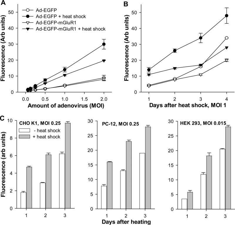
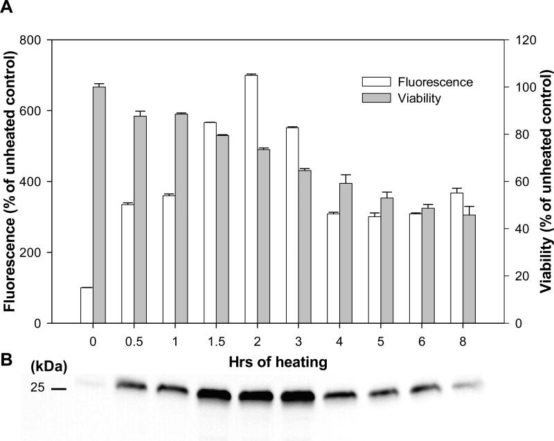
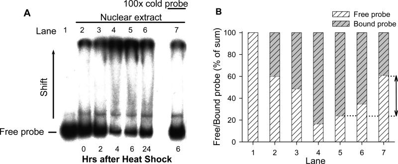
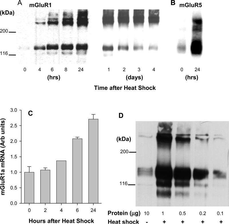
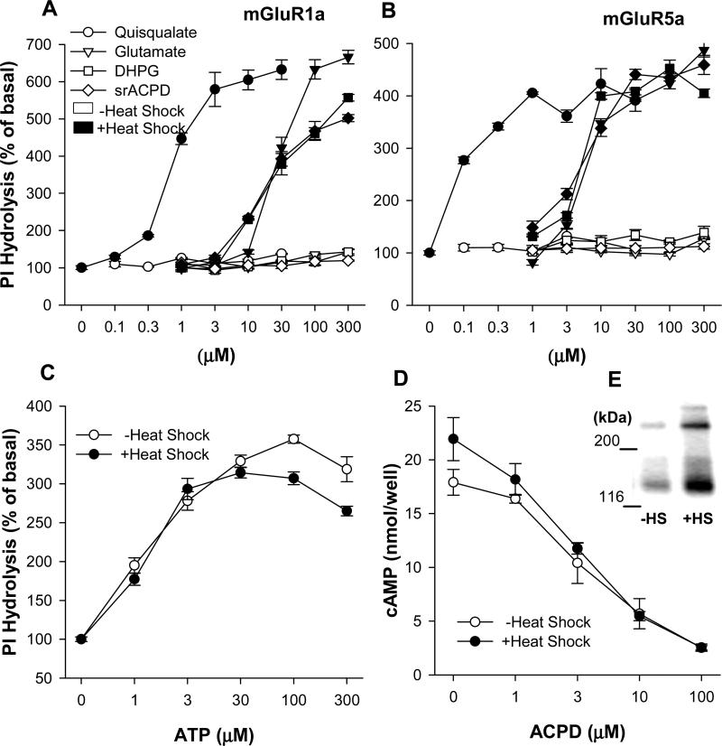
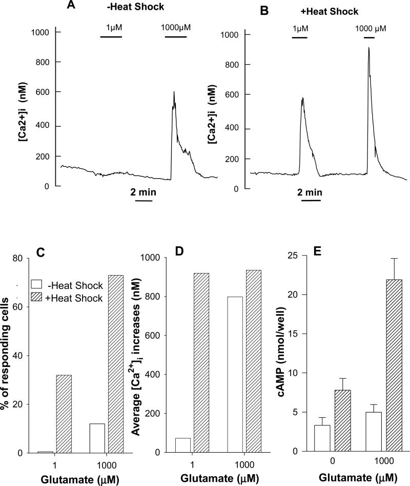
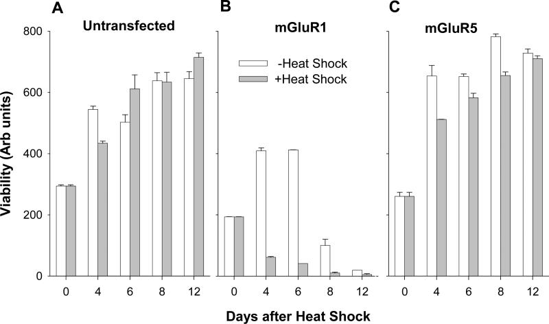
In CHO-K1 cells, heat shock strongly activated reporter-gene expression driven by the cytomegalovirus immediate-early (CMV-IE) promoter from adenoviral and plasmid vectors. Heat shock treatment (2h at 42.5 °C) significantly enhanced the promoter DNA-binding activity in nuclear extracts. In CHO cells expressing mGluR1a and mGluR5a receptors under the control of the CMV promoter, heat shock increased receptor protein expression, mRNA levels and receptor function estimated by measurement of PI hydrolysis, intracellular Ca²+ and cAMP. Hyperthermia increased average amplitudes of Ca²+ responses, the number of responding cells, and revealed the toxic properties of mGluR1a receptor. Heat shock also effectively increased the expression of EGFP. Hence, heat shock effects on mGluR expression and function in CHO cells may be attributed to the activation of the CMV promoter. Moreover, this effect was not limited to CHO cells as heat shock also increased EGFP expression in PC-12 and HEK293 cells. Heat shock treatment may be a useful tool to study the function of proteins expressed in heterologous systems under control of the CMV promoter. It may be especially valuable for increasing protein expression in transient transfections, for enhancing receptor expression in drug screening applications and to control the expression of proteins endowed with toxic properties. This article is part of a Special Issue entitled 'Trends in neuropharmacology: in memory of Erminio Costa'.


Figures

Fig. 1
Fig. 1
Effect of heat shock on expression of adenovirus-delivered EGFP and EGFP-mGluR1a. (A) CHO-K1 cells were infected one day before heating using different titers (MOI) of adenovirus and fluorescence of the expressed proteins was measured 24 hrs after HS treatment. (B) Time course of protein expression in CHO-K1 cells at different days after HS treatment, performed on day 0. (C) Effect of HS on adenovirus-mediated EGFP expression in different cell lines. CHO-K1 and PC-12 cells (infected at MOI 0.25), and HEK 293 cells (infected at MOI 0.015) were treated and monitored as in (B). Cells were heated for 2 hrs at 42.5°C.
Fig. 2
Fig. 2
Effect of heat shock on the reporter gene expression driven by the CMV-IE promoter/enhancer in transfected CHO-K1 cells. CHO-K1 cells were permanently transfected with pEGFP-C2 vector expressing EGFP as a reporter gene under control of the CMV-IE promoter/enhancer. The cells were heated at 42.5°C for the indicated time. (A) Activity of the promoter was evaluated by measurement of the reporter gene expression (i.e. EGFP fluorescence) 24 hrs later. Cell viability was measured by MTT assay in the same samples. (B) Whole cell extracts were collected next day after heating. Samples (5 μg of total protein) were resolved on 12 % PAGE. EGFP expression was measured by Western blotting with specific anti-GFP antibodies. Each protein band corresponds to the time-point indicated in (A).
Fig. 3
Fig. 3
Heat shock induces DNA-binding activity to the CMV promoter in nuclear extracts from CHO-K1 cells. (A) Nuclear extracts were obtained from the cells before heat shock (Lane 2) and 2, 4, 6 and 24 hrs after heating (Lanes 3-7). Heat shock treatment was performed for 2 hrs. Binding activity of nuclear proteins to the radiolabeled CMV promoter probe was estimated by the electromobility shift assay. The extracts were incubated with [P32]-probe for 60 min and resolved on 6% PAGE. The lower bands correspond to the free probe, the upper – to the protein-DNA complexes. Specificity of binding to the CMV promoter was examined in a competition reaction with a 100-fold excess of cold probe added to the binding reaction (Lane 7). Lane 1 contains only free [P32]-probe without nuclear extracts. (B) Quantification of ratio (in % of sum) of free and bound probe. Lanes correspond to those in (A). The arrow indicates the effect of cold probe addition (Lane 7 should be compared with Lane 5 as indicated by dotted line).
Fig. 4
Fig. 4
Heat shock induces mGluR1a and mGluR5a expression in stably transfected CHO-K1 cells. Cells were heated for 2 hrs at 42.5°C. (A) Cell membranes were collected at the indicated times after the treatment and mGluR1a expression was determined by Western blotting with specific anti-mGluR1a antibodies. The receptors appear on the blot as a combination of two bands corresponding to monomeric (145 kDa) and dimeric (290 kDa) forms of the mGluR1a. One μg of membrane protein was loaded into each well. (B) mGluR5a expression was measured similarly with anti-mGluR5a antibodies. (C) mGluR1a mRNA expression in transfected cells was measured by quantitative real-time PCR. The first bar (0 hrs) represents mRNA expression in control unheated cells. mGluR1a mRNA levels were measured in triplicates and normalized to the levels of mRNA for housekeeping gene (GAPDH). (D) Heat shock increases mGluR1a expression in CHO-K1 cells nearly 100-fold. Membrane proteins were isolated 24 hrs after heating and resolved by Western blotting. In order to estimate the magnitude of stimulation of the receptors expression by heat shock the dilution curve of the samples was done. Indicated amounts of membrane protein were loaded into the gel. Lane 1, membranes from control unheated CHO-K1 cells; lanes 2-5, membranes from heat shock-treated cells.
Fig. 5
Fig. 5
Heat shock increases PI hydrolysis in mGluR1a and mGlur5a transfected CHO-K1 cells. Permanently transfected cells cultured on 96 well plates were heated for 2 hrs at 42.5°C. Measurement of PI hydrolysis was performed on the next day. (A) mGluR1a-transfected cells were treated with mGluR agonists ACPD, DHPG, glutamate and quisqualate together with 20 mM LiCl in Locke's buffer. Open and black symbols represent IP accumulation in untreated cells and in heated cells respectively. (B) PI hydrolysis in mGluR5a-transfected cells. (C) Heat shock treatment did not induce enhancement of PI hydrolysis in untransfected CHO-K1 cells stimulated by ATP, an agonist of endogenous purinoceptors. (D and E) Heat shock increases mGluR2 expression but does not potentiate physiological response of the receptor in permanently transfected CHO-K1 cells. Adenylyl cyclase activity was measured next day after heat shock treatment. cAMP accumulation was induced by 10 μM forskolin in the presence of 300 μM IBMX. Activation of mGluR2 receptors by mGluR agonist ACPD inhibited cAMP accumulation and was measured by radioimmunoassay using a magnetic Amerlex RIA kit. The receptors expression was measured by Western blotting in control unheated (-HS) and heated (+HS) cells 24 hrs after heat shock treatment. The receptor appears on the blot as combination of two bands corresponding to monomeric and dimeric forms.
Fig. 6
Fig. 6
In mGluR1a transfected CHO cells heat shock increases both agonist potency and proportion of cells responding to agonist. (A) Average Ca2+ signals of responding cells stimulated by glutamate under control conditions (10 responding cells of 82 in the field of observation). (B) Cells were heated at 42.5°C for 2 hrs 1 day before measurements were performed (27 responding cells of 51 in the field of observation). Data of two representative experiments are shown; similar results were obtained in 3-5 other experiments. (C) Summarized data indicating proportion of cells responding to different glutamate concentrations under control conditions (17 out of 222 cells in 3 independent experiments) and after heating (141 out of 243 cells in 5 independent experiments). (D) Average [Ca2+]i signals of the same (C) responding cells. (E) Heat shock enhances cAMP formation in mGluR1a expressing CHO-K1 cells. Cells were heated for 2 hours at 42.5°C. After additional 24 hours cells were used for determination of cAMP accumulation stimulated by agonist in presence of phosphodiesterase inhibitor IBMX.
Fig. 7
Fig. 7
Heat-shock enhances toxic properties of mGluR1a in transfected CHO cell lines. Untransfected and transfected with mGluR1a or mGluR5a cells were seeded at the same density in 96 well plates. Cell viability in control and heated cultures was measured by MTT assay. The first measurement (at DIV 4) was done immediately after heat shock treatment.

Similar articles

Cited by

References

    1. Abe T, Sugihara H, Nawa H, Shigemoto R, Mizuno N, Nakanishi S. Molecular Characterization of a Novel Metabotropic Glutamate Receptor MGluR5 Coupled to Inositol Phosphate/Ca2+ Signal Transduction. J Biol Chem. 1992;267:13361–8. - PubMed
    1. Angulo A, Suto C, Heyman RA, Ghazal P. Characterization of the Sequences of the Human Cytomegalovirus Enhancer That Mediate Differential Regulation by Natural and Synthetic Retinoids. Mol Endocrinol. 1996;10:781–93. - PubMed
    1. Aramori I, Nakanishi S. Signal Transduction and Pharmacological Characteristics of a Metabotropic Glutamate Receptor, MGluR1, in Transfected CHO Cells. Neuron. 1992;8:757–65. - PubMed
    1. Boom R, Sol CJ, Minnaar RP, Geelen JL, Raap AK, van der Noordaa J. Induction of Gene Expression Under Human Cytomegalovirus Immediate Early Enhancer-Promoter Control by Inhibition of Protein Synthesis Is Cell Cycle-Dependent. J Gen Virol. 1988;69(Pt 6):1179–93. - PubMed
    1. Chan YJ, Chiou CJ, Huang Q, Hayward GS. Synergistic Interactions Between Overlapping Binding Sites for the Serum Response Factor and ELK-1 Proteins Mediate Both Basal Enhancement and Phorbol Ester Responsiveness of Primate Cytomegalovirus Major Immediate-Early Promoters in Monocyte and T-Lymphocyte Cell Types. J Virol. 1996;70:8590–605. - PMC - PubMed

Publication types

Substances