

Mefloquine neurotoxicity is mediated by non-receptor tyrosine kinase
- PMID: 21241737
- PMCID: PMC6457117
- DOI: 10.1016/j.neuro.2011.01.001
Mefloquine neurotoxicity is mediated by non-receptor tyrosine kinase
Abstract
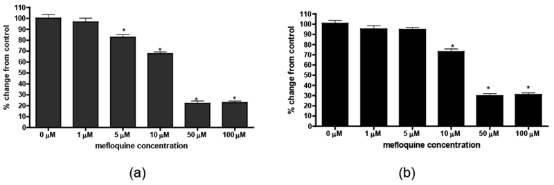
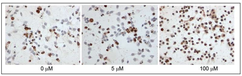
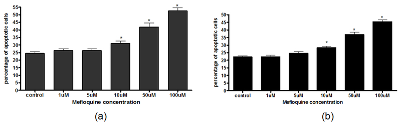
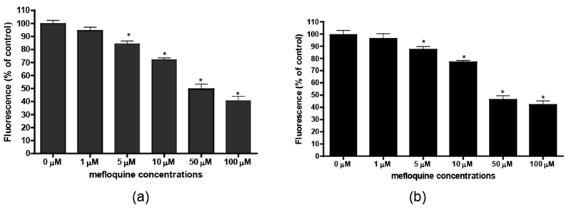
Among several available antimalarial drugs, mefloquine has proven to be effective against drug-resistant Plasmodium falciparum and remains the drug of choice for both therapy and chemoprophylaxis. However, mefloquine is known to cause adverse neurological and/or psychiatric symptoms, which offset its therapeutic advantage. The exact mechanisms leading to the adverse neurological effects of mefloquine are poorly defined. Alterations in neurotransmitter release and calcium homeostasis, the inhibition of cholinesterases and the interaction with adenosine A(2A) receptors have been hypothesized to play prominent roles in mediating the deleterious effects of this drug. Our recent data have established that mefloquine can also trigger oxidative damage and subsequent neurodegeneration in rat cortical primary neurons. Furthermore, we have utilized a system biology-centered approach and have constructed a pathway model of cellular responses to mefloquine, identifying non-receptor tyrosine kinase 2 (Pyk2) as a critical target in mediating mefloquine neurotoxicity. In this study, we sought to establish an experimental validation of Pyk2 using gene-silencing techniques (siRNA). We have examined whether the downregulation of Pyk2 in primary rat cortical neurons alters mefloquine neurotoxicity by evaluating cell viability, apoptosis and oxidative stress. Results from our study have confirmed that mefloquine neurotoxicity is associated with apoptotic response and oxidative injury, and we have demonstrated that mefloquine affects primary rat cortical neurons, at least in part, via Pyk2. The implication of these findings may prove beneficial in suppressing the neurological side effects of mefloquine and developing effective therapeutic modalities to offset its adverse effects.
Copyright © 2011 Elsevier Inc. All rights reserved.
Figures

Comment in
-
Mefloquine neurotoxicity and gap junction blockade: critical insights in drug repositioning.Neurotoxicology. 2011 Dec;32(6):986-7; author reply 987. doi: 10.1016/j.neuro.2011.05.003. Epub 2011 May 14. Neurotoxicology. 2011. PMID: 21605593 No abstract available.
References
-
- Avraham H, Park SY, Schinkmann K, Avraham S. RAFTK/Pyk2-mediated cellular signaling. Cell Signalling 2000; 12:123–133. - PubMed
-
- Chellaiah MA, Kuppuswamy D, Lasky L, Linder S. Phosphorylation of a Wiscott-Aldrich syndrome protein-associated signal complex is critical in osteoclast bone resorption. J Biol Chem 2007; 282:10104–16. - PubMed
-
- Corvol JC, Valjent E, Toutant M, Enslen H, Irinopoulou T, Lev S, Herve D, Girault JA. Depolarization Activates ERK and Proline-rich Tyrosine Kinase 2 (PYK2) Independently in Different Cellular Compartments in Hippocampal Slices. J Biol Chem 2005; 280:660–8. - PubMed
-
- Dikic I, Tokiwa G, Lev S, Courtneidge SA, Schlessinger J. A role for Pyk2 and Src in linking G-protein-coupled receptors with MAP kinase activation. Nature 1996; 383:547–550. - PubMed
Publication types
MeSH terms
Substances
Grants and funding
LinkOut - more resources
Full Text Sources
Miscellaneous
