. 2011 Apr;1812(4):507-13.
doi: 10.1016/j.bbadis.2011.01.007. Epub 2011 Jan 15.

Amyloid beta impairs mitochondrial anterograde transport and degenerates synapses in Alzheimer's disease neurons

Affiliations

Amyloid beta impairs mitochondrial anterograde transport and degenerates synapses in Alzheimer's disease neurons

Marcus J Calkins et al. Biochim Biophys Acta. 2011 Apr.

Abstract

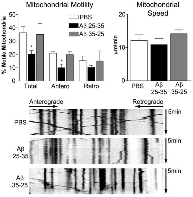
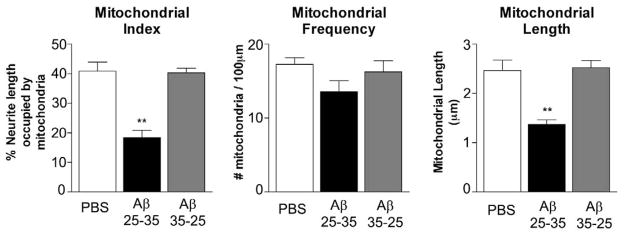
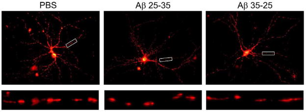
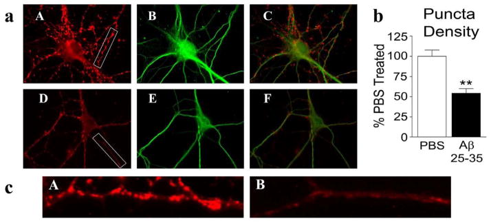
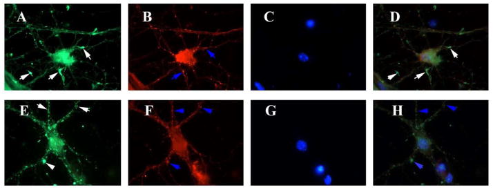
Loss of synapses and synaptic damage are the best correlates of cognitive decline identified in patients with Alzheimer's disease (AD), and mitochondrial oxidative damage and synaptic pathology have been identified as early events in the progression of AD. The progressive accumulation of amyloid beta (Aβ) in synapses and synaptic mitochondria are hypothesized to cause synaptic degeneration and cognitive decline in patients with AD. However, the precise mechanistic link between Aβ and mitochondria is not well understood. The purpose of this study was to better understand the effects of Aβ on mitochondrial axonal transport and synaptic alterations in AD. Using mouse hippocampal neurons and Aβ(25-35) peptide, we studied axonal transport of mitochondria, including mitochondrial motility, mitochondrial length and size, mitochondrial index per neurite, and synaptic alterations of the hippocampal neurons. In the PBS-treated neurons, 36.4±4.7% of the observed mitochondria were motile, with 21.0±1.3% moving anterograde and 15.4±3.4% moving retrograde and the average speed of movement was 12.1±1.8μm/min. In contrast, in the Aβ-treated neurons, the number of motile mitochondria were significantly less, at 20.4±2.6% (P<0.032), as were those moving anterograde (10.1±2.6%, P<0.016) relative to PBS-treated neurons, suggesting that the Aβ(25-35) peptide impairs axonal transport of mitochondria in AD neurons. In the Aβ-treated neurons, the average speed of motile mitochondria was also less, at 10.9±1.9μm/min, and mitochondrial length was significantly decreased. Further, synaptic immunoreactivity was also significantly less in the Aβ-treated neurons relative to the PBS-treated neurons, indicating that Aβ affects synaptic viability. These findings suggest that, in neurons affected by AD, Aβ is toxic, impairs mitochondrial movements, reduces mitochondrial length, and causes synaptic degeneration.


Figures

Figure 1
Figure 1. Amyloid beta treatment reduces mitochondrial movements
Axons from mature hippocampal neurons transfected with DsRed-mito and GFP, then treated for 24 hours with vehicle, Aβ, or reverse peptide were imaged to evaluate mitochondrial movements. Total proportion of moving mitochondria, proportion of mitochondria moving anterograde, and proportion of mitochondria moving retrograde were calculated (A). The speed of motion was also calculated for all moving mitochondria (B). Calculations were based on analysis of kymographs. Representative kymographs are shown for the three experimental groups (C). N= 4 independent cultures. * p < 0.05 compared to vehicle treated, and statistical variation is shown as mean±SE.
Figure 2
Figure 2. Amyloid beta treatment causes a reduction in mitochondrial mass within neurites
Mitochondria from DsRed-mito transfected hippocampal neurons were analyzed after vehicle, A-beta, or reverse peptide treatment. Mitochondrial index was calculated as the percent of neuritic length occupied by mitochondria. Aβ treated cultures showed pronounced reduction in mitochondrial index (A). The distribution of mitochondria was evaluated as number of mitochondria per neuritic length (B). Mitochondrial length was reduced by Aβ treatment (C). N = 4 independent cultures. ** p < 0.01. Statistical variation is shown as mean±SE.
Figure 3
Figure 3. Mitochondria are more fragmented after Amyloid beta treatment
DsRed-mito transfected hippocampal neurons were imaged after vehicle, Aβ, or reverse peptide treatment. Representative images are shown in upper panels. Enlargements of neurites are shown in the lower panels.
Figure 4
Figure 4. Double-labeling analysis of Synaptophysin and MAP2 in neurons treated with amyloid beta
Hippocampal neurons treated with PBS vehicle or Aβ were immunostained for synaptophysin and MAP2 (a). Vehicle treated cells (upper panel) showed typical strong synaptophysin (A), MAP2 (B) immunoreactivities and colocalization of synaptophysin and MAP2 (C). Aβ-treated cells (lower panel) showed reduced density of synaptophysin puncta (D), MAP2 (E) and merged (C). Image b shows quantification of synaptophysin immunoreactivity. Significantly decreased synaptophysin was found in Aβ-treated neurons (P<0.005). Image c shows enlarged portion of a neurite from PBS vehicle treated neuron (A) and Aβ-treated neuron (B).
Figure 5
Figure 5. Drp1 distribution relocates from areas of active neurite outgrowth to neuritic processes after amyloid beta treatment
Hippocampal neurons treated with PBS vehicle or Aβ were immunostained for Drp1, mitochondrial-encoded protein, Cyt. B and nuclear marker, DAPI. Vehicle treated cells (upper panel) showed intense immunoreactivities of Drp1 (A), Cyt. B (B), DAPI (C) and merged (D) in neurite growth cones (white arrows). Drp1 is colocalized with Cyt. B (D) (white arrows). After Aβ treatment (lower panel), Drp1 staining is reduced (E), and Drp1 is colocalzed with mitochondria in merged image (H).

References

    1. Selkoe DJ. Alzheimer’s disease: genes, proteins, and therapy. Physiol Rev. 2001;81:741–66. - PubMed
    1. Alzheimer Association Report. Alzheimer’s Disease Facts and Figures. 2010.
    1. Mattson MP. Pathways towards and away from Alzheimer’s disease. Nature. 2004;430:631–639. - PMC - PubMed
    1. LaFerla FM, Green KN, Oddo S. Intracellular amyloid-beta in Alzheimer’s disease. Nat Rev Neurosci. 2007:499–509. - PubMed
    1. Reddy PH, Beal MF. Amyloid beta, mitochondrial dysfunction and synaptic damage: implications for cognitive decline in aging and Alzheimer’s disease. Trends Mol Med. 2008;14:45–53. - PMC - PubMed

Publication types