. 2011 Jan 21;144(2):187-99.
doi: 10.1016/j.cell.2010.12.020.

Nuclear PTEN regulates the APC-CDH1 tumor-suppressive complex in a phosphatase-independent manner

Affiliations

Nuclear PTEN regulates the APC-CDH1 tumor-suppressive complex in a phosphatase-independent manner

Min Sup Song et al. Cell. .

Abstract

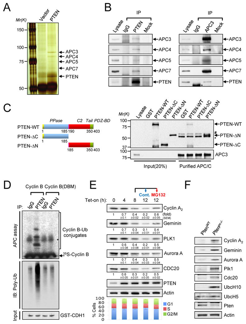
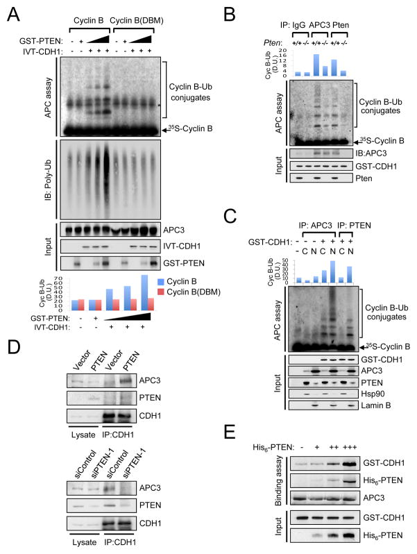
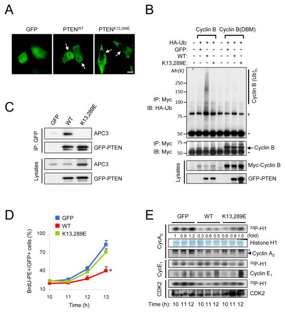
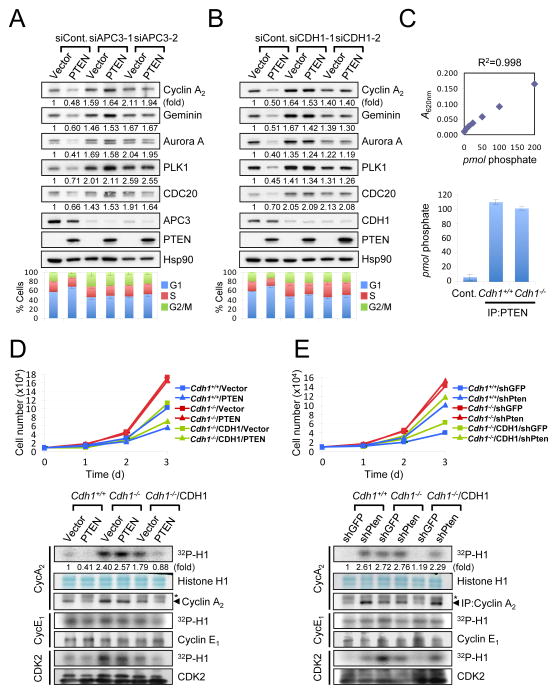
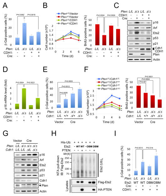
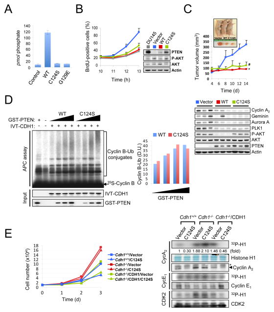
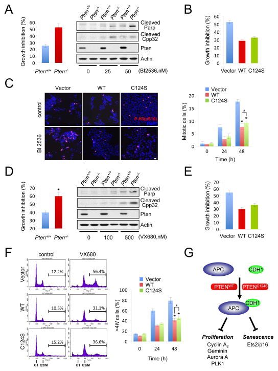
PTEN is a frequently mutated tumor suppressor gene that opposes the PI3K/AKT pathway through dephosphorylation of phosphoinositide-3,4,5-triphosphate. Recently, nuclear compartmentalization of PTEN was found as a key component of its tumor-suppressive activity; however its nuclear function remains poorly defined. Here we show that nuclear PTEN interacts with APC/C, promotes APC/C association with CDH1, and thereby enhances the tumor-suppressive activity of the APC-CDH1 complex. We find that nuclear exclusion but not phosphatase inactivation of PTEN impairs APC-CDH1. This nuclear function of PTEN provides a straightforward mechanistic explanation for the fail-safe cellular senescence response elicited by acute PTEN loss and the tumor-suppressive activity of catalytically inactive PTEN. Importantly, we demonstrate that PTEN mutant and PTEN null states are not synonymous as they are differentially sensitive to pharmacological inhibition of APC-CDH1 targets such as PLK1 and Aurora kinases. This finding identifies a strategy for cancer patient stratification and, thus, optimization of targeted therapies. PAPERCLIP:


Figures

Figure 1
Figure 1. Nuclear PTEN interacts with APC/C
(A) Nuclear extracts from PTEN-deficient PC3 cells transfected with Myc-PTEN were immunoprecipitated with an anti-Myc antibody followed by mass spectrometric peptide sequencing. APC/C components including APC3, APC4, APC5 and APC7 were identified. (B) DU145 cell lysates were immunoprecipitated (IP) without (Mock) or with anti-PTEN (left) or anti-APC3 (right) antibody followed by immunoblotting. Asterisk indicates heavy chain of IgG. (C) Recombinant GST-PTEN (aa 1–403), GST-PTEN-ΔC (aa 1–185) or GST-PTEN-ΔN (aa 185–403) proteins, as shown in top diagram, were incubated with immunopurified APC/C from PC3 cells released 3 hr from nocodazole synchronization. * and # indicate heavy chain of IgG and non-specific band, respectively. (D). Lysates from 12 hr after tetracyclin induction as in (E) were immunoprecipitated (IP) with anti-PTEN antibody and subjected to in vitro ubiquitination assay using 35S-labeled Cyclin B (WT and destruction box mutant (DBM)) in the presence of GST-CDH1 protein (50 ng) and immunoblotting with anti-polyubiquitinated proteins antibody. (E) PC3 cells were co-transfected with pcDNA4/TO/Myc-His-PTEN and pcDNA6/TR to induce PTEN expression after the addition of tetracycline (Tet-on) for the indicated times or treated with 10 μM MG132 after 8hr-Tet induction for additional 4 hr, and then subjected to immunoblotting (top) and flow cytometric analysis (bottom). The quantification of the relative immunoreactivity of each protein normalized to Actin is represented as the mean and the standard error of the mean (SEM) from three different experiments. (F) Accumulation of APC-CDH1 targets in PtenloxP/loxP;Probasin- Cre4 (Ptenpc−/−) mouse prostate epithelium. Lysates from anterior prostates in wild-type (WT-Cre) and Ptenpc−/− mice at 11 weeks age were subjected to immunoblotting. “see also Figure S1A-F”.
Figure 2
Figure 2. Nuclear PTEN regulates the activity of APC-CDH1
(A) Immunopurified APC/C from PC3 cells, released 3hr post nocodazole synchronization, were subjected to in vitro ubiquitination assay using 35S-labeled in vitro-translated (IVT) wild-type or destruction box mutated (DBM) Cyclin B in the presence of IVT-CDH1 (1 μl) and different amounts of GST-PTEN proteins (0, 50, 100ng) and immunoblotting with anti-polyubiquitinated proteins antibody. Bottom panel illustrates the density unit (D.U.) of Cyclin B-Ub conjugates analyzed by the ImageJ 1.38× software (NIH). Asterisk indicates non-specific band. (B) Lysates from wild-type and Pten−/− MEFs subjected to 48hrs of serum depletion were immunoprecipitated (IP) with anti-APC3 or anti-Pten antibody and subjected to in vitro ubiquitination assay using 35S-labeled Cyclin B in the presence of GST-CDH1 protein (50 ng). Top panel shows the density unit (D.U.) of Cyclin B-Ub conjugates analyzed by the ImageJ 1.38x software. (C) Cytoplasmic (C) and nuclear (N) extracts from DU145 cells, released 3 hr from nocodazole synchronization, were IP with anti-APC3 or anti-PTEN antibody and subjected to in vitro ubiquitination assay using 35S-labeled Cyclin B. Top panel shows the density unit (D.U.) of Cyclin B-Ub conjugates analyzed by the ImageJ 1.38x software. (D) Lysates from PTEN-deficient PC3 cells complemented with empty vector or PTEN (top) or PTEN-proficient DU145 cells transfected with siRNAs for Renilla luciferase (siControl) or PTEN (siPTEN-1) (bottom) were IP with anti-CDH1 antibody and then subjected to immunoblotting. (E) Immunopurified APC/C from PC3 cells, released 3 hr from nocodazole synchronization, were incubated with GST-CDH1 (100 ng) and different amounts of His6-PTEN proteins (0, 50, 100, 200 ng) for 1 hr followed by immunoblotting. see also Figure S1G”.
Figure 3
Figure 3. Nuclear exclusion of PTEN impairs activation of APC-CDH1
(A) Immunofluorescence analysis of GFP, GFP-tagged wild-type (PTENWT) and nuclear excluded PTEN mutant (PTENK13,289E). Arrows indicate the nucleus in PC3 cells. (B) PC3 cells were co-transfected with a combination of wild-type or destruction box mutated (DBM) Myc-Cyclin B, HA-ubiquitin (Ub), GFP, PTENWT and PTENK13,289E, and treated with the proteasome inhibitor MG132 (10 μM) for 4 hr before harvesting. Cell lysates were then immunoprecipitated (IP) with anti-Myc antibody and subjected to immunoblotting (IB). * and # indicate heavy chain of IgG and non-specific band, respectively. (C) Lysates from PC3 cells transfected with GFP, PTENWT or PTENK13,289E were IP with anti-GFP antibody and subjected to immunoblotting. (D) PC3 cells transfected with GFP, PTENWT or PTENK13,289E were synchronized by growth in nocodazole (400 nM) for 24 hr, released for the indicated times, pulsed with BrdU for 30 min and subjected to flow cytometric analysis. Data are the means from three different experiments and error bars represent the SEM. (E) Lysates from (D) were subjected to in vitro kinase assay with CDK2, Cyclin E1 or Cyclin A2. The relative kinase activities were normalized with histone H1 inputs. Asterisks indicate heavy chain of IgG. “see also Figure S2”. P value was determined by Student’s t test (*P<0.01).
Figure 4
Figure 4. Growth suppression by PTEN relies on APC-CDH1 function
A–B) PC3 cells co-transfected with empty vector or HA-PTEN, and Renilla luciferase siRNA (siControl), two independent APC3 siRNAs (siAPC3-1 and -2) (A) or CDH1 siRNAs (siCDH1-1 and -2) (B) were subjected to immunoblotting (top) and flow cytometric analysis to measure cell cycle distribution (bottom). The relative immunoreactivity of each protein was quantified by normalizing with Hsp90. (C) diC8-PtdIns(3,4,5)P3 (40 μM) was incubated with and without (control) the immunopurified Pten (1 μg) at 37°C for 30 min. The amount of free phosphate was measured Absorbance 620 nm (A620nm) and calculated using the standard curve line-fit data in top panel. Error bars represent SEM from three different experiments. (D) Growth curves (top) and in vitro kinase assay (bottom) of SV40-LT-immortalized wild-type and Cdh1−/− MEFs, infected with a retroviral combination of human CDH1 and PTEN (with selection) as indicated, and followed over a 3-day period. Error bars represent the SEM from three different experiments. Asterisks indicate heavy chain of IgG. (E) Growth curves (top) and in vitro kinase assay (bottom) of immortalized wild-type and Cdh1−/− MEFs, infected with a retroviral combination of human CDH1 and Pten shRNA (with selection) as indicated, and followed over a 3-day period. Error bars represent SEM from three different experiments. The relative kinase activities were normalized with histone H1 inputs. Asterisks indicate heavy chain of IgG. “see also Figure S3”.
Figure 5
Figure 5. Pten loss-induced cellular senescence involves APC-CDH1
Cellular senescence assay (A), growth curves and BrdU incorporation assay (B), and immunoblot (C) of primary Ptenlox/lox MEFs, infected with retroviral Cre and human CDH1 selected for 4 days. Error bars represent SEM from three different experiments. P value was determined by Student’s t test. (D) Real-time RT-PCR analysis of murine p16 expression was quantified. Error bars represent SEM. (EG) Cellular senescence assay (E), growth curves and BrdU incorporation assay (F) and immunoblotting (G) of primary conditional Ptenlox/lox;Cdh1+/+, Ptenlox/lox;Cdh1lox/+, Ptenlox/lox;Cdh1lox/lox MEFs, infected with retroviral Cre recombinase at 4 day after selection. Error bars represent the SEM from three different experiments. Asterisk indicates nonspecific band. (H) PC3 cells were co-transfected with wild-type or destruction box mutated (DBM) Flag-Ets2, His-ubiquitin (Ub) and HA-PTEN, and treated with the proteasome inhibitor MG132 (10 μM) for 4 hr before harvesting. His-Ub-conjugated Ets2 was purified from cell lysates using Ni2+-NTA spin column under denaturing conditions. (I) Cellular senescence assay of primary Ptenlox/lox MEFs, infected with a retroviral combination of wild-type or Ets2(DBM), CDH1 and Cre as indicated, at 4 day after selection. Error bars represent SEM from three different experiments. “see also Figure S4”.
Figure 6
Figure 6. PTEN regulates APC-CDH1 independently of its phosphatase activity
(A) diC8-PtdIns(3,4,5)P3 (40 μM) was incubated without (control) and with PTEN (WT), PTEN(C124S) or PTEN(G129E) immunoprecipitates (1 μg) at 37°C for 30 min and then the free phosphate was measured using the Green Reagent. Error bars represent the SEM from three different experiments. (B) PC3 cells complemented with wild-type or phosphatase-inactive PTEN(C124S) were nocodazole synchronized, released for the indicated times and pulsed with BrdU 30 min before harvesting. The proportion of BrdU+ cells was measured (left) and cell lysates were subjected to western blot (right). Error bars represent SEM from 3 different experiments. (C) PC3 cells complemented with wild-type or phosphatase-inactive PTEN(C124S) were injected subcutaneously into nude mice. Tumor volume was monitored (top) and tissue lysates at 2 week after injection were subjected to immunoblotting (bottom). Error bars represent SEM (n=6 mice/group). (D) Immunopurified APC/C from PC3 cells, released 3hrs post nocodazole synchronization, were subjected to in vitro ubiquitination assay using 35S-labeled Cyclin B in the presence of in vitro-translated (IVT)-CDH1 (2 μl) and different amounts of wild-type or PTEN(C124S) proteins (0, 50, 100, 200 ng). Right panel illustrates the density unit (D.U.) of Cyclin B-Ub conjugates analyzed by the ImageJ 1.38× software. (E) Growth curves (left) and in vitro kinase assay (right) of immortalized wild-type and Cdh1−/− MEFs, infected with a retroviral combination of human CDH1 and PTEN(C124S) (with selection) as indicated, and followed over a 3-day period. Error bars represent SEM from three different experiments. The relative kinase activities were normalized with histone H1 inputs. Asterisk indicates heavy chain of IgG. “see also Figures S5 and S6”. P value was determined by Student’s t test (*P<0.01; #P<0.05).
Figure 7
Figure 7. PTEN loss but not phosphatase inactivation results in hypersensitivity to pharmacological inhibition of APC-CDH1 targets
(A) Growth inhibition of immortalized wild-type and Pten−/− MEFs treated with the PLK1 inhibitor BI 2536 (50 nM) for 48 hr was measured (left), western blots of cell lysates after BI2536 treatment for 48 hr (right). B) Growth inhibition of PC3 cells complemented with wild-type or PTEN(C124S) and treated with the PLK1 inhibitor BI 2536 (5 nM) was measured after the 48 hr-treatment. (C) The cell population in mitosis of PC3 cells complemented with wild-type or PTEN(C124S) and treated with 5 nM BI 2536 was analyzed by immunofluorescence with anti-phospo-histone H3 (P-H3(pS10)) (left) and quantified for a 48 h-period (right). Error bars represent SEM from three different experiments. (D) Pten-null cells are hypersensitive to Aurora A inhibition. Growth inhibition of immortalized wild-type and Pten−/− MEFs treated with the Aurora A inhibitor VX680 (500 nM) was measured after the 48 hr-treatment (left), western blots of cell lysates after the treatment with different concentrations of VX680 for 48 hr (right). Error bars represent SEM from three different experiments. (E) Growth inhibition of PC3 cells complemented with wild-type or PTEN(C124S) and treated with the Aurora A inhibitor VX680 (500 nM) was measured after the 48 hr-treatment. Error bars represent SEM from three different experiments. (F) The cell population harboring >4 N DNA content of PC3 cells complemented with wild-type or PTEN(C124S) and treated with VX680 (500 nM) was analyzed by flow cytometry (left) and quantified for a 48 h-period (right). Error bars represent SEM from three different experiments. (G) A proposed model for phosphatase-independent role of nuclear PTEN towards APC-CDH1 to regulate proliferation and cellular senescence. “see also Figure S7”. P values were determined by Student’s t test (*P<0.01; #P>0.05).

Comment in

References

    1. Ali IU, Schriml LM, Dean M. Mutational spectra of PTEN/MMAC1 gene: a tumor suppressor with lipid phosphatase activity. J Natl Cancer Inst. 1999;91:1922–1932. - PubMed
    1. Alimonti A, Nardella C, Chen Z, Clohessy JG, Carracedo A, Trotman LC, Cheng K, Varmeh S, Kozma SC, Thomas G, et al. A novel type of cellular senescence that can be enhanced in mouse models and human tumor xenografts to suppress prostate tumorigenesis. J Clin Invest. 2010;120:681–693. - PMC - PubMed
    1. Bassermann F, Frescas D, Guardavaccaro D, Busino L, Peschiaroli A, Pagano M. The Cdc14B-Cdh1-Plk1 axis controls the G2 DNA-damage-response checkpoint. Cell. 2008;134:256–267. - PMC - PubMed
    1. Blanco-Aparicio C, Renner O, Leal JF, Carnero A. PTEN, more than the AKT pathway. Carcinogenesis. 2007;28:1379–1386. - PubMed
    1. Cairns P, Okami K, Halachmi S, Halachmi N, Esteller M, Herman JG, Jen J, Isaacs WB, Bova GS, Sidransky D. Frequent inactivation of PTEN/MMAC1 in primary prostate cancer. Cancer Res. 1997;57:4997–5000. - PubMed

Publication types

MeSH terms