. 2011 Jan 21;144(2):253-67.
doi: 10.1016/j.cell.2010.12.018.

RalB and the exocyst mediate the cellular starvation response by direct activation of autophagosome assembly

Affiliations

RalB and the exocyst mediate the cellular starvation response by direct activation of autophagosome assembly

Brian O Bodemann et al. Cell. .

Abstract

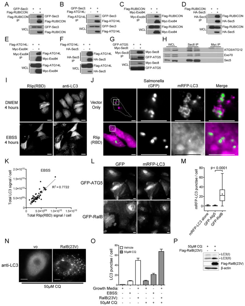
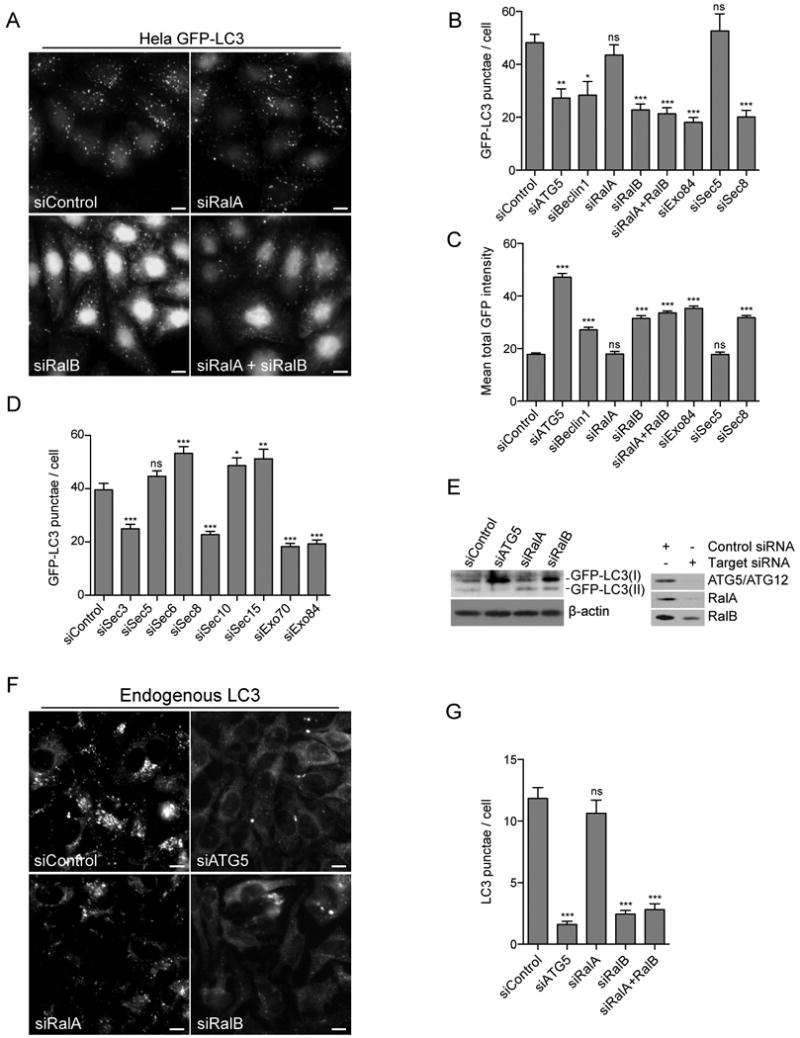
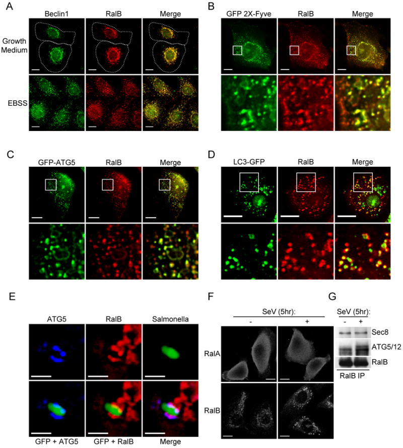
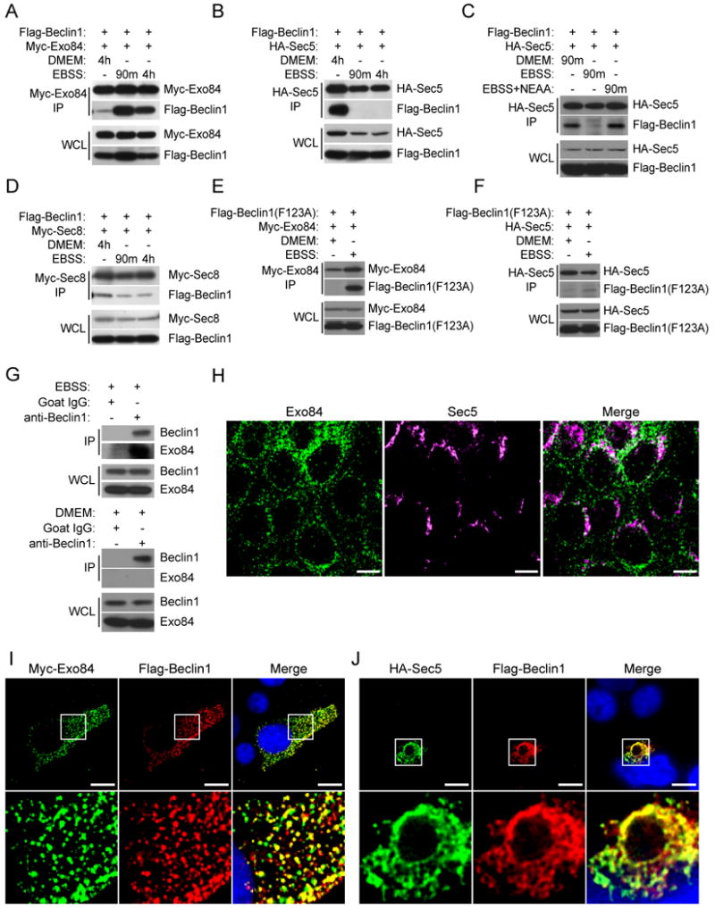
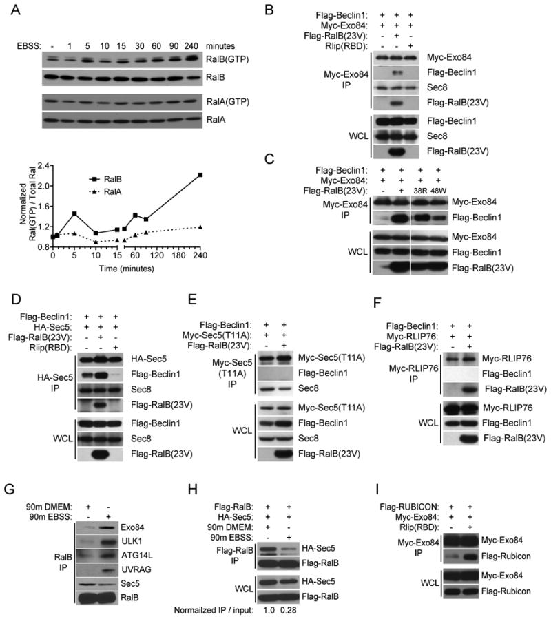
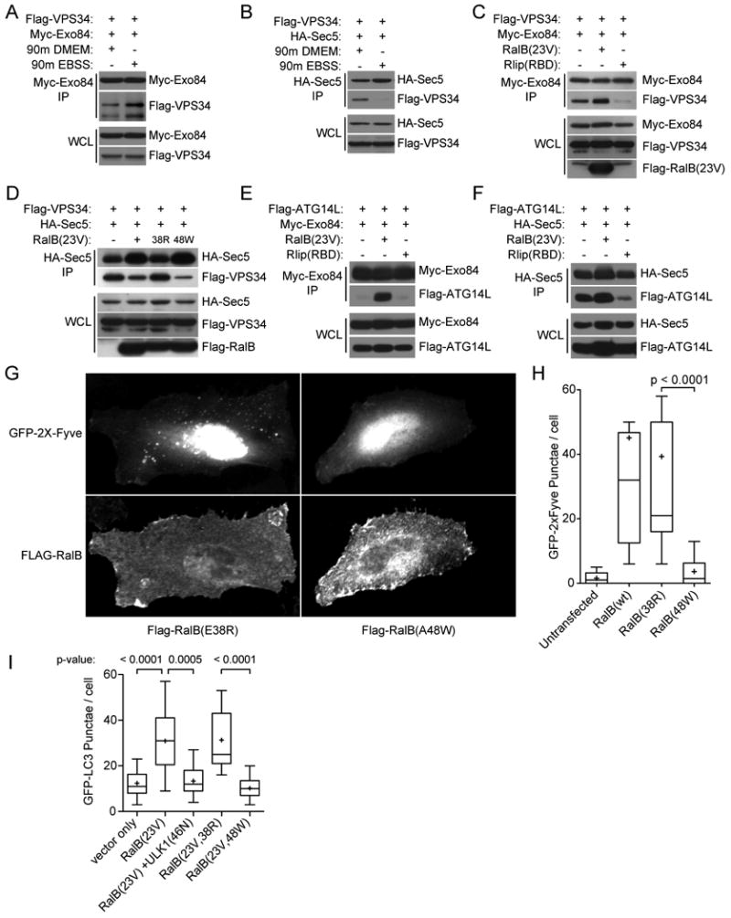
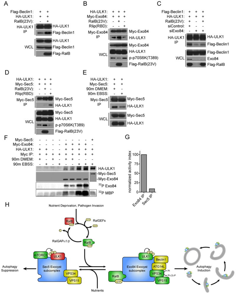
The study of macroautophagy in mammalian cells has described induction, vesicle nucleation, and membrane elongation complexes as key signaling intermediates driving autophagosome biogenesis. How these components are recruited to nascent autophagosomes is poorly understood, and although much is known about signaling mechanisms that restrain autophagy, the nature of positive inductive signals that can promote autophagy remain cryptic. We find that the Ras-like small G protein, RalB, is localized to nascent autophagosomes and is activated on nutrient deprivation. RalB and its effector Exo84 are required for nutrient starvation-induced autophagocytosis, and RalB activation is sufficient to promote autophagosome formation. Through direct binding to Exo84, RalB induces the assembly of catalytically active ULK1 and Beclin1-VPS34 complexes on the exocyst, which are required for isolation membrane formation and maturation. Thus, RalB signaling is a primary adaptive response to nutrient limitation that directly engages autophagocytosis through mobilization of the core vesicle nucleation machinery.


Figures

Figure 1
Figure 1. Physical and Functional interaction of the Ral-Exocyst complex with autophagy machinery
A-G: Exocyst subunits interact with autophagy proteins. The indicated proteins were over-expressed in HEK-293 cells, then immunoprecipitated with an antibody directed to the specified tag. Immunoprecipitates were analyzed for coprecipitation with (A,B) GFP-Sec3; (C,D) Flag-RUBICON; (E,F) Flag-ATG14L; and (G) GFP-ATG5 as indicated. Whole cell lysate (WCL), Immunoprecipitation (IP). H: Endogenous Sec8 complexes contain ATG5-12 conjugates. The endogenous exocyst complex was immunoprecipitated from HEK-293 cells with anti-Sec8 antibody and analyzed for coprecipitation of ATG5/ATG12 conjugates (Sec8 IP) using anti-ATG5 antibody. Anti-Myc immunoprecipitates served as a negative control (Myc IP). Two independent experiments are shown. Representation of the examined proteins in the input whole cell non denaturing lysates is shown (WCL). I: Inhibition of Ral signaling blocks the LC3 response to amino-acid starvation. 48 hours post-transfection with Myc-Rlip(RBD) Hela cells were incubated in DMEM or EBSS for an additional 4 hours as indicated. Myc-Rlip(RBD) and LC3 were detected by immunofluorescence using anti-myc and anti-LC3 antibodies, respectively. Vector control cells were similar to untransfected cells. Scale bar 20μm. J: Inhibition of Ral signaling blocks the LC3 response to pathogen infection. Hela cells were transfected with monomeric RFP-LC3 together with Myc-Rlip(RBD) or an empty vector control as indicated. 48 hours post-transfection, cells were infected with Salmonella typhimurium-GFP for 1 hour followed by 3 hours of post-infection selection for intracellular Salmonella. Internalized Salmonella and LC3 were visualized using their respective fluorescent fusions. High magnification of the subcellular regions indicated by the boxes are shown in the panels on the right. Dashed lines indicated cell borders as visualized in a saturated exposure. Scale bar 10μm. K: Total fluorescence intensity corresponding to Myc-Rlip(RBD) (anti-myc) and endogenous LC3 (anti-LC3) at single-cell resolution for EBSS-treated cells as shown in (I) (n=82, R2=0.7722). L: RalB is sufficient to induce accumulation of LC3 punctae. Hela cells expressing monomeric RFP-LC3 together with GFP-ATG5 or GFP-RalB are shown as indicated. Scale bar 10μm. M: mRFP-LC3 punctae in cells treated as in (L) were quantitated. The distribution of mRFP-LC3 punctae/cell is displayed as box-and-whisker plots. The three bands of the box illustrate the 25th (lower), 50th (middle), and 75th (upper) quartiles. The whiskers go 1.5 times the interquartile distance or to the highest or lowest point, whichever is shorter. The + designates the mean. P-values were calculated using the student's t-test. N: HBEC3-KT cells expressing RalB(23V) or transfected with vector control were incubated in growth medium containing 50μM Chloroquine (CQ), to prevent LC3 turnover by autophagolysosomes, for 4 hours followed by detection of endogenous LC3 with anti-LC3 antibody. O: RalB is sufficient to induce autophagic flux. HBEC3-KT cells treated as in (N) were incubated in growth media or amino-acid free EBSS (Earle's balanced salt solution) for 4 hours with or without 50μM Chloroquine (CQ), to prevent LC3 turnover in autophagolysosomes, as indicated. Immunofluorescence was performed with anti-LC3 antibody and LC3 punctae were quantitated. Data are represented as mean +/- SEM. P: Whole cell lysates from HBEC3-KT cells transfected with Flag-RalB(G23V) or vector control were analyzed for the relative accumulation of LC3(I) and LC3(II) when incubated in growth medium containing 50μM Chloroquine (CQ) for 4 hours. β-actin is shown as a loading control. See also Table S1.
Figure 2
Figure 2. RalB and an Exo84-containing subcomplex of the exocyst are necessary for amino acid starvation induced autophagy
A: RalB depletion inhibits accumulation of GFP-LC3 punctae. Hela cells stably expressing GFP-LC3 were depleted of the indicated proteins by siRNA transfection. Cells were imaged by GFP fluorescence 96 hours after transfection. Scale bar 10μm. B: Sec5 and Exo84 selectively participate in accumulation of GFP-LC3 punctae. GFP-LC3 punctae in cells treated as in (A) were quantitated. The mean distribution of GFP-LC3 punctae/cell is displayed as a bar graph, data are represented as mean +/- SEM. P-values were calculated by one-way ANOVA followed by Dunnett's Multiple Comparison Test. C: Inhibition of GFP-LC3 punctae correlates with accumulation of LC3 protein. The mean total intensity of GFP-LC3 in cells treated as in (A) was quantitated. The distribution of the mean total GFP intensity is displayed as a bar graph, data are represented as mean +/- SEM. P-values were calculated by one-way ANOVA followed by Dunnett's Multiple Comparison Test. D: A subset of exocyst subunits are limiting for accumulation of GFP-LC3 punctae. The indicated siRNAs were evaluated as in (B). E: RalB depletion inhibits accumulation of LC3-lipid conjugates. Whole cell lysates from HBEC3-KT cells stably expressing GFP-LC3 transfected with the indicated siRNAs were assayed for the relative accumulation of GFP-LC3(I) and GFP-LC3(II). β-actin is shown as a loading control. siRNA-mediated target depletion is shown 96 hours post transfection (right panels). F: RalB participates in accumulation of endogenous LC3 punctae. Hela cells were depleted of the indicated proteins by siRNA transfection. 96 hours after transfection, cells were incubated in amino acid free EBSS for 4 hours. Endogenous LC3 was imaged by anti-LC3 immunofluorescence. Scale bar 10μm. G: Endogenous LC3 punctae in cells treated as in (E) were quantitated. The mean distribution of LC3 punctae/cell is displayed as a bar graph, data are represented as mean +/- SEM. P-values were calculated by one-way ANOVA followed by Dunnett's Multiple Comparison Test. See also Figure S1.
Figure 3
Figure 3. Native RalB colocalizes with autophagy machinery
A: Beclin1 and RalB colocalize in cells pre and post induction of autophagy. Endogenous immunofluorescence of Beclin1 (anti-Beclin1) and RalB (anti-RalB) in HBEC30-KT cells incubated for 90 minutes in fresh growth medium or EBSS as indicated. Dashed line indicates cell outline. Scale bar 10μm. B-D: RalB colocalizes with early and late markers of autophagosome biogenesis. HBEC30-KT cells were transfected with (B) GFP-2X-Fyve; (C) GFP-ATG5; and (D) GFP-LC3. Cells were incubated in EBSS for (B) 30 minutes; (C) 90 minutes; or (D) 3 hours. GFP fluorescence and endogenous RalB (anti-RalB) immunofluorescence is shown. High magnification of 10μm × 10μm regions indicated by the boxes are shown in the bottom panels. Scale bar 10μm. E: ATG5 and RalB are recruited to sites of incipient isolation membrane formation. Endogenous immunofluorescence of ATG5 (anti-ATG5) and RalB (anti-RalB) in HBEC30-KT cells infected with Salmonella typhimurium-GFP. Cells were exposed to Salmonella typhimurium-GFP for 1 hour followed by 3 hours of post-infection antibiotic selection against extracellular Salmonella. Scale bar 2μm. F: SenV infection selectively alters the subcellular distribution of RalB versus RalA. Endogenous immunofluorescence of RalA (anti-RalA) and RalB (anti-RalB) in HBEC3-KT cells mock infected or infected with Sendai virus for 5 hours. Scale bar 10μm. G: SenV infection induces accumulation of endogenous RalB/ATG5-12 complexes. Endogenous RalB complexes were immunoprecipitated from mock infected or Sendai virus infected HBEC3-KT cells with anti-RalB antibodies and analyzed for coprecipitation of ATG5/ATG12 conjugates.
Figure 4
Figure 4. Nutrient deprivation drives assembly of Exo84/Beclin1 complexes
A-D: Nutrient limitation induces Beclin1/Exo84 interactions and inhibits Beclin1/Sec5 interactions. 48 hours post-transfection with tagged Beclin1 and exocyst expression constructs, HEK-293 cells were incubated in DMEM, EBSS, or EBSS with 1× Non-Essential amino acids for 90 minutes or 4 hours as shown. The indicated proteins were then immunoprecipitated with antibodies directed to the specified tag. Immunoprecipitates were analyzed for coprecipitation with Flag-Beclin1. Whole cell lysate (WCL), Immunoprecipitates (IP). E,F: Beclin1(F123A) mutant interacts with Exo84 but not Sec5. Co-expression, co-IPs with the indicated proteins were performed as in (A-D). G: Endogenous Beclin1/Exo84 complexes accumulate in response to nutrient deprivation. Endogenous Beclin1 was immunoprecipitated from HEK-293 cells incubated in EBSS (top panels) or DMEM (bottom panels) for 90 minutes and analyzed for coprecipitation of Exo84 (IP). Host species-matched non-specific IgG immunoprecipitates served as negative controls. Representation of the examined proteins in the input whole cell non-denaturing lysates is shown (WCL). H: Exo84 and Sec5 are enriched in distinct subcellular compartments. Endogenous immunofluorescence of Sec5 (anti-Sec5) and Exo84 (anti-Exo84) in MDCK cells. Scale bar 10μm. I,J: Exo84 and Sec5 can recruit Beclin1 to distinct subcellular compartments. HEK-293 cells were transfected with (F) Flag-Beclin1 and Myc-Exo84; (G) Flag-Beclin1 and HA-Sec5. Immunofluorescence of the indicated fusion tags was performed. High magnification of 10μm × 10μm regions indicated by the boxes are shown in the bottom panels. Scale bar 10μm. See also Figure S2.
Figure 5
Figure 5. RalB drives assembly of Exo84/Beclin1 complexes through direct RalB-Exo84 effector binding
A: Amino-acid depletion activates RalB. Endogenous GTP-bound RalA and RalB were collected by GST-Sec5-RBD mediated affinity purification from HEK-293 cells incubated in EBSS for the indicated times and visualized with specific anti-RalA and anti-RalB antibodies. The normalized GTP-loaded index for RalA and RalB was calculated as Ral(GTP)/Total Ral to generate the scatterplot. B-F: RalB regulates Beclin1/exocyst subcomplex interactions. The indicated proteins were expressed in HEK-293 cells and immunoprecipitated with antibodies directed to the appropriate tag. Immunoprecipitates were analyzed for coprecipitation with Flag-Beclin1, Flag RalB(23V), and endogenous Sec8 as shown. Whole cell lysate (WCL), Immunoprecipitation (IP). G: Nutrient status specifies distinct endogenous RalB/effector interactions. Endogenous RalB was immunoprecipitated with anti-RalB antibody from HEK-293 cells incubated in DMEM or EBSS for 90 minutes as indicated and analyzed for coprecipitation of Exo84, Sec5, ATG14L, UVRAG, and ULK1. H: 48 hours post-transfection, HEK-293 cells were incubated in DMEM or EBSS for 90 minutes as indicated. Flag-RalB immunoprecipitates were examined for coprecipitation of HA-Sec5. The indicated normalized IP / input ratio was calculated by dividing immunoprecipitated Sec5 by total expressed Sec5, then normalizing the calculated values to DMEM condition. I: Ral-inhibition induces accumulation of Exo84/Rubicon interactions. Co-expression, co-IPs with the indicated proteins were performed as in (B-F).
Figure 6
Figure 6. RalB expression drives assembly of Exo84/Vps34 and Exo84/ATG14L complexes
A-F: VPS34 and ATG14L/exocyst subcomplexes are regulated by nutrient limitation and RalB activation. HEK-293 cells expressing the indicated proteins were incubated in DMEM or EBSS for 90 minutes as indicated. Tagged exocyst subunits were immunoprecipitated and analyzed for coprecipitation with Flag-VPS34 and Flag-ATG14L where indicated. G: RalB/Exo84 effector interactions mobilize VPS34 activity. Hela cells expressing GFP-2X-Fyve together with RalB partial loss of function mutants Flag-RalB(E38R) or Flag-RalB(A48W) are shown as indicated. H: GFP-2X-Fyve punctae in cells treated as in (G) were quantitated. The distribution of GFP-2X-Fyve punctae/cell is displayed as box-and-whisker plots. P-values were calculated using the student's t-test. I: RalB(G23V) and RalB(G23V,E38R) are sufficient to induce accumulation of GFP-LC3 punctae and kinase dead ULK1(K46N) blocks the increase observed with RalB(G23V) expression. Hela cells stably expressing GFP-LC3 were transfected with the indicated constructs then visualized by immunofluorescence of the indicated tags. The distribution of GFP-LC3 punctae/cell is displayed as box-and-whisker plots. P-values were calculated using the student's t-test.
Figure 7
Figure 7. Active ULK1 associates with Exo84
A: RalB induces ULK1/Beclin1 Complex formation. ULK1 immunoprecipitates were analyzed for coprecipitation with Flag-Beclin1 upon RalB(23V) expression as indicated. B: ULK1/Exo84 complexes are regulated by RalB. The indicated proteins were expressed in HEK-293 cells. Myc-tagged Exo84 was immunoprecipitated and analyzed for coprecipitation with HA-ULK1. C: RalB induced ULK1/Beclin1 complexes require Exo84. HEK-293 cells were first transfected with siControl or siExo84 siRNAs before the indicated proteins were expressed 24 hours later. ULK1 immunoprecipitates were analyzed for coprecipitation with Flag-Beclin1 upon RalB(23V) expression as indicated. D: ULK1/Sec5 complexes accumulate upon Ral inhibition. Co-expression, co-IPs with the indicated proteins were performed as in (B). E: ULK1/Sec5 complexes dissociate upon nutrient deprivation. Co-expression, co-IPs with the indicated proteins were performed as in (B) with the addition of 90 minute incubation in DMEM or EBSS as indicated. F: Amino-acid starvation induces association of Exo84 with catalytically active ULK1. Exo84 and Sec5 complexes were assayed for coprecipitation with ULK1 and for associated protein kinase activity as indicated. G: The normalized activity ratio for EBSS stimulated Exo84 and Sec5 precipitates was calculated by the division of MBP 32P incorporation by the HA-ULK1 signal coprecipitated from (F). H: Working model of RalB/exocyst dependent mobilization of autophagy. See also Figure S3.

Comment in

References

    1. Amar N, Lustig G, Ichimura Y, Ohsumi Y, Elazar Z. Two newly identified sites in the ubiquitin-like protein Atg8 are essential for autophagy. EMBO Rep. 2006;7:635–642. - PMC - PubMed
    1. Balakireva M, Rosse C, Langevin J, Chien YC, Gho M, Gonzy-Treboul G, Voegeling-Lemaire S, Aresta S, Lepesant JA, Bellaiche Y, et al. The Ral/exocyst effector complex counters c-Jun N-terminal kinase-dependent apoptosis in Drosophila melanogaster. Mol Cell Biol. 2006;26:8953–8963. - PMC - PubMed
    1. Bhuvanakantham R, Li J, Tan TT, Ng ML. Human Sec3 protein is a novel transcriptional and translational repressor of flavivirus. Cell Microbiol. 2010;12:453–472. - PubMed
    1. Bodemann BO, White MA. Ral GTPases and cancer: linchpin support of the tumorigenic platform. Nat Rev Cancer. 2008;8:133–140. - PubMed
    1. Cascone I, Selimoglu R, Ozdemir C, Del Nery E, Yeaman C, White M, Camonis J. Distinct roles of RalA and RalB in the progression of cytokinesis are supported by distinct RalGEFs. EMBO J. 2008;27:2375–2387. - PMC - PubMed

Publication types

MeSH terms