Modern fluorescent proteins and imaging technologies to study gene expression, nuclear localization, and dynamics
- PMID: 21242078
- PMCID: PMC3143818
- DOI: 10.1016/j.ceb.2010.12.004
Modern fluorescent proteins and imaging technologies to study gene expression, nuclear localization, and dynamics
Abstract
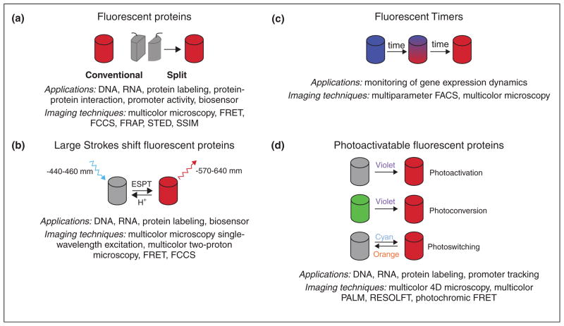
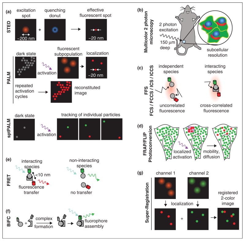
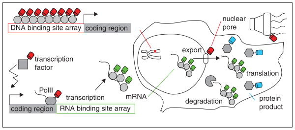
Recent developments in reagent design can address problems in single cells that were not previously approachable. We have attempted to foresee what will become possible, and the sorts of biological problems that become tractable with these novel reagents. We have focused on the novel fluorescent proteins that allow convenient multiplexing, and provide for a time-dependent analysis of events in single cells. Methods for fluorescently labeling specific molecules, including endogenously expressed proteins and mRNA have progressed and are now commonly used in a variety of organisms. Finally, sensitive microscopic methods have become more routine practice. This article emphasizes that the time is right to coordinate these approaches for a new initiative on single cell imaging of biological molecules.
Copyright © 2010 Elsevier Ltd. All rights reserved.
Figures


Similar articles
-
Q&A: Single-molecule localization microscopy for biological imaging.BMC Biol. 2010 Aug 11;8:106. doi: 10.1186/1741-7007-8-106. BMC Biol. 2010. PMID: 20707882 Free PMC article. No abstract available.
-
Super-resolution localization microscopy with photoactivatable fluorescent marker proteins.Protoplasma. 2014 Mar;251(2):349-62. doi: 10.1007/s00709-013-0566-z. Epub 2013 Oct 27. Protoplasma. 2014. PMID: 24162869 Review.
-
New tools for in vivo fluorescence tagging.Curr Opin Plant Biol. 2005 Dec;8(6):565-73. doi: 10.1016/j.pbi.2005.09.011. Epub 2005 Sep 26. Curr Opin Plant Biol. 2005. PMID: 16188488 Review.
-
Examining intracellular organelle function using fluorescent probes: from animalcules to quantum dots.Circ Res. 2004 Aug 6;95(3):239-52. doi: 10.1161/01.RES.0000137875.42385.8e. Circ Res. 2004. PMID: 15297386 Review.
-
Application perspectives of localization microscopy in virology.Histochem Cell Biol. 2014 Jul;142(1):43-59. doi: 10.1007/s00418-014-1203-4. Epub 2014 Mar 11. Histochem Cell Biol. 2014. PMID: 24614971
Cited by
-
Optical control and study of biological processes at the single-cell level in a live organism.Rep Prog Phys. 2013 Jul;76(7):072601. doi: 10.1088/0034-4885/76/7/072601. Epub 2013 Jun 14. Rep Prog Phys. 2013. PMID: 23764902 Free PMC article. Review.
-
Amino acid residue at the 165th position tunes EYFP chromophore maturation. A structure-based design.Comput Struct Biotechnol J. 2021 May 11;19:2950-2959. doi: 10.1016/j.csbj.2021.05.017. eCollection 2021. Comput Struct Biotechnol J. 2021. PMID: 34136094 Free PMC article.
-
Peculiarities of the Super-Folder GFP Folding in a Crowded Milieu.Int J Mol Sci. 2016 Oct 28;17(11):1805. doi: 10.3390/ijms17111805. Int J Mol Sci. 2016. PMID: 27801849 Free PMC article.
-
Fluorescence Imaging Methods to Investigate Translation in Single Cells.Cold Spring Harb Perspect Biol. 2019 Apr 1;11(4):a032722. doi: 10.1101/cshperspect.a032722. Cold Spring Harb Perspect Biol. 2019. PMID: 30082468 Free PMC article. Review.
-
Structure of the red fluorescent protein from a lancelet (Branchiostoma lanceolatum): a novel GYG chromophore covalently bound to a nearby tyrosine.Acta Crystallogr D Biol Crystallogr. 2013 Sep;69(Pt 9):1850-60. doi: 10.1107/S0907444913015424. Epub 2013 Aug 17. Acta Crystallogr D Biol Crystallogr. 2013. PMID: 23999308 Free PMC article.
References
-
- Konig K. Multiphoton microscopy in life sciences. J Microsc. 2000;200:83–104. - PubMed
-
- Chudakov DM, Matz MV, Lukyanov S, Lukyanov KA. Fluorescent proteins and their applications in imaging living cells and tissues. Physiol Rev. 2010;90:1103–1163. - PubMed
-
- Tsutsui H, Karasawa S, Okamura Y, Miyawaki A. Improving membrane voltage measurements using FRET with new fluorescent proteins. Nat Methods. 2008;5:683–685. - PubMed
Publication types
MeSH terms
Substances
Grants and funding
LinkOut - more resources
Full Text Sources