Review
. 2011 Feb 27;366(1564):492-503.
doi: 10.1098/rstb.2010.0186.

Neuronal mechanisms for visual stability: progress and problems

Affiliations
Review

Neuronal mechanisms for visual stability: progress and problems

Robert H Wurtz et al. Philos Trans R Soc Lond B Biol Sci. .

Abstract

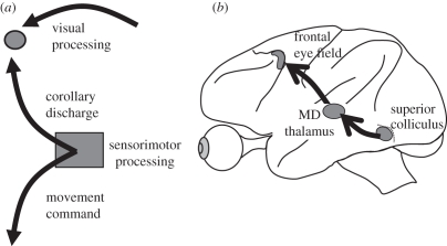
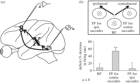
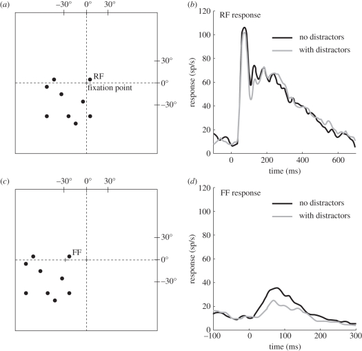
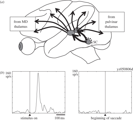
How our vision remains stable in spite of the interruptions produced by saccadic eye movements has been a repeatedly revisited perceptual puzzle. The major hypothesis is that a corollary discharge (CD) or efference copy signal provides information that the eye has moved, and this information is used to compensate for the motion. There has been progress in the search for neuronal correlates of such a CD in the monkey brain, the best animal model of the human visual system. In this article, we briefly summarize the evidence for a CD pathway to frontal cortex, and then consider four questions on the relation of neuronal mechanisms in the monkey brain to stable visual perception. First, how can we determine whether the neuronal activity is related to stable visual perception? Second, is the activity a possible neuronal correlate of the proposed transsaccadic memory hypothesis of visual stability? Third, are the neuronal mechanisms modified by visual attention and does our perceived visual stability actually result from neuronal mechanisms related primarily to the central visual field? Fourth, does the pathway from superior colliculus through the pulvinar nucleus to visual cortex contribute to visual stability through suppression of the visual blur produced by saccades?


Figures

Figure 1.
Figure 1.
A corollary discharge (CD) in the monkey brain. (a) The CD originates from the same sensorimotor processing area as does the motor command to produce the saccadic eye movement. The CD projects to other regions of the brain including those devoted to visual processing. (b) A pathway conveying a CD extends from the intermediate layers of the superior colliculus (SC), through the medial dorsal (MD) nucleus of the thalamus, to the frontal eye field (FEF) in the frontal cortex.
Figure 2.
Figure 2.
Neurons with shifting receptive fields (RFs) in frontal and parietal cortex. (a) The defining characteristic of a neuron showing a shifting RF is that just before the onset of a saccade, a region of the visual field becomes sensitive to visual stimulation. This region has the same spatial relation to the target of the saccade as the RF has to the present fixation point. The region is referred to as the future field (FF) of the neuron. (b) An example of an FEF neuron with a shifting RF. The left column shows the response in a spike density plot over multiple trials to a 50 ms probe stimulus in the RF of the neuron (upper record) and the lack of response at that time in the FF (lower record). The right column shows the increase in activity to the probe stimulus in the FF flashed just before the onset of the saccade and the lack of response in the RF field at that time. Adapted from Sommer & Wurtz [8].
Figure 3.
Figure 3.
Dependence of FEF shifting RFs on the CD input from MD thalamus. (a) FEF neurons were identified as receiving from or projecting to the SC by using stimulation of the SC. Those FEF neurons with both visual RFs and shifting RFs were studied before and during inactivation of the relay region of MD. (b) The per cent decrease (mean and s.e.m.) in the activity of eight FEF neurons with shifting RFs during MD inactivation. Bar graph shows (i) the lack of decrease in the RF response, (ii) over 50% decrease in the FF with saccades directed to the visual field contralateral to the brain that was inactivated, (iii) the lack of decrease in the FF with saccades to the ipsilateral visual field. Asterisks indicate a significant difference with p < 0.0001, t-test. Adapted from Sommer & Wurtz [8].
Figure 4.
Figure 4.
Reduction of FF activity when onset attention is reduced by the presence of distractors. (a) The location of the flashed stimulus falling in the RF of an FEF neuron (RF) and the configuration of eight flashed distractor stimuli outside the RF. (b) Response to the RF stimulus and to the RF stimulus with distractors. (c) Location of the flashed FF stimulus flashed just before the saccade and the eight distractors flashed at the same time. (d) Activity following the FF stimulus and following the FF stimulus with distractors.
Figure 5.
Figure 5.
Contribution of the SC—pulvinar—cortical pathway to the visual suppression with saccades. (a) Symmetry of projections from MD to frontal cortex and from pulvinar to parietal and occipital cortex. (b) Increased activity of a pulvinar neuron in the path from SC to MT to a visual stimulus (left), and decrease in activity of that neuron following a saccade (right). Adapted from Berman & Wurtz [53].

References

    1. Wurtz R. H. 2008. Neuronal mechanisms of visual stability. Vision Res. 48, 2070–208910.1016/j.visres.2008.03.021 (doi:10.1016/j.visres.2008.03.021) - DOI - DOI - PMC - PubMed
    1. von Helmholtz H. 1925. In Helmholtz's treatise on physiological optics, 3rd edn (transl. J. P. C. Southall, 1910) New York, NY: Optical Society of America
    1. Sperry R. W. 1950. Neural basis of the spontaneous optokinetic response produced by visual inversion. J. Comp. Physiol. Psychol. 43, 482–48910.1037/h0055479 (doi:10.1037/h0055479) - DOI - DOI - PubMed
    1. von Holst E., Mittelstaedt H. 1950. Das Reafferenzprinzip. Wechselwirkungen zwischen Zentralnervensystem und Peripherie. Naturwissenschaften 37, 464–476
    1. Sommer M. A., Wurtz R. H. 2002. A pathway in primate brain for internal monitoring of movements. Science 296, 1480–148210.1126/science.1069590 (doi:10.1126/science.1069590) - DOI - DOI - PubMed

LinkOut - more resources