Comparative Study
. 2011 Mar 15;589(Pt 6):1349-66.
doi: 10.1113/jphysiol.2010.203257. Epub 2011 Jan 17.

L-Dopa activates histaminergic neurons

Affiliations
Comparative Study

L-Dopa activates histaminergic neurons

Yevgenij Yanovsky et al. J Physiol. .

Abstract

L-Dopa is the most effective treatment of early and advanced stages of Parkinson's disease (PD), but its chronic use leads to loss of efficiency and dyskinesia. This is delayed by lower dosage at early stages, made possible by additional treatment with histamine antagonists. We present here evidence that histaminergic tuberomamillary nucleus (TMN) neurons, involved in the control of wakefulness, are excited under L-Dopa (EC50 15 μM), express Dopa decarboxylase and show dopamine immunoreactivity. Dopaergic excitation was investigated with patch-clamp recordings from brain slices combined with single-cell RT-PCR analysis of dopamine receptor expression. In addition to the excitatory dopamine 1 (D1)-like receptors, TMN neurons express D2-like receptors, which are coupled through phospholipase C (PLC) to transient receptor potential canonical (TRPC) channels and the Na+/Ca2+ exchanger. D2 receptor activation enhances firing frequency, histamine release in freely moving rats (microdialysis) and wakefulness (EEG recordings). In histamine deficient mice the wake-promoting action of the D2 receptor agonist quinpirole (1 mg kg⁻¹, I.P.) is missing. Thus the histamine neurons can, subsequent to L-Dopa uptake, co-release dopamine and histamine from their widely projecting axons. Taking into consideration the high density of histaminergic fibres and the histamine H3 receptor heteromerization either with D1 or with D2 receptors in the striatum, this study predicts new avenues for PD therapy.


Figures

Figure 1
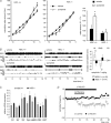
Figure 1. TMN histaminergic neurons are excited by l-Dopa, express dopamine (D) receptors and Dopa decarboxylase
A, excitatory action of l-Dopa depends on l-amino acid uptake and can be blanked by the preincubation with l-histidine, but not with d-histidine. B, effect of l-Dopa is abolished by the combined application of D1/5 and D2-like receptor antagonists. C, l-Dopa dose–response curve fitting with the Hill equation yielded EC50= 15.4 ± 5 μm, Hill coefficient (nH) = 0.6 ± 0.7. Potentiation of firing frequency over the control level was normalized on potentiation by 100 μm of l-Dopa. Number of cells tested with each concentration is given in brackets. D, floating bar histogram illustrating co-localization of histaminergic markers: histidine decarboxylase (HDC), vesicular monoamine transporter 2 (VMAT2), peripherin (Prph), the dopaminergic marker Dopa decarboxylase (DDC) and 5 dopamine receptors (D2L for long splice variant) in 29 TMN neurons (single-cell RT-PCR). Digital images of two neurons (scale bar 20 μm) and corresponding gels documenting RT-PCR analysis of dopamine receptor expression as well as cellular markers in these neurons (no. 13 and no. 22) and in positive control (TMN whole) are given below. M: DNA size marker (100 b.p. ladder). Expected size of PCR products is given above the upper gel. E, co-localization of DDC (AF488, green) with histidine decarboxylase (cy3, red) in rat TMN. Scale bar 100 μm.
Figure 2
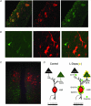
Figure 2. Co-localization of tyrosine hydroxylase- and histamine-containing neurons in the posterior hypothalamus
A, coronal rat (P25) brain slice as used for the electrophysiological recordings from ventral TMN (TMv). 3V: third ventricle. B and C, two levels of horizontal slice sections (R-C for rostro-caudal axis) are indicated with red and blue lines. Double immunostaining for histamine (revealed with cy3, red) and tyrosine hydroxylase (TH, revealed with AF488, green). White dotted line indicates location of third ventricle. Scale bar: 200 μm. D, schematic presentation of coronal sections containing TMN (TMd-TMN dorsal; TMv-TMN ventral; TMm-TMN medial, adopted from the Atlas of Swanson (1992). Areas containing dopaminergic/dopaergic cells are marked green. Abbreviations: cpd, cerebral peduncle; fr, fasciculus retroflexus; ml, medial lemniscus; mm, medial mamillary nucleus; mp, mamillary peduncle; PV, posterior periventricular nucleus of hypothalamus; PMv, ventral premamillary nucleus; VTA, ventral tegmental area; SNc, substantia nigra compacta; SNr, substantia nigra reticulata; sum, supramamillary nucleus.
Figure 3
Figure 3. Dopaminergic excitation of TMN histaminergic neurons
A, dopamine excites rat TMN neurons (n = 5), and the effect is partially blocked by the D2-selective antagonist sulpiride (n = 4). B, the D1/5 dopamine receptor type agonist SKF 38393 increases the firing rate of TMN neurons (n = 6). C, quinpirole (100 μm) increases firing frequency in TMN neurons (n = 12). Sulpiride abolishes the action of quinpirole (n = 4). D, left graph, increase in firing rate of TMN neurons is independent of glutamate, ATP/ADP, growth hormones (IP3-dependent signalling) and nitric oxide (possible messengers of glia–neuron signalling). The following antagonists were used: d-AP5 (NMDA receptors), CNQX (AMPA receptors), LY367385 (mGluR1), MPEP (mGluR5), cibacron blue (broad spectrum antagonist at P2Y receptors), LY294002 (IP3-kinase inhibitor), PTX (picrotoxin), CGP 55845 (GABAB receptor antagonist) and NL-Arg (=l-NAME, NOS inhibitor). Right graph, quinpirole action is not affected by PKA inhibitor H-89 (10 μm), but significantly reduced by an inhibitor of phospholipase C (PLC), U73122 (5 μm), by the Na+/Ca2+ exchanger antagonist benzamil (10 μm) and at room temperature. Four to six TMN neurons were tested with each experimental protocol, except for the pooled control experiments (quinpirole 100 μm, n = 7 in left graph, n = 12 in right graph).
Figure 4
Figure 4. Dose dependence and mechanisms of quinpirole action
A, dose–response curve of quinpirole-induced increase in firing rate of TMN neurons recorded in cell-attached mode (each point is an average obtained from 4–6 neurons. B, quinpirole depolarizes TMN neurons recorded under TTX in whole-cell current clamp mode, indicating a direct postsynaptic effect. C, time course diagram for the averaged whole-cell currents recorded from TMN neurons. Replacement of Na+ in the extracellular solution with NMDG+ or appliction of the TRPC channel antagonist SKF 93635 blocked quinpirole-induced inward current. Arrows indicate application of ramps. D, current–voltage relationship of the quinpirole-induced net current (mean, black line ± SEM, dotted grey line, n = 4).
Figure 5
Figure 5. D2-like receptor activation increases histamine release in vivo in accordance with increased firing of TMN neurons despite increasing GABAergic tone in the posterior hypothalamus in vitro
A, quinpirole (quin) increases frequency of GABAergic mIPSCs recorded from TMN neurons in slices. Example of cumulative fraction histograms for the mIPSCs interevent interval (i.e.i) and amplitude (ampl) and corresponding original mIPSC recordings in control, under quinpirole (quin) and after drug withdrawal in one TMN neuron. B, time course diagram for the averaged frequency of IPSCs (normalized to the control levels, n = 5). C, quinpirole enhances histamine release in the hypothalamus, which reaches a maximal value after 1 h of administration. Shown are means ± SEM of 6 experiments normalized on the last sample before drug administration.
Figure 6
Figure 6. Quinpirole-induced arousal is partially mediated through histamine
A, histograms for the cumulative amount of waking show that HDC−/− mice (n = 15) display a similar decrease in waking during the first 3 h after quinpirole intake when compared to the WT (n = 15) mice but lack subsequent increase in waking. B, typical example of polygraphic recording and corresponding hypnograms showing the effect of systemically applied quinpirole on the cortical EEG and EMG and sleep–wake cycle, illustrating the increase in waking in HDC+/+ (n = 16) and HDC−/− (n = 14) mice. The moment of injection is indicated with array. Diagram (at the right) illustrates sleep–wake changes relative to baseline for the mean values (min ± SEM) spent in each sleep–wake stage during 3 h after injection of quinpirole at 10.00 h. Note that quinpirole (1 mg kg−1) causes a significant increase in waking (W) and a decrease in slow wave sleep (SWS) in WT but the opposite effects in HDC−/− mice. C, real-time RT-PCR analysis of the relative expression of transcripts of dopamine receptors and cellular markers in HDC KO and WT mice shows no difference between the two genotypes. D, in rat but not in mouse slices, the dopamine reuptake inhibitor nomifensine (1 μm) causes suppression of TMN firing, which is prevented by the D4 receptor antagonist L-745,870 (1 μm).
Figure 7
Figure 7. Dopamine–histamine interaction in the posterior hypothalamus
A, group of neurons in TMNv stained for histamine (revealed with cy3, red) and dopamine (revealed with AF488, green). B, dorsal TMN bordering dopaminergic neurons of posterior periventricular nucleus of the hypothalamus (slice in Fig. 2Da). This is an enlarged image from elliptic field indicated in C. Scale bars: A and B, 20 μm; C, 65 μm. Schematic drawing of dopaminergic and GABAergic inhibitory (–) inputs to the TMN neuron, which are overwhelmed by excitation under l-Dopa. Juxtacellular unit recordings are given for control and l-Dopa supplemented medium.

References

    1. Anaclet C, Parmentier R, Ouk K, Guidon G, Buda C, Sastre JP, Akaoka H, Sergeeva OA, Yanagisawa M, Ohtsu H, Franco P, Haas HL, Lin JS. Orexin/hypocretin and histamine: distinct roles in the control of wakefulness demonstrated using knock-out mouse models. J Neurosci. 2009;29:14423–14438. - PMC - PubMed
    1. Anichtchik OV, Rinne JO, Kalimo H, Panula P. An altered histaminergic innervation of the substantia nigra in Parkinson's disease. Exp Neurol. 2000;163:20–30. - PubMed
    1. Arbuthnott G, Wright AK. Some non-fluorescent connections of the nigro-neostriatal dopamine neurones. Brain Res Bull. 1982;9:367–368. - PubMed
    1. Arnulf I. Results of clinical trials of tiprolisant in narcolepsy and Parkinson's disease. Eur Neuropsychopharmacology. 2009;19:S204.
    1. Arnulf I, Leu-Semenescu S. Sleepiness in Parkinson's disease. Parkinsonism Relat Disord. 2009;15(Suppl 3):S101–S104. - PubMed

Publication types

LinkOut - more resources