

Review
doi: 10.1503/cmaj.091740.
Epub 2011 Jan 17.
Benign spotted bones: a diagnostic dilemma
Affiliations
- PMID: 21242270
- PMCID: PMC3050950
- DOI: 10.1503/cmaj.091740
Item in Clipboard
Review
Benign spotted bones: a diagnostic dilemma
CMAJ.
.
No abstract available
Figures
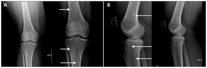
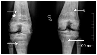
(A) Anteroposterior and (B) lateral radiographs of the right knee in a young woman after a fall. Sclerotic foci of variable size (arrows) appear in the femur and tibia. Note the periarticular distribution and predominant meta-epiphyseal location (sites of endochondral bone formation) characteristic of osteopoikilosis.
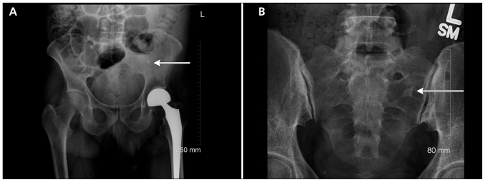
(A) Anteroposterior view of the pelvis showing a single sclerotic focus in the left iliac bone (arrow) typical of a bone island or enostosis. (B) Anteroposterior view of the sacrum showing a single dense sclerotic focus in the left ala. Spiculated margins (arrow) merge with underlying normal trabeculae, which is characteristic of bone islands.
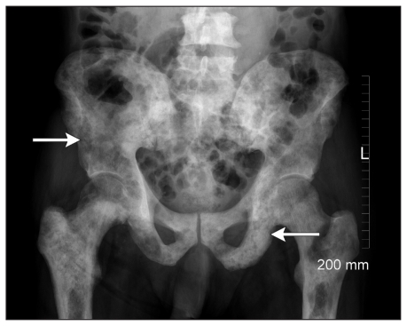
Anteroposterior view of the pelvis showing patchy, poorly defined areas of sclerosis over the entire pelvis and proximal femora. Subtle, more focal areas and diffuse areas of sclerosis are visible. This pattern is most typical of sclerotic bone metastasis (e.g., as a result of prostate cancer in this patient). A subtle underlying pattern of small, rounded lesions can also be seen (arrows).
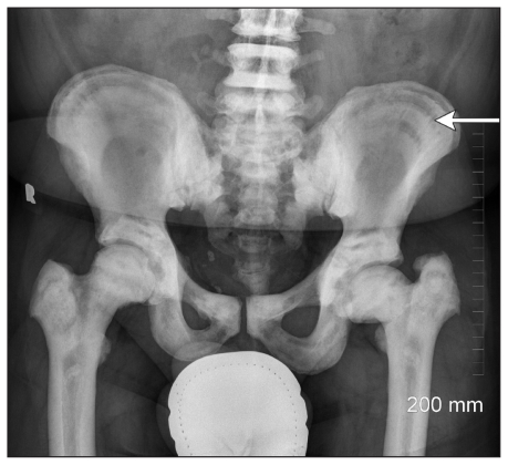
Anteroposterior view of the pelvis showing a pattern of layered sclerosis (arrow) described as bone within bone and characteristic of osteopetrosis.
Anteroposterior view of a patient’s knees showing a pattern of diffuse sclerosis similar to that seen in osteopetrosis. However, vertical striations (arrows) shown in the distal femora and proximal tibia are consistent with the pattern seen in osteopathia striata.
References
-
- Albers-Schönberg H. Eine seltene, bischer nicht bekannte strukturanomalie des skelettes. Fortschr Roentgenstrb 1915;73:174–5
-
- Jonash E. 12 falle von soteopoikilie. Fortschr Roentgenstr 1955;82:344–53 - PubMed
-
- Szabo AD. Osteopoikilosis in a twin. Clin Orthop Relat Res 1971; 79:156–63 - PubMed
-
- Melnick JC. Osteopathia condensas disseminata (osteopoikilosis). Study of a family of 4 generations. Am J Roentgen 1959;82:344–53 - PubMed
-
- Vanhoenacker FM, De Beuckeleer LH, Van Hul W, et al. Sclerosing bone dysplasias: genetic and radioclinical features. Eur Radiol 2000;10:1423–33 - PubMed
Publication types
MeSH terms
LinkOut - more resources
Full Text Sources