. 2011 Jan 24;192(2):307-19.
doi: 10.1083/jcb.201007095. Epub 2011 Jan 17.

The Drosophila blood brain barrier is maintained by GPCR-dependent dynamic actin structures

Affiliations

The Drosophila blood brain barrier is maintained by GPCR-dependent dynamic actin structures

Meital Hatan et al. J Cell Biol. .

Abstract

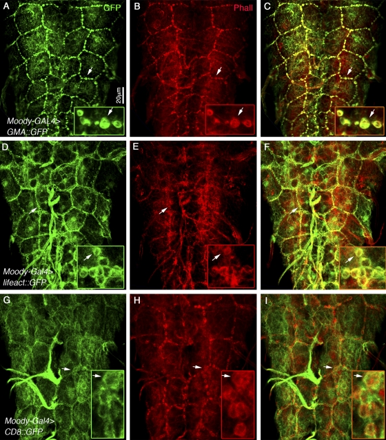
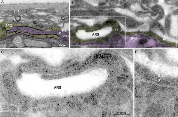
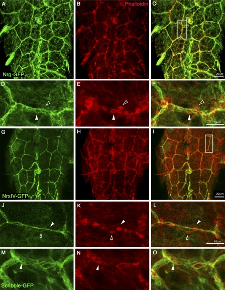
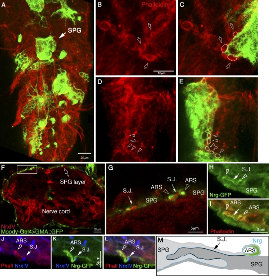
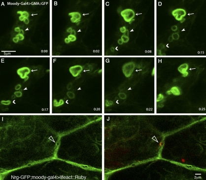
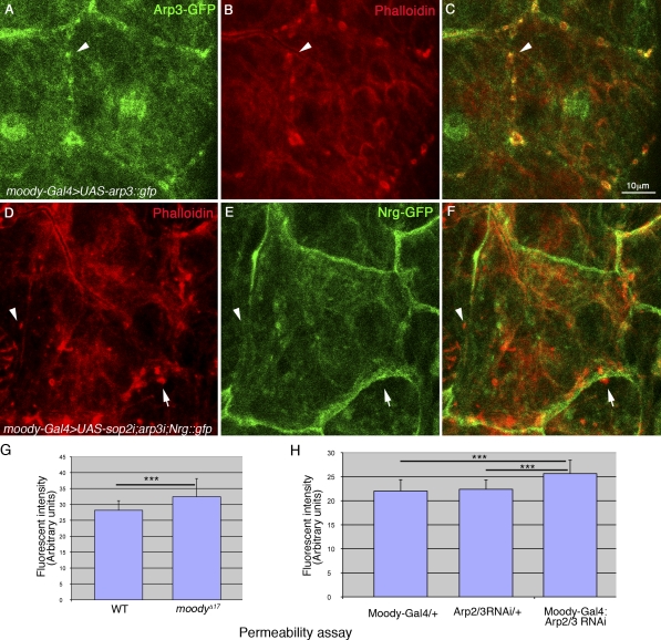
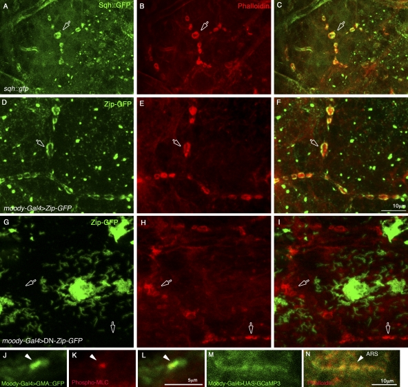
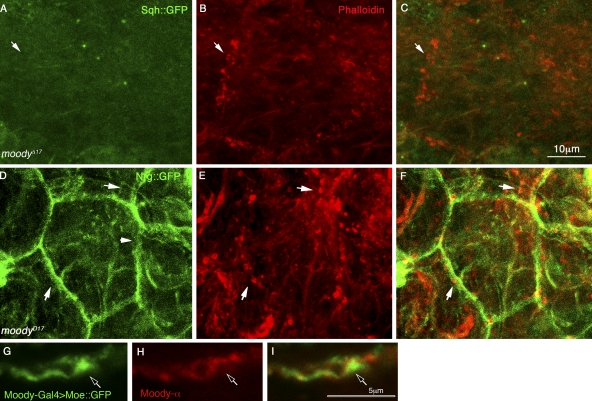
The blood brain barrier (BBB) is essential for insulation of the nervous system from the surrounding environment. In Drosophila melanogaster, the BBB is maintained by septate junctions formed between subperineurial glia (SPG) and requires the Moody/G protein-coupled receptor (GPCR) signaling pathway. In this study, we describe novel specialized actin-rich structures (ARSs) that dynamically form along the lateral borders of the SPG cells. ARS formation and association with nonmuscle myosin is regulated by Moody/GPCR signaling and requires myosin activation. Consistently, an overlap between ARS localization, elevated Ca(2+) levels, and myosin light chain phosphorylation is detected. Disruption of the ARS by inhibition of the actin regulator Arp2/3 complex leads to abrogation of the BBB. Our results suggest a mechanism by which the Drosophila BBB is maintained by Moody/GPCR-dependent formation of ARSs, which is supported by myosin activation. The localization of the ARSs close to the septate junctions enables efficient sealing of membrane gaps formed during nerve cord growth.


Figures

Figure 1.
Figure 1.
ARSs are distributed along the SPG intercellular borders. (A–I) Nerve cords were dissected from third instar larvae, labeled with moesin-GFP (A–C), Lifeact-GFP (D–F), or CD8-GFP (G–I), driven to be expressed in the SPG cells, and then double labeled with phalloidin (Phall). The GFP labeling is shown in A, D, and G, phalloidin in B, E, and H, and their merged images in C, F, and I. The insets in each panel represent high magnification of the region marked by an arrow in the nerve cords. Note that the ARSs are distributed along the borders of the large SPG cells. Overlap between GFP and phalloidin is observed.
Figure 2.
Figure 2.
Ultrastructure of the ARSs labeled with gold-conjugated anti-GFP. (A–D) Cross sections of nerve cords dissected from third instar larvae carrying moesin-GFP in the SPG cells and processed either by chemical fixation (A) or by HPF followed by freeze substitution (B–D). The perineurial (PN) and subperineurial (SPG) cells (two neighboring SPG cells are marked by yellow or by pink), septate junctions (S.J.), and neurons (N) are indicated in A and B. The nerve cords shown in B–D were labeled with anti-GFP antibody and secondary 10-nm gold–conjugated antibody to visualize moesin-GFP. Low magnification (similar to that in A) of the nerve cord is shown in B, and the two corresponding regions marked in B are shown in higher similar magnification in C and D. The immunogold labeling in C (arrows) is detected very close to the membrane indentation that corresponds to the ARSs. No GFP labeling was detected at the nearby septate junction shown in D (arrow).
Figure 3.
Figure 3.
The ARSs are associated with Nrg-containing membrane loops. (A–O) Nerve cords dissected from third instar larvae carrying Nrg-GFP (A–F), Nrx-GFP (G–L), or Scribble-GFP (M–O). GFP labeling is shown in A, D, G, J, and M, and the corresponding phalloidin staining is shown in B, E, H, K, and N. The corresponding merged images are shown in C, F, I, L, and O. D–F and J–L show high magnifications of the corresponding area marked with a white rectangle in C and I. Note that the ARSs often overlap the Nrg-GFP labeling (white arrowheads in D–F). In rare cases, we detected an ARS distal from the SPG border that does not overlap Nrg-GFP (open arrowheads in D–F). NrxIV does not overlap the ARSs (white and open arrowheads in J–L). Partial overlap is also noted between Scribble and phalloidin staining (arrowheads in M–O).
Figure 4.
Figure 4.
The relative distribution of the ARSs within a single SPG cell. Nerve cord dissected from third instar larvae in which single glia cells were labeled with GFP and with phalloidin (e.g., arrow in A). High magnification of the borders between GFP-positive and -negative SPG cells labeled with phalloidin (B and D) or their merged images (C and E) are shown. Arrows in B and C indicate three ARSs that belong to the GFP-negative cell marked by white lines in C. Arrowheads in D and E indicate five ARSs, all of which belong to the GFP-positive SPG cell, marked by white lines in E. (F) A cross section in the nerve cord of third instar larvae carrying moesin-GFP, driven by Moody-Gal4 and stained with anti-NrxIV (red; F and G) and anti-GFP (green; F and G; arrow in F indicates the SPG layer). (G) High magnification of the region marked by the rectangle in F. The septate junction (S.J.) formed between neighboring SPG cells is marked by NrxIV staining (red; arrows). The ARSs are marked by arrowheads. (H and I) High magnification of a cross section of nerve cord dissected from larvae expressing Nrg-GFP (green; H) labeled with phalloidin (red; I). The ARSs are indicated by arrowheads, and the septate junction between neighboring SPG is marked by arrows. J–L show a cross section in Nrg-GFP–expressing larvae labeled with anti-GFP (green; K and L), phalloidin (Phall; red; J and L), and NrxIV (blue; J–L). Arrowheads indicate the ARSs, and arrows show the septate junction. (M) A scheme of the relative distribution of the ARSs, membrane convoluted loop, and septate junction deduced from our analysis.
Figure 5.
Figure 5.
ARS dynamics in live third instar larvae. (A–H) GFP images of the ARSs taken at distinct time intervals (in minutes) of the nerve cord of third instar larvae immobilized between two coverslips carrying the moesin-GFP under Moody-Gal4. The arrows and arrowheads indicate to the same ARSs at the different time points. I and J are images of live larvae carrying Nrg-GFP and Lifeact-Ruby driven by Moody-Gal4. An Nrg loop and the ARS associated with it, labeled with Lifeact-Ruby, are indicated (empty arrowheads).
Figure 6.
Figure 6.
The Arp2/3 complex is required for ARS formation and for maintenance of BBB function. (A–C) Nerve cord of third instar larvae expressing Arp3-GFP fusion protein driven by Moody-Gal4. The Arp3-GFP (A) colocalizes with the ARSs labeled with phalloidin (B). The merged image is shown in C. Arrowheads in A–C indicate ARSs colabeled with phalloidin and Arp3-GFP. (D–F) Nerve cord from larvae expressing RNAi for both Arp2 and Arp3 proteins labeled for phalloidin (D). The larvae also carried the Nrg-GFP protein trap. Nrg-GFP is shown in E, and the merged image is shown in F. Arrows show aberrant ARSs that no longer associate with Nrg-GFP labeling. Arrowheads indicate sites lacking Nrg-GFP continuity. (G) Dye penetration to the nerve cord was measured by the fluorescent intensity of nerve cords dissected from wild-type (WT) larvae and compared with moody mutant larvae (moodyΔ17). The difference between the averaged fluorescent intensity of the two groups was calculated by Student’s t test and was found to be significant (***, P < 0.0066). (H) Dye penetration was compared between three groups: control larvae carrying RNAi to the Arp2/3 components alone (UAS-sop2i;arp3i/+), control larvae carrying moody-gal4 alone (moody-gal4/+), or an experimental group of Arp2/3 knockdown larvae (moody-gal4:UAS-sop2i;arp3i). One-way ANOVA test with Dunnett’s test (using SAS program) was used to determine the statistical significance of the difference between the experimental and the control groups (Moody-gal4/+ or Sop2i;Arp3i/+). In both cases, the difference between the experimental group and each of the control groups was statistically significant (at α = 0.05; indicated by asterisks). (G and H) Error bars indicate standard deviation.
Figure 7.
Figure 7.
Activation of nonmuscle myosin is essential for correct morphology of the ARSs. (A–I) Nerve cords were dissected from third instar larvae carrying MLC Sqh (Sqh-GFP) under its own promoter (A–C) and myosin heavy chain Zipper (Zip-GFP; D–F) or a dominant-negative form of Zipper fused to GFP (DNZip-GFP; G–I) both driven by the Moody-Gal4 driver. A, D, and G show GFP labeling, B, E, and H show phalloidin labeling, and C, F, and I are the corresponding merged images. The arrows in A–F show the ARSs and indicate overlap staining between phalloidin and myosin labeling. The arrows in G show dissociation of Zip-GFP from the ARSs and in H show abnormal morphology of the ARSs. J–L show a cross section of third instar larvae expressing moesin-GFP in SPG cells labeled with anti–P-MLC (red) and GFP. Their merged image is shown in L. An overlap between the ARSs and the P-MLC staining is observed (arrowheads). M and N show the border between two SPG cells of nerve cord dissected from larvae expressing the Ca2+ indicator GCaMP3 in SPG cells (M) and labeled with phalloidin (red; N). An overlap between the phalloidin staining representing the ARSs and the fluorescence of the Ca2+ indicator is observed (arrowhead).
Figure 8.
Figure 8.
The Moody/GPCR pathway regulates myosin association with the ARSs. (A–F) Nerve cords dissected from moodyΔ17 mutant larvae and also carrying either Sqh-GFP (A–C) or Nrg-GFP (D–F). GFP labeling is shown in A and D, and phalloidin labeling is shown in B and E. C and F are their corresponding merged images. The arrow in A shows a complete dissociation of Sqh-GFP from the ARS and in B abnormal morphology of the ARSs. The arrowhead in D shows discontinuity of Nrg-GFP labeling, and the arrows in D and E show corresponding locations of abnormal ARS morphology. (G–I) Cross section of third instar larvae nerve cord expressing moesin-GFP and labeled with Moody-α (red). A partial overlap between the moesin-GFP and Moody-α is detected. Moody staining wraps the ARS (arrows).

Similar articles

Cited by

References

    1. Abbott N.J., Rönnbäck L., Hansson E. 2006. Astrocyte-endothelial interactions at the blood-brain barrier. Nat. Rev. Neurosci. 7:41–53 10.1038/nrn1824 - DOI - PubMed
    1. Bainton R.J., Tsai L.T., Schwabe T., DeSalvo M., Gaul U., Heberlein U. 2005. moody encodes two GPCRs that regulate cocaine behaviors and blood-brain barrier permeability in Drosophila. Cell. 123:145–156 10.1016/j.cell.2005.07.029 - DOI - PubMed
    1. Banerjee S., Bhat M.A. 2007. Neuron-glial interactions in blood-brain barrier formation. Annu. Rev. Neurosci. 30:235–258 10.1146/annurev.neuro.30.051606.094345 - DOI - PMC - PubMed
    1. Baumgartner S., Littleton J.T., Broadie K., Bhat M.A., Harbecke R., Lengyel J.A., Chiquet-Ehrismann R., Prokop A., Bellen H.J. 1996. A Drosophila neurexin is required for septate junction and blood-nerve barrier formation and function. Cell. 87:1059–1068 10.1016/S0092-8674(00)81800-0 - DOI - PubMed
    1. Behr M., Riedel D., Schuh R. 2003. The claudin-like megatrachea is essential in septate junctions for the epithelial barrier function in Drosophila. Dev. Cell. 5:611–620 10.1016/S1534-5807(03)00275-2 - DOI - PubMed

Publication types

MeSH terms