. 2011 Jan 24;192(2):263-76.
doi: 10.1083/jcb.201008005. Epub 2011 Jan 17.

RAD21L, a novel cohesin subunit implicated in linking homologous chromosomes in mammalian meiosis

Affiliations

RAD21L, a novel cohesin subunit implicated in linking homologous chromosomes in mammalian meiosis

Jibak Lee et al. J Cell Biol. .

Abstract

Cohesins are multi-subunit protein complexes that regulate sister chromatid cohesion during mitosis and meiosis. Here we identified a novel kleisin subunit of cohesins, RAD21L, which is conserved among vertebrates. In mice, RAD21L is expressed exclusively in early meiosis: it apparently replaces RAD21 in premeiotic S phase, becomes detectable on the axial elements in leptotene, and stays on the axial/lateral elements until mid pachytene. RAD21L then disappears, and is replaced with RAD21. This behavior of RAD21L is unique and distinct from that of REC8, another meiosis-specific kleisin subunit. Remarkably, the disappearance of RAD21L at mid pachytene correlates with the completion of DNA double-strand break repair and the formation of crossovers as judged by colabeling with molecular markers, γ-H2AX, MSH4, and MLH1. RAD21L associates with SMC3, STAG3, and either SMC1α or SMC1β. Our results suggest that cohesin complexes containing RAD21L may be involved in synapsis initiation and crossover recombination between homologous chromosomes.


Figures

Figure 1.
Figure 1.
RAD21L is the third kleisin subunit of cohesins conserved among vertebrate species. (A) A phylogenetic tree of kleisin subunits of cohesins among vertebrates was constructed by using ClustalW and TreeView programs. The GenBank/EMBL/DDBJ accession nos. or Ensemble database nos. are as follows; hsRAD21 (BC050381), mmRAD21 (AK163992), ggRAD21 (AJ719731), xlRAD21 (BC077991), olRAD21 (AB115697), hsRAD21L (BC157891), mmRAD21L (AB574185), ggRAD21L (ENSGALG00000006186), olRAD21L (ENSORLT00000018324), hsREC8 (BC010887), mmREC8 (BC052155), and xlREC8 (BC087346). The nomenclatures of species are as follows; hs, Homo sapiens; mm, Mus musculus; gg, Gallus gallus (chicken); xl, Xenopus laevis; ol, Oryzias latipes (medaka fish). (B) Schematic representation of the primary structures of three different kleisin subunits of cohesins in the mouse. Gray boxes indicate winged-helix domains conserved among the kleisin family of proteins.
Figure 2.
Figure 2.
Specific expression of RAD21L in both male and female gonads. (A) Relative transcript levels of RAD21, RAD21L, REC8, and GAPDH (an internal control) in mouse liver, spleen, and testis were assessed by real-time RT-PCR. For each gene, 100-fold diluted, 10-fold diluted, or undiluted cDNAs from testis were used for making a standard curve, and all values are shown in comparison with values obtained in the control samples using the undiluted cDNAs from testis. (B) Immunoblot analysis of a testis extract using rat and rabbit antibodies (Ab) raised against RAD21L or preimmune sera (P). (C) Immunoblot analysis of extracts from various mouse organs with rabbit polyclonal antibodies against the different kleisin subunits and SMC3. (D) Immunofluorescent labeling of frozen sections of fetal ovary (18.5 d post-coitum) (a–d) and adult testis (e–h) with mouse polyclonal anti-SYCP3 (b and f) and rat polyclonal anti-RAD21L (c and g) antibodies. DNA was counterstained with Hoechst 34580 (a and e) and merged images are shown (d and h). Bar, 100 µm.
Figure 3.
Figure 3.
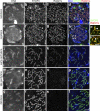
RAD21 and RAD21L are detected in a mutually exclusive manner in mouse testicular cells. (A) Frozen sections of adult testis were immunofluorescently labeled with rat polyclonal anti-RAD21L (b and f) and rabbit polyclonal anti-RAD21 (c and d) antibodies. Note that spermatocytes located near the fibrous capsule (a and e; green dashed lines) of seminiferous tubules were RAD21L-positive cells (f and h; white arrowheads), whereas spermatocytes located relatively close to the center of the tubules are RAD21-positive cells (g and h; yellow arrowheads). In e and h, sg and st indicate spermatogonium and spermatid, respectively. DNA was counterstained with Hoechst 34580 (a and e) and merged images are shown (d and h). An overview image (a–d) and a magnified image (e–h) of different testis sections are shown. Bars: (d) 50 µm; (h) 10 µm. (B) Nuclear spreads from testicular cells were immunofluorescently labeled with rat polyclonal anti-RAD21L (b and f) and rabbit polyclonal anti-RAD21 (c and g) antibodies. Note that spermatocyte nuclei were exclusively labeled with either anti-RAD21L (b and d; white arrowheads) or anti-RAD21 (c and d; yellow arrowheads) antibody, whereas a nucleus (f–h; white arrow) was faintly labeled with both antibodies. DNA was counterstained with Hoechst 34580 (a and e) and merged images are shown (d and h). Also shown are magnified images of a chromosome colabeled with the two antibodies (f′–h′). Bars: (h) 10 µm; (h′) 5 µm.
Figure 4.
Figure 4.
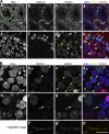
The appearance of RAD21L on meiotic chromosomes is strictly confined in early stages of meiotic prophase I. Nuclear spreads from testicular cells were prepared and immunofluorescently labeled with rat polyclonal anti-RAD21L (c, g, k, o, and s) and mouse polyclonal anti-SYCP3 (b, f, j, n, and r) antibodies. DNA was counterstained with Hoechst 34580 (a, e, i, m, and q) and merged images are shown (d, h, l, p, and t). Some of the bivalents in h are enlarged in h′ and h′′. Bars: (x) 5 µm; (h′′) 2 µm.
Figure 5.
Figure 5.
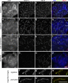
Differential distributions of RAD21L and REC8 on the synaptonemal complex during meiotic prophase I. Nuclear spreads from testicular cells were prepared and immunofluorescently labeled with rat polyclonal anti-RAD21L (b, f, j, n, f′, and j′) and rabbit polyclonal anti-REC8 (c, g, k, o, g′, and k′) antibodies. The regions indicated in h and l are magnified in f′–h′ and j′–k′, respectively. DNA was counterstained with Hoechst 34580 (a, e, i, and m) and merged images are shown (d, h, l, p, h′, and l′). Bar, 5 µm.
Figure 6.
Figure 6.
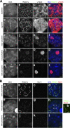
Temporal correlation between the disappearance of RAD21L and other pachytene events. (A) Nuclear spreads from testicular cells were prepared and subjected to immunofluorescent labeling with rat polyclonal anti-RAD21L (b, f, j, n, and r) and rabbit polyclonal anti–γ-H2AX (c, g, k, o, and s) antibodies. DNA was counterstained with Hoechst 34580 (a, e, i, m, and q) and merged images are shown (d, h, l, p, and t). Bar, 5 µm. (B) Nuclear spreads from testicular cells were immunofluorescently labeled with rat polyclonal anti-RAD21L (b, f, and j) and mouse monoclonal anti-MLH1 (c, g, and k) antibodies. DNA was counterstained with Hoechst 34580 (a, e, and i) and merged images are shown (d, h, and l). The region indicated in h is magnified in h′. Bars: (l) 5 µm; (h′) 2 µm.
Figure 7.
Figure 7.
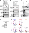
Comprehensive immunoprecipitation analysis with antibodies against different cohesin subunits. (A–C) Testis extracts were subjected to immunoprecipitations using antibodies against kleisin subunits (A), SMC subunits (B), and SA–STAG subunits (C). The precipitates, along with aliquots of the input fractions, were analyzed by immunoblotting with the antibodies indicated. The arrow in A indicates the phosphorylated form of STAG3 coprecipitated with RAD21L. (D) Immunoprecipitates obtained with anti-RAD21L antibody were treated with (+) or without (−) λ-PPase, and were analyzed by immunoblotting with antibodies indicated. (E) Putative subunit compositions of the cohesin complexes as revealed by our immunoprecipitation assays. The number assigned for each complex is also used in Fig. 8. STAG3 might not be included in the third cohesin complex (therefore shown in parentheses) because the association between SMC1α and STAG3 was hardly detectable in our assays.
Figure 8.
Figure 8.
Spatiotemporal distribution of multiple cohesin complexes in the processes of paring, synapsis, and recombination of homologous chromosomes during meiotic prophase I in mammals. RAD21-, RAD21L-, and REC8-containing cohesins are indicated by the blue, red, and orange ovals, respectively. Circled numbers correspond to the different cohesin complexes summarized in Fig. 7 E. Although the intriguing possibility is depicted here that cohesinRAD21L could embrace two duplexes from homologous chromosomes whereas cohesinREC8 could do so from sister chromatids (inset), our current results by no means exclude other models.

References

    1. Anderson L.K., Reeves A., Webb L.M., Ashley T.. 1999. Distribution of crossing over on mouse synaptonemal complexes using immunofluorescent localization of MLH1 protein. Genetics. 151:1569–1579. - PMC - PubMed
    1. Anderson L.K., Royer S.M., Page S.L., McKim K.S., Lai A., Lilly M.A., Hawley R.S.. 2005. Juxtaposition of C(2)M and the transverse filament protein C(3)G within the central region of Drosophila synaptonemal complex. Proc. Natl. Acad. Sci. USA. 102:4482–4487. 10.1073/pnas.0500172102 - DOI - PMC - PubMed
    1. Barchi M., Mahadevaiah S., Di Giacomo M., Baudat F., de Rooij D.G., Burgoyne P.S., Jasin M., Keeney S.. 2005. Surveillance of different recombination defects in mouse spermatocytes yields distinct responses despite elimination at an identical developmental stage. Mol. Cell. Biol. 25:7203–7215. 10.1128/MCB.25.16.7203-7215.2005 - DOI - PMC - PubMed
    1. Bellani M.A., Romanienko P.J., Cairatti D.A., Camerini-Otero R.D.. 2005. SPO11 is required for sex-body formation, and Spo11 heterozygosity rescues the prophase arrest of Atm−/− spermatocytes. J. Cell Sci. 118:3233–3245. 10.1242/jcs.02466 - DOI - PubMed
    1. Bhalla N., Dernburg A.F.. 2008. Prelude to a division. Annu. Rev. Cell Dev. Biol. 24:397–424. 10.1146/annurev.cellbio.23.090506.123245 - DOI - PMC - PubMed

Publication types

MeSH terms