. 2011 Feb 14;208(2):217-25.
doi: 10.1084/jem.20100370. Epub 2011 Jan 17.

Growth differentiation factor 15 deficiency protects against atherosclerosis by attenuating CCR2-mediated macrophage chemotaxis

Affiliations

Growth differentiation factor 15 deficiency protects against atherosclerosis by attenuating CCR2-mediated macrophage chemotaxis

Saskia C A de Jager et al. J Exp Med. .

Abstract

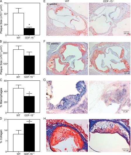
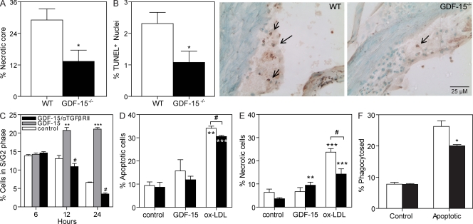
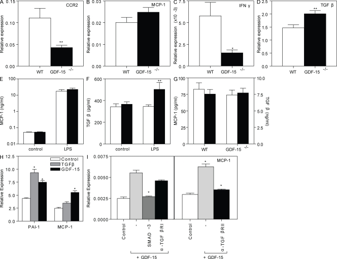
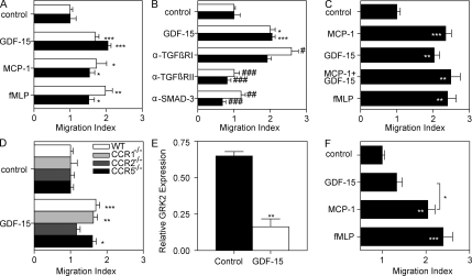
Growth differentiation factor (GDF) 15 is a member of the transforming growth factor β (TGF-β) superfamily, which operates in acute phase responses through a currently unknown receptor. Elevated GDF-15 serum levels were recently identified as a risk factor for acute coronary syndromes. We show that GDF-15 expression is up-regulated as disease progresses in murine atherosclerosis and primarily colocalizes with plaque macrophages. Hematopoietic GDF-15 deficiency in low density lipoprotein receptor(-/-) mice led to impaired initial lesion formation and increased collagen in later lesions. Although lesion burden in GDF-15(-/-) chimeras was unaltered, plaques had reduced macrophage infiltrates and decreased necrotic core formation, all features of improved plaque stability. In vitro studies pointed to a TGFβRII-dependent regulatory role of GDF-15 in cell death regulation. Importantly, GDF-15(-/-) macrophages displayed reduced CCR2 expression, whereas GDF-15 promoted macrophage chemotaxis in a strictly CCR2- and TGFβRII-dependent manner, a phenomenon which was not observed in G protein-coupled receptor kinase 2(+/-) macrophages. In conclusion, GDF-15 deletion has a beneficial effect both in early and later atherosclerosis by inhibition of CCR2-mediated chemotaxis and by modulating cell death. Our study is the first to identify GDF-15 as an acute phase modifier of CCR2/TGFβRII-dependent inflammatory responses to vascular injury.


Figures

Figure 1.
Figure 1.
GDF-15 is progressively expressed in atherosclerotic lesions in a pattern similar to that of macrophages. (A–D) Temporal expression of GDF-15 (A), CD68 (B), Smoothelin (C) and PECAM-1 (D) during atherogenesis was assessed by whole genome microarray. Values are expressed as fold induction compared with time point zero. The experiment was performed twice, with n = 3 (each containing pooled plaque material of three mice) per time point. *, P < 0.05; ***, P < 0.001, compared with the 0-wk timepoint. Error bars are depicted as SEM. (E and F) Immunohistochemistry for GDF-15 in human (E) and murine (F) atherosclerotic lesions. Arrows represent intimal cells (based on nuclear staining) that express GDF-15.
Figure 2.
Figure 2.
Effects of GDF-15 deficiency on atherogenesis and plaque cellularity. Irradiated LDLr−/− recipients were reconstituted with WT or GDF-15−/− bone marrow. (A and B) Plaque size after 4 wk (A) or 12 wk (B; representative pictures in E and F). (C) Macrophages were stained with α–MoMa-2 and depicted as percentage of MoMa-2+ cells among total plaque area (representative pictures in G). (D) Collagen was visualized by Masson’s trichrome staining and depicted as percentage of collagen among total plaque area (representative pictures in H). *, P < 0.05, compared with WT controls. n = 9 animals per group. The experiment was independently performed two times. Error bars are depicted as SEM.
Figure 3.
Figure 3.
Effects of GDF-15 deficiency on plaque stability. (A) Necrotic core size depicted as percentage among total plaque area. (B) Cellular apoptosis was visualized by TUNEL staining and depicted as TUNEL+ cells among all mononuclear cells (including representative pictures) in week-12 plaques. *, P < 0.05, compared with WT controls (n = 9 per group). Arrows indicate TUNEL-positive nuclei. (C) S/G2 phase arrest (depicted as percentage among total cells) in RAW 264.7 macrophages after treatment with 10 ng/ml GDF-15 (gray bars) and 100 ng/ml α-TGFβRII (black bars). **, P < 0.01; ***, P < 0.001, compared with untreated controls (white bars); #, P < 0.001, compared with GDF-15 treatment. Studies were performed four times per condition and repeated in three separate experiments. (D and E) Rate of macrophage apoptosis (D; percentage annexin V+/PI cells) and necrosis (E; percentage annexin V+/PI+ cells) after treatment with 10 ng/ml GDF-15 or 50 µg/ml ox-LDL in both GDF-15−/− (black bars) and WT (white bars) macrophages. **, P < 0.01; ***, P < 0.001 when compared with control; #, P < 0.05, compared with WT. (F) Phagocytosis capacity in WT (white bars) and GDF-15−/− (black bars) macrophages. *, P < 0.05 when compared with WT. Bone marrow–derived macrophages from WT and GDF-15 chimeras were pooled and used for apoptosis and phagocytosis assays. Each experiment was performed four times. Error bars are depicted as SEM.
Figure 4.
Figure 4.
Pro- and antiinflammatory mediators in GDF-15−/− cells and chimeras. (A–D) Relative mRNA expression of CCR2 (A), MCP-1 (B), IFN-γ (C), and TGF-β (D) in WT (white bars) and GDF-15−/− (black bars) macrophages. Values are expressed relative to average expression of GAPDH and HPRT reference genes. (E and F) MCP-1 (E) and TGF-β (F) production in WT (white bars) and GDF-15−/− (black bars) macrophages after LPS treatment. (G) Basal levels of MCP-1 (white bars) and TGF-β (black bars) in WT and GDF-15−/− chimeras after 4 wk of Western type diet. (H) Relative mRNA expression of PAI-1 and MCP-1 in response to 10 ng/ml GDF-15– and 15 ng/ml TGF-β1–treated WT macrophages. (I) Relative MCP-1 mRNA expression after SMAD-3 inhibition (SIS3; 3 µM) and α-TGFβRI and α-TGFβRII treatment (100 ng/ml) in WT macrophages. Bone marrow–derived macrophages from WT and GDF-15 were pooled and used for RNA expression. Each experiment was done four times. *, P < 0.05; **, P < 0.01. Error bars are depicted as SEM.
Figure 5.
Figure 5.
GDF-15 sensitizes CCR2-mediated chemotactic response. (A) Migration response of WT (white bars) and GDF-15−/− (black bars) macrophages to GDF-15, MCP-1, and fMLP. (B) Migratory response of WT (white bars) and GDF-15−/− (black bars) macrophages to GDF-15 after treatment with α-TGFβRI, α-TGFβRII, and SMAD-3 inhibition. (C) Migratory response of WT peritoneal macrophages after combined GDF-15 and MCP-1 treatment. (D) Macrophage migration toward GDF-15 in WT and CCR1-, CCR2-, and CCR5-deficient macrophages. (E) Relative GRK-2 mRNA expression in WT bone marrow–derived macrophages after exposure to GDF-15. (F) Migratory response toward GDF-15 and MCP-1 of GRK-2+/− macrophages. *, P < 0.05; **, P < 0.01; ***, P < 0.001, when compared with control; and #, P < 0.05; ##, P < 0.01; ###, P < 0.001, when compared with GDF-15. Migration and mRNA expression assays were performed with pooled bone marrow–derived macrophages from WT and GDF-15. Each experiment was repeated six (migration) and four (mRNA) times. Error bars are depicted as SEM.

References

    1. Ait-Oufella H., Pouresmail V., Simon T., Blanc-Brude O., Kinugawa K., Merval R., Offenstadt G., Lesèche G., Cohen P.L., Tedgui A., Mallat Z. 2008. Defective mer receptor tyrosine kinase signaling in bone marrow cells promotes apoptotic cell accumulation and accelerates atherosclerosis. Arterioscler. Thromb. Vasc. Biol. 28:1429–1431 10.1161/ATVBAHA.108.169078 - DOI - PubMed
    1. Aragay A.M., Mellado M., Frade J.M., Martin A.M., Jimenez-Sainz M.C., Martinez-A C., Mayor F., Jr 1998. Monocyte chemoattractant protein-1-induced CCR2B receptor desensitization mediated by the G protein-coupled receptor kinase 2. Proc. Natl. Acad. Sci. USA. 95:2985–2990 10.1073/pnas.95.6.2985 - DOI - PMC - PubMed
    1. Bootcov M.R., Bauskin A.R., Valenzuela S.M., Moore A.G., Bansal M., He X.Y., Zhang H.P., Donnellan M., Mahler S., Pryor K., et al. 1997. MIC-1, a novel macrophage inhibitory cytokine, is a divergent member of the TGF-beta superfamily. Proc. Natl. Acad. Sci. USA. 94:11514–11519 10.1073/pnas.94.21.11514 - DOI - PMC - PubMed
    1. Bouzas-Mosquera A., Peteiro J., Vázquez-Rodríguez J.M., Alvarez-García N. 2008. Growth-differentiation factor-15 for risk stratification in patients with acute chest pain. Eur. Heart J. 29:2947, author reply :2947–2948 10.1093/eurheartj/ehn457 - DOI - PubMed
    1. Brown D.A., Moore J., Johnen H., Smeets T.J., Bauskin A.R., Kuffner T., Weedon H., Milliken S.T., Tak P.P., Smith M.D., Breit S.N. 2007. Serum macrophage inhibitory cytokine 1 in rheumatoid arthritis: a potential marker of erosive joint destruction. Arthritis Rheum. 56:753–764 10.1002/art.22410 - DOI - PubMed

Publication types

MeSH terms