. 2011 Feb;137(2):155-72.
doi: 10.1085/jgp.201010470. Epub 2011 Jan 17.

Mechanistic analysis of massive endocytosis in relation to functionally defined surface membrane domains

Affiliations

Mechanistic analysis of massive endocytosis in relation to functionally defined surface membrane domains

Donald W Hilgemann et al. J Gen Physiol. 2011 Feb.

Abstract

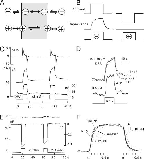
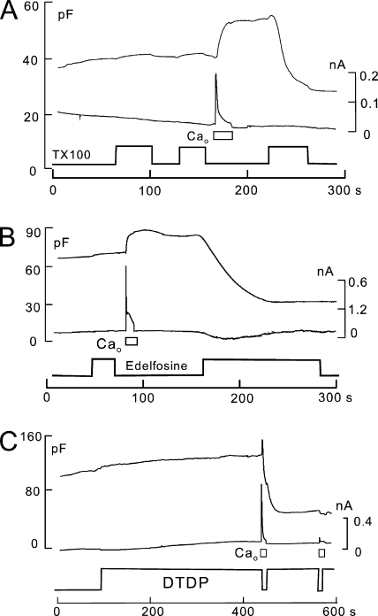
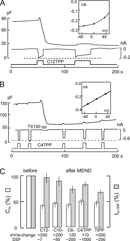
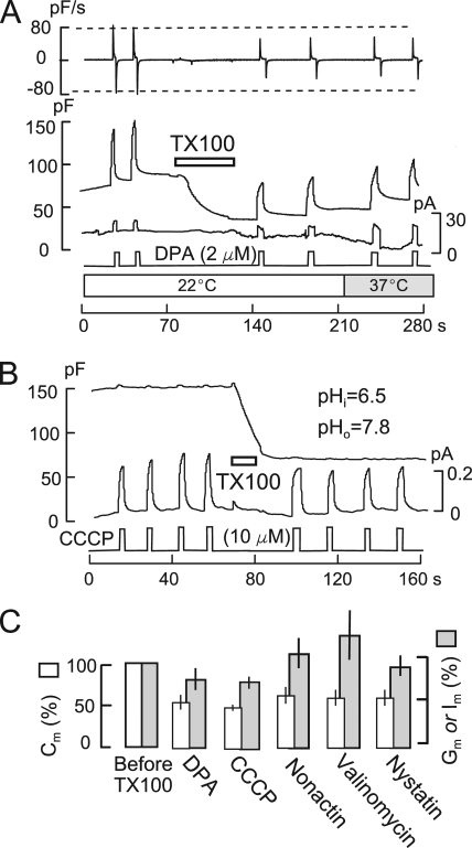
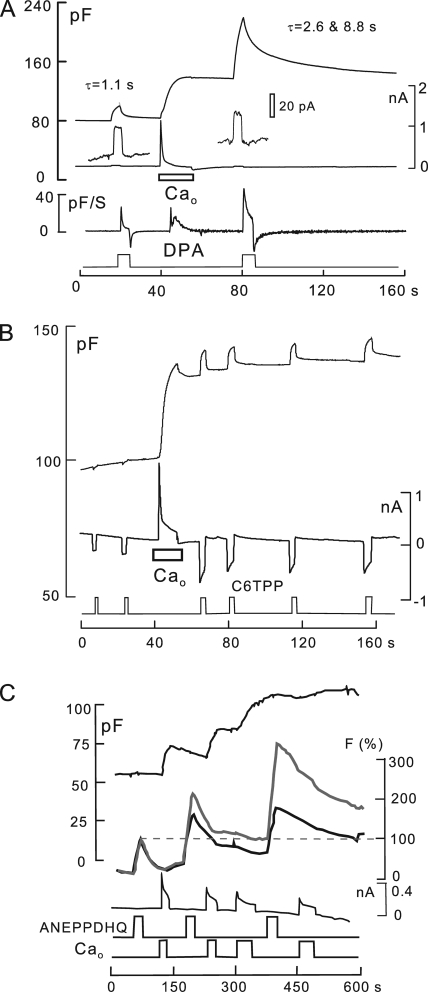
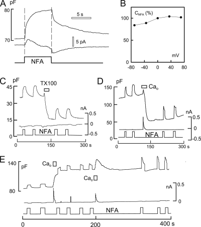
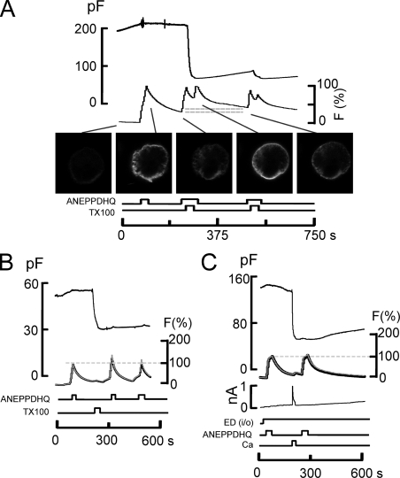
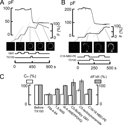
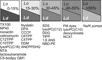
A large fraction of endocytosis in eukaryotic cells occurs without adaptors or dynamins. Here, we present evidence for the involvement of lipid domains in massive endocytosis (MEND) activated by both large Ca transients and amphipathic compounds in baby hamster kidney and HEK293 cells. First, we demonstrate functional coupling of the two MEND types. Ca transients can strongly facilitate detergent-activated MEND. Conversely, an amphipath with dual alkyl chains, ditridecylphthalate, is without effect in the absence of Ca transients but induces MEND to occur within seconds during Ca transients. Ca transients, like amphipaths, enhance the extraction of lipids from cells by β-cyclodextrins. Second, we demonstrate that electrical and/or optical signals generated by selected membrane probes are nearly insensitive to MEND, suggesting that those probes segregate into membrane domains that are not taken up by MEND. Triphenylphosphoniums are increasingly excluded from domains that internalize as the carbon chain length increases from 4 to 12. The small cationic membrane dye, FM 4-64, binds well to domains that internalize, whereas a closely related dye with a larger hydrophobic moiety, di-4-ANEPPDHQ (ANEPPDHQ) is excluded. Multiple carrier-type ionophores and a small amphipathic anion, niflumic acid, are also excluded. Probes with modest MEND sensitivity include the hydrophobic anion, dipicrylamine, carbonyl cyanide m-chlorophenylhydrazone, and NBD-phosphatidylethanolamine. Third, we demonstrate that large Ca transients can strongly enhance the extracellular binding of several membrane probes, monitored electrically or optically, consistent with a more disordered membrane with more amphipath-binding sites. Fluorescence shifts of ANEPPDHQ report increased disorder of the extracellular monolayer after large Ca transients, consistent with an increased propensity of the membrane to phase separate and vesiculate. Collectively, the results indicate that >50% of the outer monolayer is ordered and can be selectively internalized during MEND responses initiated by two very different cell perturbations.


Figures

Figure 1.
Figure 1.
The function of hydrophobic anions and cations. (A) Hydrophobic anions translocate reversibly through the hydrophobic core of the membrane much faster than they dissociate from the membrane. Hydrophobic cations translocate slowly across the membrane in relation to their dissociation rates from the membrane. A positive dipole potential within the membrane is assumed to promote anion binding over cation binding by several hundredfold. (B) Predictions of simple models of hydrophobic ion function. When applied rapidly to membranes, hydrophobic anions give rise to a capacitive signal that develops with the time course of their binding/dissociation from the membrane. The net current generated by their passage through the membrane field (“flux”) develops immediately. In contrast, hydrophobic cations do not generate a capacitive signal. (C) DPA-capacitive signals and currents in BHK cells using standard solutions with 2 mM ATP and 0.2 mM GTP on the cytoplasmic side. Rapid application and removal of 2 µM DPA causes an immediate outward current and a slowly rising capacitive signal, which decays toward an increased baseline upon DPA removal. The major time constant of the rising and falling signals is 0.35 s. The DSF, i.e., the ratio of minimum diffusional flux to current, is 1.8. (D) DPA-capacitive signals upon applying and removing different DPA concentrations in the same BHK cell. The signals are scaled to allow comparison of the wave forms. With low DPA concentrations, capacitive signals come to a steady state more rapidly than with high concentrations (>2 µM), and slowly decaying signal components become more pronounced with high DPA concentrations. (E) Currents and capacitance records for the application and removal of the hydrophobic cation, C6TPP (0.5 mM). Capacitance changes are negligible in relation to those evoked by DPA. (F) Normalized current records showing the time courses of current activation and deactivation upon applying and removing 2 µM DPA, 0.5 mM C6TPP, and 25 µM C12TPP. The gray curve shows the expected time course for diffusion through a 10-µm solution layer, with a diffusion coefficient of 0.5 × 10⁻⁵ cm2/s.
Figure 2.
Figure 2.
Amphipath-induced MEND is facilitated by Ca transients associated with exocytosis. Cytoplasmic solutions are ATP and GTP free. (A) As with many other amphipaths that induce MEND, the threshold concentrations needed to induce MEND by TX100 are reduced by a Ca transient associated with exocytosis. In this example, 80 µM TX100 was applied twice before the activation of Ca influx, and it was without effect. After Ca influx and exocytosis, the same TX100 concentration causes a rapid 50% MEND response. (B) Similar to TX100, a low concentration of edelfosine (30 µM) is without effect before the activation of Ca influx. After a Ca influx episode, the same edelfosine concentration causes a 65% MEND response. (C) DTDP-saturated extracellular solution (5 µM; sonicated for 5 min) is without effect when applied for 5 min before the activation of reverse Na/Ca exchange. Subsequent activation of Ca influx by NCX1 causes a rapid 60% MEND response.
Figure 3.
Figure 3.
Insensitivity of TPP currents to MEND. BHK cells using standard solutions with 2 mM ATP and 0.2 mM GTP on the cytoplasmic side. (A) The rapid application of extracellular solution containing 40 µM C12TPP causes an immediate 0.2-nA inward current and a MEND response amounting to 70% of Cm within 15 s. Thereafter, the renewed application of C12TPP generates an inward current of similar magnitude but no further loss of Cm. DSF, 6.6. (B) The rapid application of 10 mM C4TPP induces an inward current of 0.6 nA. After the induction of a 63% MEND response with 200 µM TX100, the C4TPP current is reduced by only 12% on average. DSF, 1,017. (C) Normalized results (n = 5) for the same protocols using 40 µM C12TPP, 0.12 mM C10TPP, 0.25 mM C6TPP, 10 mM C4TPP, and 0.5 mM TtPP. The average MEND responses ranged from 45 to 57%. The average current decrease was 10% for C12TPP, 15% for C10TPP, 26% for C6TPP, 17% for C4TPP, and 31% for TtPP. Slopes of current–voltage relations and average DSP values are given below the bar graphs for each agent.
Figure 4.
Figure 4.
DPA and multiple ionophores show low sensitivity to TX100-promoted MEND. BHK cells with standard solutions containing 2 mM ATP and 0.2 mM GTP on the cytoplasmic side. (A) 2 µM DPA is applied twice before and twice after the induction of MEND by 200 µM TX100 at 22°C. DSF, 4.4. MEND amounts to 54% of Cm, whereas the capacitive DPA signals decrease by 28%. DPA currents are unchanged by MEND but double upon warming to 37°C. (B) 10 µM CCCP is applied and removed four times before and four times after inducing MEND with 200 µM TX100. To generate outward proton transport, the cytoplasmic solution was set to pH 6.5 and the extracellular solution was set to 7.8. A 51% MEND response results in a 13% decrease of the CCCP current. (C) Composite results for several electrogenic membrane probes. In all cases, TX100 causes an ∼50% loss of Cm. DPA-capacitive signals (n = 5) and CCCP currents (n = 6) are decreased by 25% on average. The conductance induced by 12 µM nonactin (n = 4) and 25 µM valinomycin (n = 4) is increased on average by 15 and 27%, respectively, after TX100-induced MEND. The conductance induced by the application and removal of 65 µM nystatin (n = 4) is decreased by 15% (n = 4).
Figure 5.
Figure 5.
C6TPP currents are nearly unaffected by Ca-induced MEND. 0.3 mM C6TPP is applied briefly multiple times in each experiment, as indicated. (A) 2 mM of the polyamine, EDA, is included in both the cytoplasmic and extracellular solutions to promote rapid MEND (50% in 5 s) during Ca influx by NCX1. Thereafter, C6TPP current is decreased by 12% on average. DSF, 50. (B) Composite results for four experiments similar to A. Ca-activated MEND causes a 50% loss of Cm, whereas C12TPP currents decrease by just 10%. (C) Using a cytoplasmic solution with 6 mM ATP and no polyamine, Ca influx by NCX1 is activated four times. Cm decreases substantially in a delayed fashion after each of the Ca influx episodes, resulting finally in a 45% decrease of Cm. 0.3-mM C6TPP currents are unchanged. DSF, 41.
Figure 6.
Figure 6.
Analysis of DPA-capacitive signals before and after MEND induced by a Ca transient in the presence of 1 mM spermidine and 6 mM ATP. MEND amounts to 68% of Cm. DSF, 4.0. (A) 2 µM DPA is applied twice before MEND and twice after MEND for times of 6–10 s. During each application of DPA, voltage is stepped to −150 mV and then ramped to +80 mV to determine the capacitance–voltage relation of DPA. (B and C) Capacitance records indicated by “before” and “after” in A. The magnitudes of DPA-capacitive signal and the time constants of the rising and falling signals (0.67–0.82 s) are not detectably changed. (D) The magnitudes of DPA capacitance–voltage relations are not detectably changed; the peak capacitance undergoes a small 8-mV shift to more negative potentials.
Figure 7.
Figure 7.
Steady-state DPA-capacitive signal changes in response to TX100- and Ca-induced MEND. BHK cells with standard solutions, 8 mM of cytoplasmic ATP, 2 µM extracellular DPA, and 6 µM of cytoplasmic DPA. The DPA- and membrane-related capacitance components are determined by applying voltage pulses to +120 mV for 1.3 s. Membrane capacitance does not depend on voltage, whereas the DPA capacitance becomes negligible at +120 mM. Membrane capacitance is demarcated by a dotted line. (A) TX100-induced MEND. Upon the first application of TX100, MEND amounts to just 50% of the initial cell area. The DPA-capacitive signal, defined by the downward deflections of capacitance, is unchanged by MEND. After a second application of TX100, DPA-capacitive signals increase slowly by 20% with respect to the pre-MEND magnitude. (B) Ca-induced MEND with high (8 mM) ATP and no polyamine in response to two Ca transients. The first Ca transient causes exocytosis without subsequent endocytosis. Cell capacitance increases by 20%, whereas the DPA-capacitive signal increases by nearly threefold. The second Ca transient causes little further exocytosis and a delayed MEND response amounting to 60% of the cell surface. The DPA-capacitive signal decreases in parallel by 20%.
Figure 8.
Figure 8.
Summary of changes of DPA signals in response to a single Ca influx episode and multiple Ca-dependent MEND protocols. Examples of each experiment type are presented in Figs. S12–S14. Each bar set gives give the average of three or more experiments for cell capacitance, DPA capacitance, and DPA currents, normalized to their initial experimental values (leftmost bar set). From left to right, Ca influx causes on average a threefold greater increase of DPA capacitance than cell capacitance, and a small increase of DPA current. After Ca influx, the presence of high ATP in the cytoplasm causes a return of DPA capacitance and current to baseline, whereas cell capacitance decreased to 50% below baseline. Polyamine/Ca-induced MEND causes on average an increase of DPA capacitance and current, whereas cell capacitance decreases to 55% below baseline. When cells are enriched with cholesterol, Ca influx without polyamines causes a 58% decrease of cell capacitance, whereas DPA capacitance and current decrease on averaged by 18 and 15%. Cholesterol enrichment after a Ca transient has increased cell capacitance by 25% results on average in a decrease of cell capacitance by 66%, whereas DPA capacitance and current remain 18 and 25% greater than baseline.
Figure 9.
Figure 9.
Ca transients associated with exocytosis cause dramatic changes of the surface membrane, as reported by electrogenic and optical membrane probes. Standard solutions with no ATP or polyamines. (A) Effects on DPA signals. 2 µM DPA was applied for 4 s and removed before and after activating reverse Na/Ca exchange in the absence of cytoplasmic ATP. Cm increases by 60%, and the DPA-capacitive signal increases by a factor of 4. The DPA current is unchanged, whereas the rate of rise of the capacitive DPA signal (dCm/dt, measured in pF/S) is increased by 70%. DSP, 3.3. (B) Effects on C6TPP signals. 300 µM C6TPP is applied and removed six times. Ca influx associated with a 40% increase of membrane area causes a 2.4-fold increase of C6TPP currents, causes decay phases of the currents, and induces C6TPP to develop a capacitive signal component. These changes are consistent with decreased dissociation rates from the membrane, similar to the effects of DPA. DSF, 670. (C) Effects on ANEPPDHQ optical signals. Ca transients with membrane fusion cause large shifts in fluorescence spectra of ANEPPDHQ (8 µM) binding. The large relative increase of emission above 640 nm (gray line) is similar to changes described for cholesterol depletion and presumably increased membrane disorder (Jin et al., 2006). Electrophysiological parameters were recorded in parallel with time-lapse confocal imaging at emission bandwidths of 500–580 nm (black) and 640 nmLP (gray).
Figure 10.
Figure 10.
Capacitive binding signals for the hydrophobic Cl channel blocker, NFA (0.2 mM), are nearly unchanged by MEND. BHK cells with standard solutions. (A) The application and removal of NFA causes a robust increase of Cm. Decay of the Cm signal occurs in two distinct phases. Currents associated with NFA application are very small, or absent, and show a delay from the onset of the rise of Cm. (B) The capacitance–voltage relation of the NFA signal is nearly flat, indicating that capacitive signals do not arise from translocation of the probe across the membrane. (C) The capacitive signal induced by NFA is unaffected by TX100-induced MEND. (D) MEND induced by Ca influx in the presence of 2 mM EDA in both cytoplasmic and extracellular solutions. The capacitive signal induced by NFA rises and falls faster after Ca-induced MEND, but its magnitude is not affected. (E) Effect of Ca influx associated with exocytosis without MEND on NFA-capacitive signals. Cytoplasmic solutions contain no ATP and no polyamine. The Ca transient results in a 60% increase of Cm and a smaller increase of the NFA signal. After a second Ca transient, the NFA signal is nearly doubled and becomes faster.
Figure 11.
Figure 11.
The styryl dye, ANEPPDHQ, does not occupy the membrane that internalizes during TX100- and Ca-activated MEND in BHK cells. (A) 10 µM ANEPPDHQ was applied before MEND and removed during the induction of MEND with 150 µM TX100 and again after MEND. The apparent binding rates of ANEPPDHQ are unchanged by the removal of >70% of the cell surface by MEND, and the amount of fluorescence internalized during MEND (i.e., does not wash off) amounts to no more than 15% of the initial labeling (see horizontal gray lines). (B) 8 µM ANEPPDHQ was applied and removed before and after TX100 (150 µM)–induced MEND while imaging at bandwidths of 500–580 nm (black) and 640 nmLP (gray). The optical records are scaled. Neither the apparent binding rates of ANEPPDHQ nor its spectral properties are changed after TX100-induced MEND. (C) Same as B, using Ca influx in the presence of polyamine, here EDA, to induce MEND. Dye signals, which approach a steady state in these records, are unchanged by MEND.
Figure 12.
Figure 12.
Optically determined binding rates of multiple lipid probes in BHK cells before and after inducing MEND with 150 µM TX100. Binding rates of dyes are assumed to be proportional to the rate of fluorescence increase over 20 s upon applying dyes. The gray dashed lines alongside of the fluorescence records indicate the average rate of fluorescence increase. (A) An experiment using 1 µM C5-Bodipy-Gm1. (B) An experiment using 5 µM C16:0-NBD-PE in the presence of 10% DMSO to promote lipid solubility. (C) Summary of results showing capacitance loss in white and binding rates of fluorescent probes in gray. Only the styryl FM dye and the C16:0-NBD-PE are significantly affected by TX100-induced MEND.
Figure 13.
Figure 13.
Summary of membrane probes studied in this paper and our companion paper by Fine et al. (2011) in relation to MEND responses. The figure represents best estimates of their distribution between membrane domains that internalize and those that do not internalize during MEND, interpreted to represent Lo and Ld domains, respectively. From left to right, the group with the highest relative affinity for disordered membrane is the largest group. It includes nonionic detergents, carrier-type ionophores, long-chain TPPs, NFA, and lactosylceramide. The second group, which is modestly affected by MEND, includes nystatin, DPA, CCCP, short-chain TPPs, dodecylmaltoside (DDM), and ANEPPDHQ. The third group, which is substantially but still under-proportionally affected by MEND, includes SDS, dodecylglucoside (DDG), TtPP, 1-anilinonaphthalene-8-sulfonic acid (1,8 ANS), and NBD-PE. Similar to TPPs, lyso-PCs appear to associate more effectively with membrane that internalizes as the carbon chain length is decreased. FM dyes, deoxychoate, and NCX1 are affected proportionally by MEND, or nearly proportionally. From all probes and transporters examined to date, only Na/K pump activities are preferentially reduced by MEND, with the implication that pumps may be regulated by Lo domain–dependent endocytosis.

Comment in

References

    1. Andersen P.S., Fuchs M. 1975. Potential energy barriers to ion transport within lipid bilayers. Studies with tetraphenylborate. Biophys. J. 15:795–830 10.1016/S0006-3495(75)85856-5 - DOI - PMC - PubMed
    1. Artalejo C.R., Elhamdani A., Palfrey H.C. 1996. Calmodulin is the divalent cation receptor for rapid endocytosis, but not exocytosis, in adrenal chromaffin cells. Neuron. 16:195–205 10.1016/S0896-6273(00)80036-7 - DOI - PubMed
    1. Benz R., Läuger P., Janko K. 1976. Transport kinetics of hydrophobic ions in lipid bilayer membranes. Charge-pulse relaxation studies. Biochim. Biophys. Acta. 455:701–720 10.1016/0005-2736(76)90042-0 - DOI - PubMed
    1. Brown D.A. 2006. Lipid rafts, detergent-resistant membranes, and raft targeting signals. Physiology (Bethesda). 21:430–439 - PubMed
    1. Bruner L.J. 1975. The interaction of hydrophobic ions with lipid bilayer membranes. J. Membr. Biol. 22:125–141 10.1007/BF01868167 - DOI - PubMed

Publication types