Review
. 2011 Feb 1;124(Pt 3):299-310.
doi: 10.1242/jcs.072728.

Intravital microscopy: new insights into metastasis of tumors

Affiliations
Review

Intravital microscopy: new insights into metastasis of tumors

Evelyne Beerling et al. J Cell Sci. .

Abstract

Metastasis, the process by which cells spread from the primary tumor to a distant site to form secondary tumors, is still not fully understood. Although histological techniques have provided important information, they give only a static image and thus compromise interpretation of this dynamic process. New advances in intravital microscopy (IVM), such as two-photon microscopy, imaging chambers, and multicolor and fluorescent resonance energy transfer imaging, have recently been used to visualize the behavior of single metastasizing cells at subcellular resolution over several days, yielding new and unexpected insights into this process. For example, IVM studies showed that tumor cells can switch between multiple invasion strategies in response to various densities of extracellular matrix. Moreover, other IVM studies showed that tumor cell migration and blood entry take place not only at the invasive front, but also within the tumor mass at tumor-associated vessels that lack an intact basement membrane. In this Commentary, we will give an overview of the recent advances in high-resolution IVM techniques and discuss some of the latest insights in the metastasis field obtained with IVM.


Figures

Fig. 1.
Fig. 1.
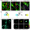
IVM of individual steps of metastasis. The schematic illustrations aim to provide a simplified overview of the metastatic process. To metastasize, tumor cells (green) have to escape from the primary tumor and colonize a distant site. During this process, cells employ traits, such as invasiveness, motility, attachment, chemosensing and survival, that allow them to detach from the primary tumor, invade the interstitial matrix (purple), overcome the barrier of the BM (blue), enter the blood (red), be transported to a distant site, exit the blood and finally grow out to form metastatic foci. IVM can be used to image metastatic processes, as illustrated by the IVM images of tumor cells (green), type I collagen (purple) and blood (red). IVM images A and B represent different time points of invasion of a polyoma middle T (PyMT) mammary tumor. IVM image C shows the tumor cells present in a vessel that collects blood from a C26 colorectal tumor and is one time point of supplementary material Movie 1. Scale bar: 10 μm.
Fig. 2.
Fig. 2.
Multicolor and FRET IVM. Caspase-3 activity and cell–cell interactions can be imaged using multicolor and FRET IVM. (A) Schematic representation (left) and corresponding multicolor IVM image (zoom and overview on the right) of a colorectal C26 tumor. Merged images of Dendra2-expressing tumor cells (green), 70 kDa Texas-Red-labeled macrophages (red) and type I collagen (purple; SHG) illustrate the interaction between macrophages and tumor cells. (B) Cartoon of a caspase-3 FRET biosensor to measure caspase-3 activity during apoptosis induction. Here, CFP (C) and YFP (Y) are in close proximity, resulting in efficient FRET. When caspase-3 is activated in the cell by induction of apoptosis, the sensor is cleaved, leading to increased distance between CFP and YFP with a subsequent loss of FRET. This loss is observed as increased CFP fluorescence and decreased YFP fluorescence. (C) The IVM image shows a large overview of the skin of a mouse in which single keratinocytes are transfected by DNA tattooing with the caspase-3 FRET biosensor and a chemical-sensitive inducer of apoptosis. The arrows point to examples of cells that are transfected with the caspase-3 sensor, in which the apoptosis status can be imaged using FRET. (D) Using the system shown in B, IVM images of CFP and YFP are acquired upon CFP excitation and FRET is expressed as a ratio of YFP over CFP (see color bar on the right). A striking drop in FRET is observed upon chemical induction of apoptosis (lower row of images), illustrating the measurement of signaling events in vivo. All scale bars: 10 μm.
Fig. 3.
Fig. 3.
IVM over several days using an MIW. Experimental setup for repeated IVM imaging of live mice over several days using an MIW. (A) Schematic illustration of an MIW consisting of a plastic frame and a cover glass (top image), which is surgically placed on a mammary tumor (lower image). (B) After implantation of the MIW at day 0, the mouse recovers for two days. This is followed by repeated IVM sessions in the subsequent days. (C) IVM images of a Dendra2-expressing colorectal C26 tumor, in which green represents the non-switched Dendra2 and red the switched Dendra2. At day 3, tumor cells that are present either close to (upper images) or surrounding (lower images) a blood vessel are photolabeled in a square region by photoswitching Dendra2 from green to red using violet illumination. Note that the combination of using a MIW and photomarking of tumor cells allows imaging regions to be retraced in subsequent imaging sessions, and thus the motility and intravasation to be visualized. Also note the high intratumoral motility (top images) and the loss of red-shifted tumor cells by intravasation (lower images). Scale bars: 10 μm.
Fig. 4.
Fig. 4.
Single moving tumor cells of MTLn3 tumors use the hematogenous route to metastasize to other organs. (A) Simplified schematic overview of spreading routes from a breast tumor to the lung (L). At the primary tumor site, tumor cells can enter the blood either directly (blue route) or through lymphatic vessels (purple route), which drain into the blood at a merging point located just before the heart (H), from where the blood is then transported into the lung. Using IVM of the invasive MTLn3 tumor model, it has been shown that cells move either as single cells, which leave the primary tumor using the hematogenous route, or as collective chains using the lymphatic route. (B) Inhibition of TFGβ signaling leads to inhibition of invasion by single moving cells and a subsequent drop in the number of tumor cells in the heart, whereas the collective movement to lymphatic vessels remains unaltered. These experiments show that, in this invasive mammary carcinoma model, tumor cells use the hematogenous (blood) route to metastasize to other organs.
Fig. 5.
Fig. 5.
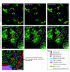
Direct entry of tumor cells into tumor-associated vessels as shown by IVM. Tumor cell migration and intravasation take place not only at the invasive front, but also deep within the tumor mass. The upper row of IVM images, in which the mouse invasive lobular carcinoma tumor cells are in green and the SHG signal is in purple, shows high motility within the tumor mass. The arrows point to the position of motile cells at time (t) zero (see also supplementary material Movie 3). The row of images below shows maximum projections of Z-stacks with a total depth of 50 μm. In the IVM images, green represents C26 colorectal tumor cells and purple represents SHG. This time series of the maximum projections shows the disappearance of a tumor cell, which is no longer present in the Z-stack because it enters a tumor-associated vessel and is transported out of sight by blood (see circle; supplementary material Movie 2). The cartoon shows a simplified model of leaky tumor-associated vessels within tumors, which lack an intact BM and through which tumor cells can enter the blood. Scale bars: 10 μm.

References

    1. Abdul-Karim M.-A., Al-Kofahi K., Brown E. B., Jain R. K., Roysam B. (2003). Automated tracing and change analysis of angiogenic vasculature from in vivo multiphoton confocal image time series. Microvasc. Res. 66, 113-125 - PubMed
    1. Alexander S., Koehl G., Hirschberg M., Geissler E., Friedl P. (2008). Dynamic imaging of cancer growth and invasion: a modified skin-fold chamber model. Histochem. Cell Biol. 130, 1147-1154 - PubMed
    1. Ando R., Hama H., Yamamoto-Hino M., Mizuno H., Miyawaki A. (2002). An optical marker based on the UV-induced green-to-red photoconversion of a fluorescent protein. Proc. Natl. Acad. Sci. USA 99, 12651-12656 - PMC - PubMed
    1. Bajénoff M., Egen J. G., Koo L. Y., Laugier Jean P., Brau F., Glaichenhaus N., Germain R. N. (2006). Stromal cell networks regulate lymphocyte entry, migration, and territoriality in lymph nodes. Immunity 25, 989-1001 - PMC - PubMed
    1. Baluk P., Morikawa S., Haskell A., Mancuso M., McDonald D. M. (2003). Abnormalities of basement membrane on blood vessels and endothelial sprouts in tumors. Am. J. Pathol. 163, 1801-1815 - PMC - PubMed

Publication types

MeSH terms