. 2011 Feb 15;186(4):2444-53.
doi: 10.4049/jimmunol.1001567. Epub 2011 Jan 17.

A2B adenosine receptor blockade enhances macrophage-mediated bacterial phagocytosis and improves polymicrobial sepsis survival in mice

Affiliations

A2B adenosine receptor blockade enhances macrophage-mediated bacterial phagocytosis and improves polymicrobial sepsis survival in mice

Bryan G Belikoff et al. J Immunol. .

Abstract

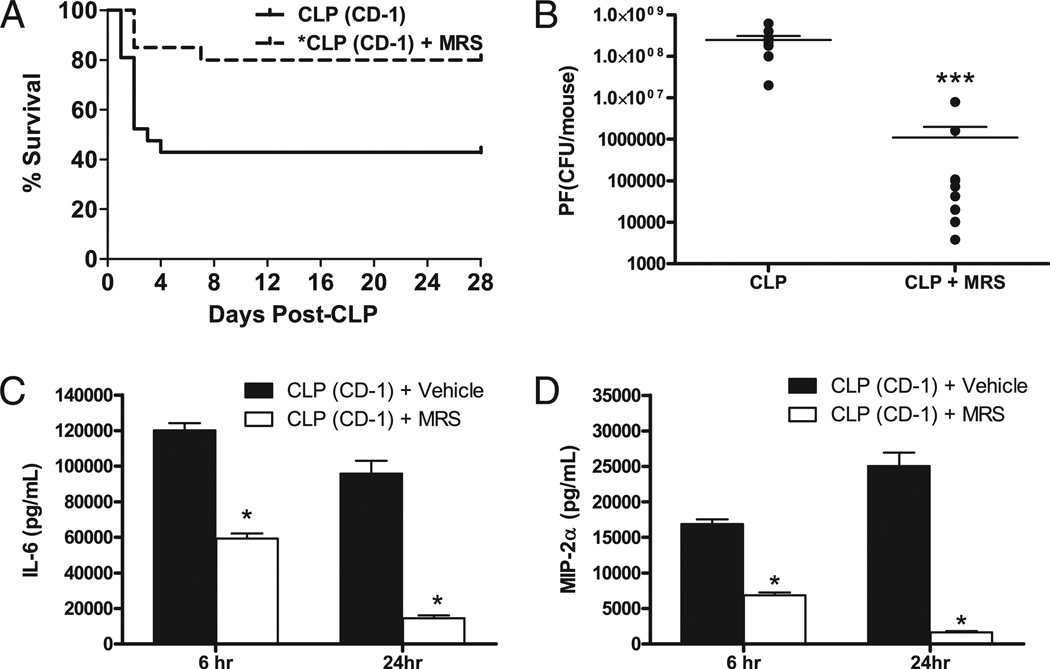
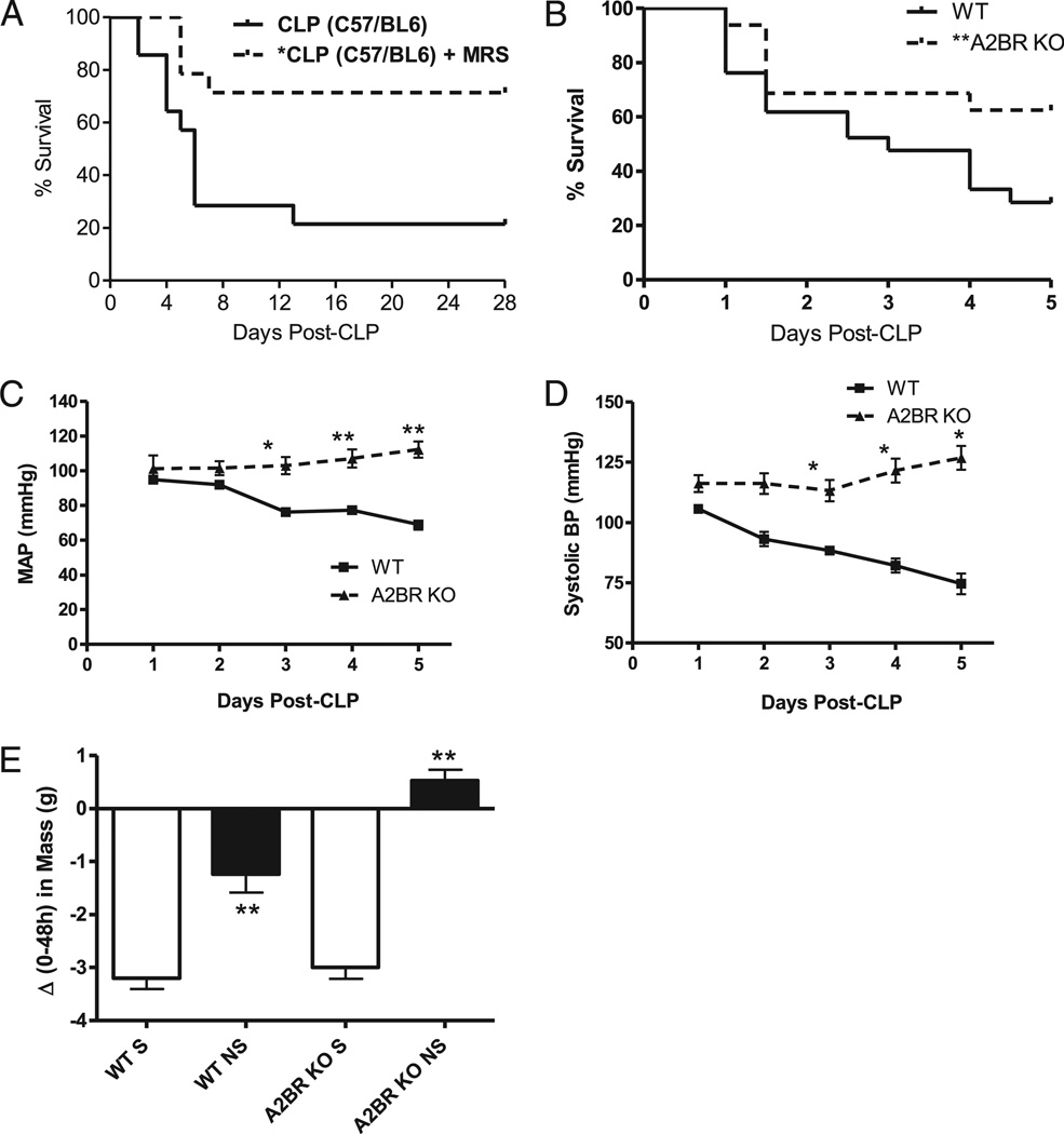
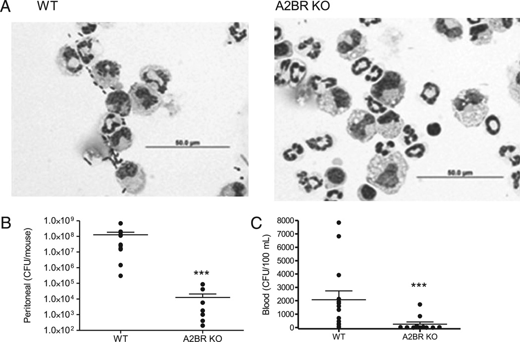
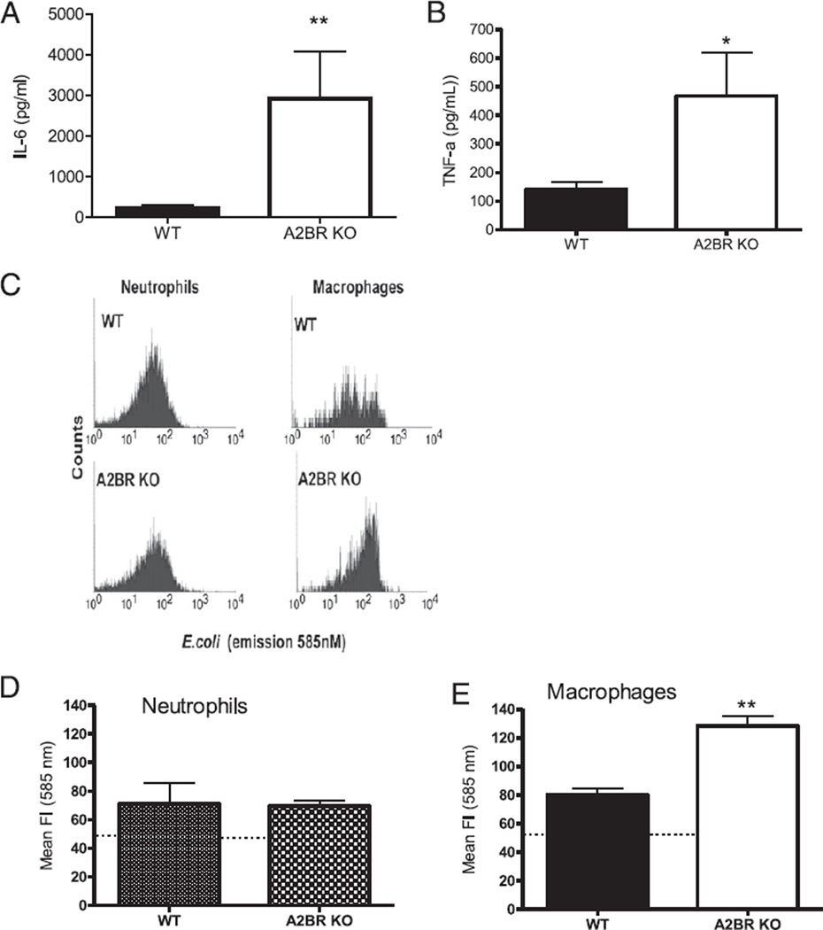
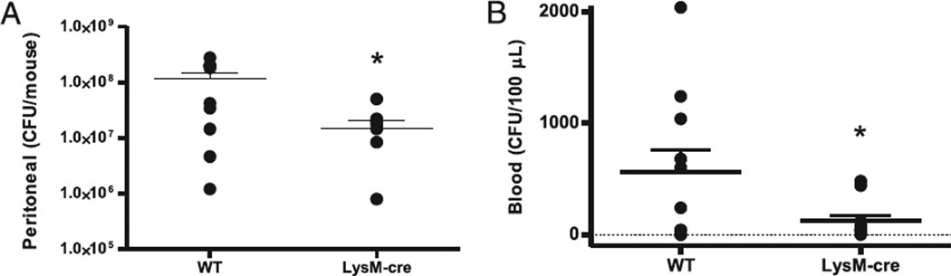
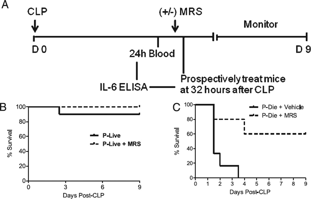
Antimicrobial treatment strategies must improve to reduce the high mortality rates in septic patients. In noninfectious models of acute inflammation, activation of A2B adenosine receptors (A2BR) in extracellular adenosine-rich microenvironments causes immunosuppression. We examined A2BR in antibacterial responses in the cecal ligation and puncture (CLP) model of sepsis. Antagonism of A2BR significantly increased survival, enhanced bacterial phagocytosis, and decreased IL-6 and MIP-2 (a CXC chemokine) levels after CLP in outbred (ICR/CD-1) mice. During the CLP-induced septic response in A2BR knockout mice, hemodynamic parameters were improved compared with wild-type mice in addition to better survival and decreased plasma IL-6 levels. A2BR deficiency resulted in a dramatic 4-log reduction in peritoneal bacteria. The mechanism of these improvements was due to enhanced macrophage phagocytic activity without augmenting neutrophil phagocytosis of bacteria. Following ex vivo LPS stimulation, septic macrophages from A2BR knockout mice had increased IL-6 and TNF-α secretion compared with wild-type mice. A therapeutic intervention with A2BR blockade was studied by using a plasma biomarker to direct therapy to those mice predicted to die. Pharmacological blockade of A2BR even 32 h after the onset of sepsis increased survival by 65% in those mice predicted to die. Thus, even the late treatment with an A2BR antagonist significantly improved survival of mice (ICR/CD-1) that were otherwise determined to die according to plasma IL-6 levels. Our findings of enhanced bacterial clearance and host survival suggest that antagonism of A2BRs offers a therapeutic target to improve macrophage function in a late treatment protocol that improves sepsis survival.


Conflict of interest statement

Disclosures

The authors have no financial conflicts of interest.

Figures

FIGURE 1
FIGURE 1
A2BR antagonism improves CLP survival and bacterial clearance, and decreases plasma cytokine levels. Outbred CD-1 mice were given antibiotics (25 mg/kg imipenem) twice daily with fluid resuscitation (Lactated Ringer + 5% dextrose) for 4 d to evaluate the ability of the A2BR antagonist (10 mg/kg MRS 1754) to improve outcome after CLP-induced sepsis. A, 28-d survival in mice treated with the A2BR antagonist (MRS 1754) (broken line; n = 20) had significantly (p < 0.05) better survival than vehicle-treated mice (solid line; n = 21). MRS 1754 therapy was initiated 1.5 h after CLP and given once daily for 3 d. B, The effect of MRS 1754 on PF bacterial load 24 h after CLP. Mice were subjected to CLP and given vehicle with or without MRS 1754 via s.c. injection. Each symbol is an individual animal. Only one dose of MRS 1754 therapy was given to mice that received the antagonist. A2BR antagonism significantly reduced peritoneal bacterial counts. Plasma cytokine levels for IL-6 (C) or TNF (D). Plasma was collected 6 and 24 h after CLP. A2BR antagonism significantly reduced plasma levels of these inflammatory mediators. Data are expressed as mean ± SEM of at least two independent experiments. The survival study data were pooled from three independent studies. *p < 0.01, ***p < 0.001, comparing untreated to treated mice.
FIGURE 2
FIGURE 2
A2BR-deficient mice are resistant to CLP mortality and cardiovascular dysfunction. A, The ability of MRS 1754 (0.5 mg/kg) to improve CLP survival in C57BL/6 mice given antibiotics plus fluid resuscitation was confirmed in this mouse strain (n = 14 per group; p < 0.01). B, Survival curve for WT (solid line; n = 21) and A2BR KO (broken line; n = 27) mice subjected to CLP. Antibiotics were withheld from this group to evaluate A2BR’s role in bacterial clearance. The CLP survival graph depicts data pooled from at least three independent studies. Log rank analysis shows a significant improvement in survival, p < 0.05. A2BR deficiency improves cardiovascular parameters in CLP mice. CLP was performed in WT (n = 6) and A2BR KO (n = 3) mice previously implanted with a radiotelemetry transmitter. Cardiovascular parameters including MAP (C), and systolic BP (D) were collected over a 24-h time period and quantified. E, Body mass was recorded at the time of CLP, and every 24 h thereafter, for 5 d. The graph depicts a post hoc analysis of the change in body mass over 48 h after CLP of mice stratified into either 5-d survivors (S) or nonsurvivors (NS). Data are expressed as mean ± SEM of at least three independent experiments. *p < 0.05, **p < 0.01 compared with control.
FIGURE 3
FIGURE 3
A2BR deficiency improves local and systemic bacterial clearance. A, Photomicrographs of peritoneal cells from WT or A2BR KO mice showing the presence of neutrophils, macrophages, and bacteria. Fewer bacteria are visible in the A2BR KO mice. Scale bar, 50 µm. B, Peritoneal bacterial load was decreased in A2BR-deficient mice. The local bacterial load was determined in the peritoneal lavage collected from CLP mice 24 h after CLP. C, Bacteremia was decreased 24 h after CLP in A2BR-deficient mice. Data in B and C are expressed as mean ± SEM of 11 or more mice in at least three independent experiments, and each symbol is an individual animal. ***p < 0.001 WT versus A2BR KO mice.
FIGURE 4
FIGURE 4
A2BR deficiency enhances macrophage function. Peritoneal cells used in these experiments were analyzed from either A2BR KO or WT mice subjected to CLP and harvested 24 h later. LPS stimulation enhanced cytokine release from macrophages isolated from septic A2BR-deficient mice. Isolated peritoneal macrophages from septic mice were stimulated ex vivo with 100 ng/ml LPS for 24 h. Supernatants were measured for IL-6 (A) and TNF-α (B) by ELISA. Data are expressed as mean ± SEM of three independent experiments. Gated neutrophil and macrophage populations were identified as Gr-1highF4/80negative and Gr-1lowF4/80positive to discriminate between neutrophil and macrophage bacteria phagocytosis activity, respectively, from other cell types. C, Histogram of gated neutrophil and macrophage populations showing the phagocytic/endocytic capacity of phagocytes from septic WT and A2BR KO mice preincubated with the pH-sensitive fluorogenic E. coli. E. coli present inside the phagolysosome are highly fluorescent relative to nonendocytosed E. coli. Quantitative analysis of the phagocytic activity for neutrophils (D) (Gr-1highF4/80negative) and macrophages (E) (Gr-1lowF4/80positive) from septic WT and A2BR KO mice. *p < 0.05, **p < 0.01 compared with WT CLP mice.
FIGURE 5
FIGURE 5
A2BR deficiency in myeloid cells improves local and systemic bacteria. The contribution of A2BRs on myeloid cells to bacterial clearance during sepsis was analyzed in A2BRflox/flox LysM-cre+/− mice that are selectively lacking the A2BR in myeloid cells. A, Bacteria in the peritoneum were decreased in A2BRflox/flox LysM-cre+/− mice. In the same mice, blood was analyzed for the presence of bacteria. B, Bacteremia was decreased in A2BRflox/flox LysM-cre+/− mice. Bacterial counts were determined 24 h after CLP. Data are expressed as mean ± SEM of 11 or greater mice in two independent experiments, and each symbol is an individual animal. *p < 0.05, WT versus A2BRflox/flox LysM-cre+/− mice.
FIGURE 6
FIGURE 6
A2BR antagonism saves mice predicted to die of CLP-induced lethality. A, Experimental protocol for the identification and treatment strategy for mice P-live and mice P-die based on plasma IL-6 levels 24 h after CLP. Mice are predicted to live or die during the first 5 d after CLP. B, Survival curve of CD-1 mice P-live treated 32 h post-CLP with the A2BR antagonist MRS 1754, 10 mg/kg (broken line; n = 10) or vehicle (solid line; n = 10). C, Survival curve of mice P-die treated 32 h post-CLP with MRS 1754 (broken line; n = 9) or vehicle (solid line; n = 7). p < 0.01 untreated versus treated mice (P-die) by log rank test for C.

Similar articles

Cited by

References

    1. Bernard GR, Margolis BD, Shanies HM, Ely EW, Wheeler AP, Levy H, Wong K, Wright TJ Extended Evaluation of Recombinant Human Activated Protein C United States Investigators. Extended evaluation of recombinant human activated protein C United States Trial (ENHANCE US): a single-arm, phase 3B, multicenter study of drotrecogin alfa (activated) in severe sepsis . Chest. 2004;125:2206–2216. - PubMed
    1. Laterre PF, Levy H, Clermont G, Ball DE, Garg R, Nelson DR, Dhainaut JF, Angus DC. Hospital mortality and resource use in subgroups of the Recombinant Human Activated Protein C Worldwide Evaluation in Severe Sepsis (PROWESS) trial. Crit. Care Med. 2004;32:2207–2218. - PubMed
    1. Remick DG. Pathophysiology of sepsis. Am. J. Pathol. 2007;170:1435–1444. - PMC - PubMed
    1. Buras JA, Holzmann B, Sitkovsky M. Animal models of sepsis: setting the stage. Nat. Rev. Drug Discov. 2005;4:854–865. - PubMed
    1. Osuchowski MF, Connett J, Welch K, Granger J, Remick DG. Stratification is the key: inflammatory biomarkers accurately direct immunomodulatory therapy in experimental sepsis. Crit. Care Med. 2009;37:1567–1573. - PMC - PubMed

Publication types

MeSH terms