Comparative Study
. 2011 Feb 15;186(4):2613-22.
doi: 10.4049/jimmunol.1003162. Epub 2011 Jan 17.

IL-1F5, -F6, -F8, and -F9: a novel IL-1 family signaling system that is active in psoriasis and promotes keratinocyte antimicrobial peptide expression

Affiliations
Comparative Study

IL-1F5, -F6, -F8, and -F9: a novel IL-1 family signaling system that is active in psoriasis and promotes keratinocyte antimicrobial peptide expression

Andrew Johnston et al. J Immunol. .

Abstract

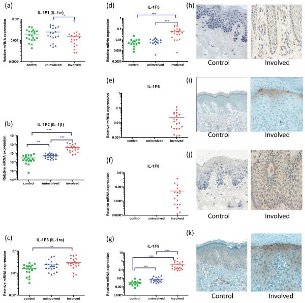
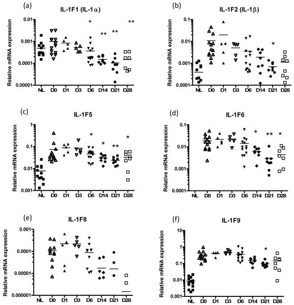
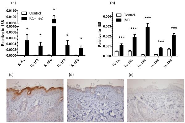
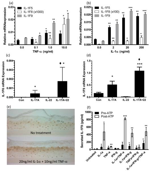
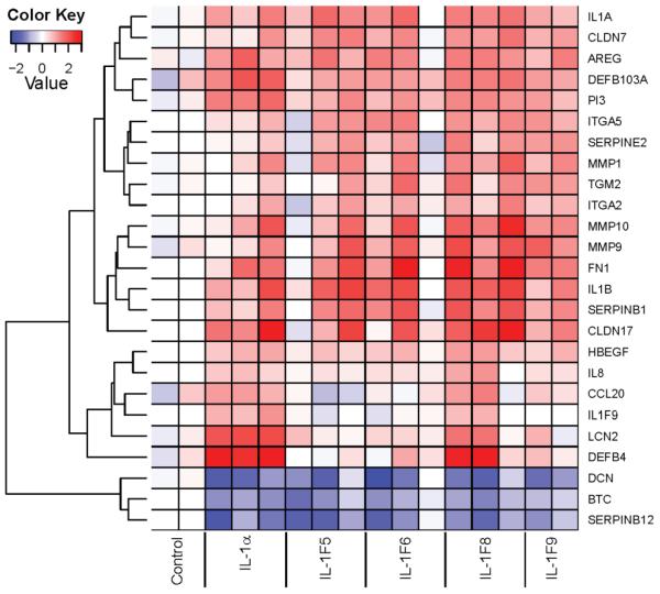
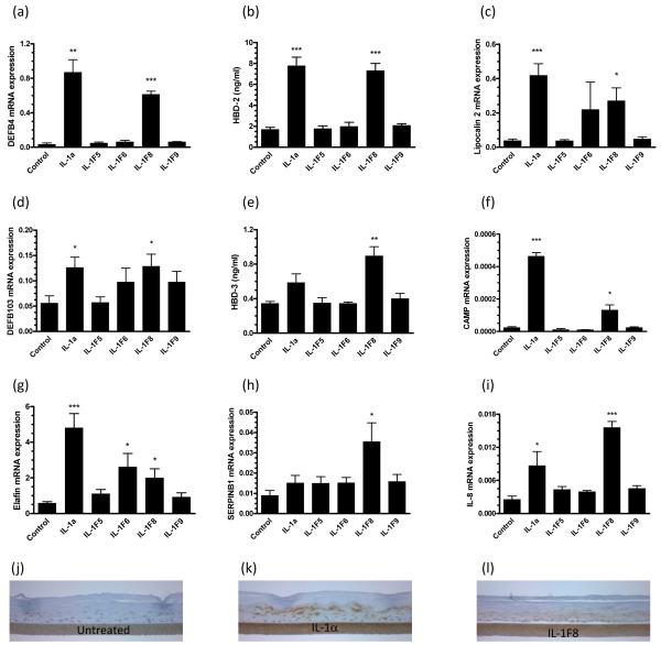
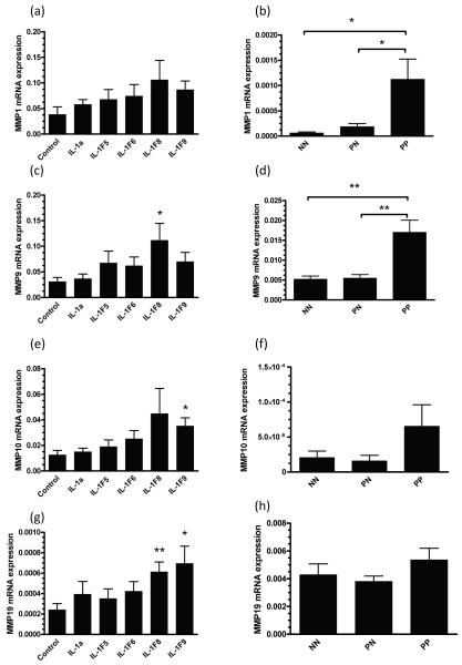
IL-1F6, IL-1F8, and IL-1F9 and the IL-1R6(RP2) receptor antagonist IL-1F5 constitute a novel IL-1 signaling system that is poorly characterized in skin. To further characterize these cytokines in healthy and inflamed skin, we studied their expression in healthy control, uninvolved psoriasis, and psoriasis plaque skin using quantitative RT-PCR and immunohistochemistry. Expression of IL-1F5, -1F6, -1F8, and -1F9 were increased 2 to 3 orders of magnitude in psoriasis plaque versus uninvolved psoriasis skin, which was supported immunohistologically. Moreover, treatment of psoriasis with etanercept led to significantly decreased IL-1F5, -1F6, -1F8, and -1F9 mRNAs, concomitant with clinical improvement. Similarly increased expression of IL-1F5, -1F6, -1F8, and -1F9 was seen in the involved skin of two mouse models of psoriasis. Suggestive of their importance in inflamed epithelia, IL-1α and TNF-α induced IL-1F5, -1F6, -1F8, and -1F9 transcript expression by normal human keratinocytes. Microarray analysis revealed that these cytokines induce the expression of antimicrobial peptides and matrix metalloproteinases by reconstituted human epidermis. In particular, IL-1F8 increased mRNA expression of human β-defensin (HBD)-2, HBD-3, and CAMP and protein secretion of HBD-2 and HBD-3. Collectively, our data suggest important roles for these novel cytokines in inflammatory skin diseases and identify these peptides as potential targets for antipsoriatic therapies.


Figures

Figure 1
Figure 1. The IL-1 family members IL-1F5, -1F6, -1F8 and -1F9 are over-expressed in plaque psoriasis skin
Quantitative real-time RT-PCR revealed significant over-expression of IL-1β, IL-1ra, IL-1F5, -1F6, -1F8 and -1F9 mRNA in psoriasis skin plaques (a-g). This was confirmed by immunohistochemical detection showing diffuse cytoplasmic expression of IL-1F5 (h), -1F6 (i), heavy peri-nuclear staining of IL-1F8 (j) and heavy up-regulation of IL-1F9 in the upper spinous layers (k) of psoriasis plaques compared with healthy control skin. RT-PCR values expressed relative to the housekeeping gene RPLP0 (36B4). Statistical significance indicated * p<0.05, ** p<0.01, *** p<0.001 (n=18-20, 2-tailed t-test or Mann Whitney test as appropriate). Images 600x final magnification.
FIGURE 2
FIGURE 2. Treatment of chronic plaque psoriasis with the TNF-α scavenger etanercept (a human TNF receptor fusion protein) leads to a significant decrease in IL-1α (p<0.001), IL-1β (p<0.001), IL-1F5 (p<0.01), -1F6 (p<0.001), -1F8 (p<0.001) and -1F9 (p<0.001) mRNA expression in lesional skin (a-f, 1-way ANOVA with Dunnett’s test)
These changes in IL-1 family expression co-ordinate with decreases in disease severity. NL, non-lesional skin, D0-D28, day 0 - day 28 post-treatment lesional skin biopsy.
FIGURE 3
FIGURE 3. IL-1F5, -1F6, -1F8 and -1F9 are over-expressed in the involved skin of 2 mouse models of psoriasis
mRNA was isolated from the involved skin of the of bi-transgenic KC-Tie2 mice and mono-transgenic controls (a, n=10), and imiquimod (IMQ)-treated C57BL/6 mice and untreated controls (b, n=6). Transcripts for IL-1F5, -1F6, -1F8 and -1F9 were all significantly increased in the involved skin of both mice compared with their corresponding controls. Statistical significance indicated * p<0.05, ** p<0.01, *** p<0.001 (2-tailed t-test or Mann Whitney test as appropriate). IL-1F6 protein was heavily expressed in the involved skin of the of bi-transgenic KC-Tie2 mice (c) compared with mono-transgenic controls (d) as assessed by immunohistochemistry. No staining was detected in the corresponding isotype control (e). Representative photomicrographs of n=6 bi-transgenic and mono-transgenic mice.
FIGURE 4
FIGURE 4. The inflammatory cytokines IL-1a, TNF-a and IL-17A induce expression of IL-1 family members by cultured keratinocytes
Post-confluent NHK were treated as indicated for 24h and qRT-PCR analyses revealed induction of IL-1F5, -1F8, -1F9 by TNF-a (a) and IL-1a treatment (b). IL-1F6 (c) and -1F9 (d) transcripts were induced by IL-17A which could be augmented by IL-22. IL-1α and TNF-α promote keratinocyte IL-1F9 expression (e) but extracellular ATP (1mM, 2h) is required for optimal secretion of IL-1F9 into the culture medium as analyzed by IL-1F9 ELISA (f). IL-1F9 was expressed in the upper spinous layers of cultures of reconstituted human epidermis as manifested by immunohistochemical staining with antibodies against IL-1F9 (e). Bars show mean ± SD (n=3). Statistical significance indicated * p<0.05, ** p<0.01, *** p<0.001 versus no treatment, and ◆ p<0.05 for IL17A+IL-22 versus IL-17A alone.
FIGURE 5
FIGURE 5. Microarray analysis revealed the induction of anti-microbial peptides and molecules involved in the remodeling and barrier function of the epidermis
Reconstituted human epidermal cultures were stimulated for 24h with 25ng/ml recombinant IL-1a, 5μg/ml IL-1F5, -1F6, -1F8 or -1F9 and analyzed using Affymetrix Human Gene ST 1.0 microarrays. X-axis contains data from replicate RHE cultures. Color key: Dark red, 3-fold increased expression; dark blue 2.5-fold decreased expression compared with the average expression values of the untreated (control) cultures.
FIGURE 6
FIGURE 6. IL-1F8 induces expression of epithelial defense proteins by cultured human keratinocytes
Reconstituted human epidermal cultures were stimulated for 24h with 25ng/ml recombinant IL-1α, 5μg/ml IL-1F5, -1F6, -1F8 or -1F9. QRT-PCR shows IL-1F8 increased expression of the anti-microbial peptides DEFB4 (a), lipocalin 2 (c) DEFB103 (d), CAMP (f), elafin (g), serpin B1 (h) and IL-8 (i). ELISA of the conditioned culture medium revealed respective 4-fold and 2.6-fold increases in secreted HBD-2 (b) and HBD-3 peptides (e) in response to IL-1F8. Induction of HBD-2 could also be visualized immunohistochemically (j-l). Bars, mean ± S.D (n=6). Statistical significance indicated * p<0.05, ** p<0.01, *** p<0.001.
FIGURE 7
FIGURE 7. IL-1F8 induces MMP expression by reconstituted human epidermal (RHE) cultures
Reconstituted human epidermal cultures were stimulated for 24h with 25ng/ml recombinant IL-1a or 5μg/ml IL-1F5, -1F6, -1F8 or -1F9. QRT-PCR revealed significant increases in MMP9 (3.6-fold, c), MMP10 (3.6-fold, e) and MMP19 (2.6-fold, g) transcripts in response to IL-1F8, while IL-1F9 induced 2.8-fold and 2.9-fold increases in MMP10 (e) and MMP9 (g) respectively. MMP1 (b) and MMP9 (d) were both significantly over-expressed in psoriasis lesions (PP) compared with symptomless skin (NN or PN). Bars, mean ± S.D (n=9). Statistical significance indicated * p<0.05, ** p<0.01, *** p<0.001.

References

    1. Dunn E, Sims JE, Nicklin MJ, O’Neill LA. Annotating genes with potential roles in the immune system: six new members of the IL-1 family. Trends Immunol. 2001;22:533–536. - PubMed
    1. Dinarello CA. Immunological and inflammatory functions of the interleukin-1 family. Annu Rev Immunol. 2009;27:519–550. - PubMed
    1. Sims JE, Smith DE. The IL-1 family: regulators of immunity. Nat Rev Immunol. 2010;10:89–102. - PubMed
    1. Mulero JJ, Pace AM, Nelken ST, Loeb DB, Correa TR, Drmanac R, Ford JE. IL1HY1: A novel interleukin-1 receptor antagonist gene. Biochem Biophys Res Commun. 1999;263:702–706. - PubMed
    1. Kumar S, McDonnell PC, Lehr R, Tierney L, Tzimas MN, Griswold DE, Capper EA, Tal-Singer R, Wells GI, Doyle ML, Young PR. Identification and initial characterization of four novel members of the interleukin-1 family. J Biol Chem. 2000;275:10308–10314. - PubMed

Publication types

MeSH terms

Associated data