

STAT6 expression in multiple cell types mediates the cooperative development of allergic airway disease
- PMID: 21242523
- PMCID: PMC3139332
- DOI: 10.4049/jimmunol.1002567
STAT6 expression in multiple cell types mediates the cooperative development of allergic airway disease
Abstract
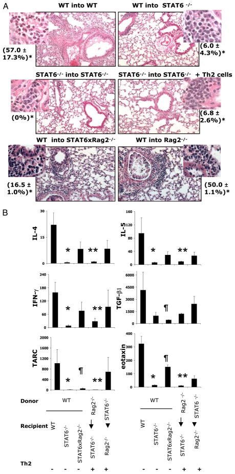
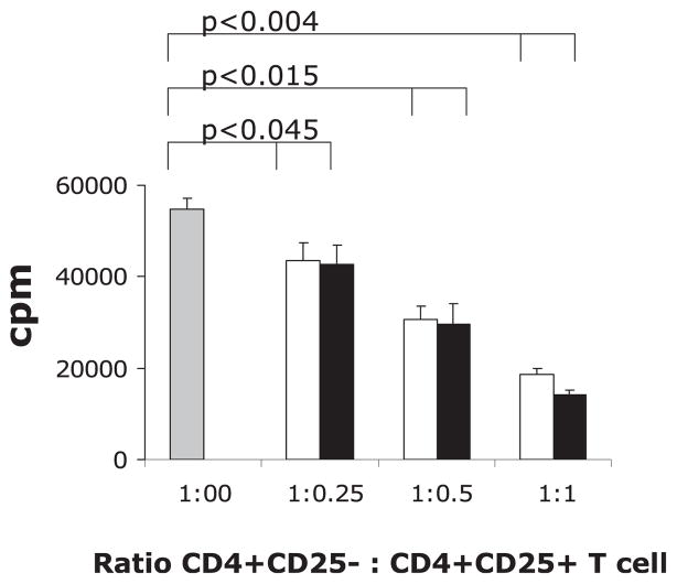
Th2 cells induce asthma through the secretion of cytokines. Two such cytokines, IL-4 and IL-13, are critical mediators of many features of this disease. They both share a common receptor subunit, IL-4Rα, and signal through the STAT6 pathway. STAT6(-/-) mice have impaired Th2 differentiation and reduced airway response to allergen. Transferred Th2 cells were not able to elicit eosinophilia in response to OVA in STAT6(-/-) mice. To clarify the role of STAT6 in allergic airway inflammation, we generated mouse bone marrow (BM) chimeras. We observed little to no eosinophilia in OVA-treated STAT6(-/-) mice even when STAT6(+/+) BM or Th2 cells were provided. However, when Th2 cells were transferred to STAT6×Rag2(-/-) mice, we observed an eosinophilic response to OVA. Nevertheless, the expression of STAT6 on either BM-derived cells or lung resident cells enhanced the severity of OVA-induced eosinophilia. Moreover, when both the BM donor and recipient lacked lymphocytes, transferred Th2 cells were sufficient to induce the level of eosinophilia comparable with that of wild-type (WT) mice. The expression of STAT6 in BM-derived cells was more critical for the enhanced eosinophilic response. Furthermore, we found a significantly higher number of CD4(+)CD25(+)Foxp3(+) T cells (regulatory T cells [Tregs]) in PBS- and OVA-treated STAT6(-/-) mouse lungs compared with that in WT animals suggesting that STAT6 limits both naturally occurring and Ag-induced Tregs. Tregs obtained from either WT or STAT6(-/-) mice were equally efficient in suppressing CD4(+) T cell proliferation in vitro. Taken together, our studies demonstrate multiple STAT6-dependent and -independent features of allergic inflammation, which may impact treatments targeting STAT6.
Conflict of interest statement
The authors have no financial conflicts of interest.
Figures


Similar articles
-
STAT6 controls the number of regulatory T cells in vivo, thereby regulating allergic lung inflammation.J Immunol. 2013 Aug 15;191(4):1517-28. doi: 10.4049/jimmunol.1300486. Epub 2013 Jul 3. J Immunol. 2013. PMID: 23825312 Free PMC article.
-
CD11b+ myeloid cells are the key mediators of Th2 cell homing into the airway in allergic inflammation.J Immunol. 2009 Jan 1;182(1):623-35. doi: 10.4049/jimmunol.182.1.623. J Immunol. 2009. PMID: 19109196 Free PMC article.
-
Complex role of the IL-4 receptor alpha in a murine model of airway inflammation: expression of the IL-4 receptor alpha on nonlymphoid cells of bone marrow origin contributes to severity of inflammation.J Immunol. 2004 Apr 1;172(7):4545-55. doi: 10.4049/jimmunol.172.7.4545. J Immunol. 2004. PMID: 15034072
-
Transfer of in vivo primed transgenic T cells supports allergic lung inflammation and FIZZ1 and Ym1 production in an IL-4Rα and STAT6 dependent manner.BMC Immunol. 2011 Oct 20;12:60. doi: 10.1186/1471-2172-12-60. BMC Immunol. 2011. PMID: 22014099 Free PMC article.
-
IL-4 promotes airway eosinophilia by suppressing IFN-gamma production: defining a novel role for IFN-gamma in the regulation of allergic airway inflammation.J Immunol. 2001 Feb 15;166(4):2760-7. doi: 10.4049/jimmunol.166.4.2760. J Immunol. 2001. PMID: 11160342
Cited by
-
STAT6 and lung inflammation.JAKSTAT. 2013 Oct 1;2(4):e25301. doi: 10.4161/jkst.25301. Epub 2013 Jun 10. JAKSTAT. 2013. PMID: 24416647 Free PMC article. Review.
-
Neuroimmune semaphorin 4D is necessary for optimal lung allergic inflammation.Mol Immunol. 2013 Dec;56(4):480-7. doi: 10.1016/j.molimm.2013.05.228. Epub 2013 Aug 1. Mol Immunol. 2013. PMID: 23911404 Free PMC article.
-
Molecular Mechanisms of Airway Hyperresponsiveness in a Murine Model of Steroid-Resistant Airway Inflammation.J Immunol. 2016 Feb 1;196(3):963-77. doi: 10.4049/jimmunol.1501531. Epub 2016 Jan 4. J Immunol. 2016. PMID: 26729801 Free PMC article.
-
IL-4 and IL-13 Receptor Signaling From 4PS to Insulin Receptor Substrate 2: There and Back Again, a Historical View.Front Immunol. 2018 May 15;9:1037. doi: 10.3389/fimmu.2018.01037. eCollection 2018. Front Immunol. 2018. PMID: 29868002 Free PMC article. Review.
-
Adoptive transfer of IL-4Rα+ macrophages is sufficient to enhance eosinophilic inflammation in a mouse model of allergic lung inflammation.BMC Immunol. 2012 Jan 31;13:6. doi: 10.1186/1471-2172-13-6. BMC Immunol. 2012. PMID: 22292924 Free PMC article.
References
-
- Robinson DS, Hamid Q, Ying S, Tsicopoulos A, Barkans J, Bentley AM, Corrigan C, Durham SR, Kay AB. Predominant TH2-like bronchoalveolar T-lymphocyte population in atopic asthma. N Engl J Med. 1992;326:298–304. - PubMed
-
- Coyle AJ, Le Gros G, Bertrand C, Tsuyuki S, Heusser CH, Kopf M, Anderson GP. Interleukin-4 is required for the induction of lung Th2 mucosal immunity. Am J Respir Cell Mol Biol. 1995;13:54–59. - PubMed
Publication types
MeSH terms
Substances
Grants and funding
LinkOut - more resources
Full Text Sources
Molecular Biology Databases
Research Materials
Miscellaneous
