. 2011 Mar;25(3):261-7.
doi: 10.1038/eye.2010.201. Epub 2011 Jan 14.

Anterior chamber angle imaging with optical coherence tomography

Affiliations

Anterior chamber angle imaging with optical coherence tomography

C K-S Leung et al. Eye (Lond). 2011 Mar.

Abstract

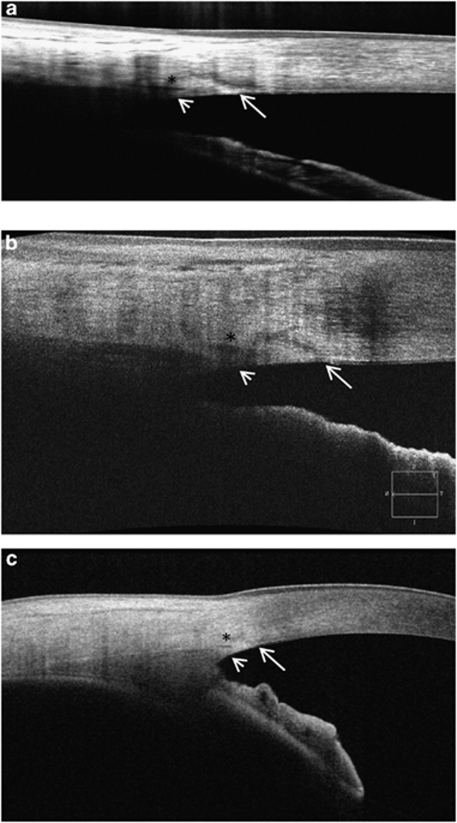
The technology of optical coherence tomography (OCT) has evolved rapidly from time-domain to spectral-domain and swept-source OCT over the recent years. OCT has become an important tool for assessment of the anterior chamber angle and detection of angle closure. Improvement in image resolution and scan speed of OCT has facilitated a more detailed and comprehensive analysis of the anterior chamber angle. It is now possible to examine Schwalbe's line and Schlemm's canal along with the scleral spur. High-speed imaging allows evaluation of the angle in 360°. With three-dimensional reconstruction, visualization of the iris profiles and the angle configurations is enhanced. This article summarizes the development and application of OCT for anterior chamber angle measurement, detection of angle closure, and investigation of the pathophysiology of primary angle closure.


Figures

Figure 1
Figure 1
Imaging of the anterior chamber angle with the Stratus OCT (Carl Zeiss Meditec) in an eye with primary angle closure before (a) and right after (b) laser iridotomy. The resulting anterior chamber inflammation after laser iridotomy can be visualized as scattering reflective signals. Although the configuration of the iris and the angle can be examined with the Stratus OCT, the scleral spur could not be detected. With the advent of the anterior segment OCT, visualization of the scleral spur is improved. Examples of clear appearance of the scleral spur in the Visante OCT (Carl Zeiss Meditec; c) and the SL-OCT (Heidelberg Engineering, GmbH; d) images are illustrated (N, nasal; T, temporal). Arrows indicate the locations of the scleral spur. (Modified from Figure 1 in Leung et al and Figure 1 in Leung et al.)
Figure 2
Figure 2
Measurement of trabecular iris angle (TIA; a), angle-opening distance (AOD; b), trabecular iris space area (TISA; c), and angle recess area (ARA; d). TIA 500 is defined as an angle measured with the apex in the iris recess and the arms of the angle passing through a point on the trabecular meshwork 500 μm from the scleral spur and the point on the iris perpendicularly. AOD 500 is calculated as the perpendicular distance measured from the trabecular meshwork at 500 μm anterior to the scleral spur to the anterior iris surface. The TISA 500 is an area bounded anteriorly by the AOD 500; posteriorly by a line drawn from the scleral spur perpendicular to the plane of the inner scleral wall to the opposing iris; superiorly by the inner corneoscleral wall; and inferiorly by the iris surface. ARA 500 is the area of the angle recess bounded anteriorly by the AOD 500. (Modified from Figure 2 in Leung et al.)
Figure 3
Figure 3
An open anterior chamber angle imaged by the RTVue FD-OCT (2 × 2 mm) (Optovue; a), Cirrus HD-OCT (3 × 1 mm) (Carl Zeiss Meditec; b) and the CASIA OCT (8 × 4 mm) (Tomey; c). Multiple B-scan averaging was performed in RTVue FD-OCT (16 B-scans) and CASIA OCT (3 B-scans). Schwalbe's line-long arrow, scleral spur-short arrow, Schlemm's canal-*.
Figure 4
Figure 4
The CASIA OCT (Tomey) is a swept-source OCT with a scan-speed of 30 000 A scans per s. The whole anterior segment can be radially imaged in 64 cross-sections in 1.2 s. A closed angle (a, b) and an open angle (c, d) in cross-section (a, c) and in three-dimensional display (b, d) are illustrated.

References

    1. Huang D, Swanson EA, Lin CP, Schuman JS, Stinson WG, Chang W, et al. Optical coherence tomography. Science. 1991;254:1178–1181. - PMC - PubMed
    1. Izatt JA, Hee MR, Swanson EA, Lin CP, Huang D, Schuman JS, et al. Micrometer-scale resolution imaging of the anterior eye in vivo with optical coherence tomography. Arch Ophthalmol. 1994;112:1584–1589. - PubMed
    1. Leung CK, Chan WM, Ko CY, Chui SI, Woo J, Tsang MK, et al. Visualization of anterior chamber angle dynamics using optical coherence tomography. Ophthalmology. 2005;112:980–984. - PubMed
    1. Ang GS, Wells AP. Changes in Caucasian eyes after laser peripheral iridotomy: an anterior segment optical coherence tomography study. Clin Experiment Ophthalmol. 2010;38:778–785. - PubMed
    1. See JL, Chew PT, Smith SD, Nolan WP, Chan YH, Huang D, et al. Changes in anterior segment morphology in response to illumination and after laser iridotomy in Asian eyes: an anterior segment OCT study. Br J Ophthalmol. 2007;91:1485–1489. - PMC - PubMed

Publication types

MeSH terms