

Progress in the Development of a new Angiography Suite including the High Resolution Micro-Angiographic Fluoroscope (MAF), a Control, Acquisition, Processing, and Image Display System (CAPIDS), and a New Detector Changer Integrated into a Commercial C-Arm Angiography Unit to Enable Clinical Use
- PMID: 21243037
- PMCID: PMC3021378
- DOI: 10.1117/12.844909
Progress in the Development of a new Angiography Suite including the High Resolution Micro-Angiographic Fluoroscope (MAF), a Control, Acquisition, Processing, and Image Display System (CAPIDS), and a New Detector Changer Integrated into a Commercial C-Arm Angiography Unit to Enable Clinical Use
Abstract
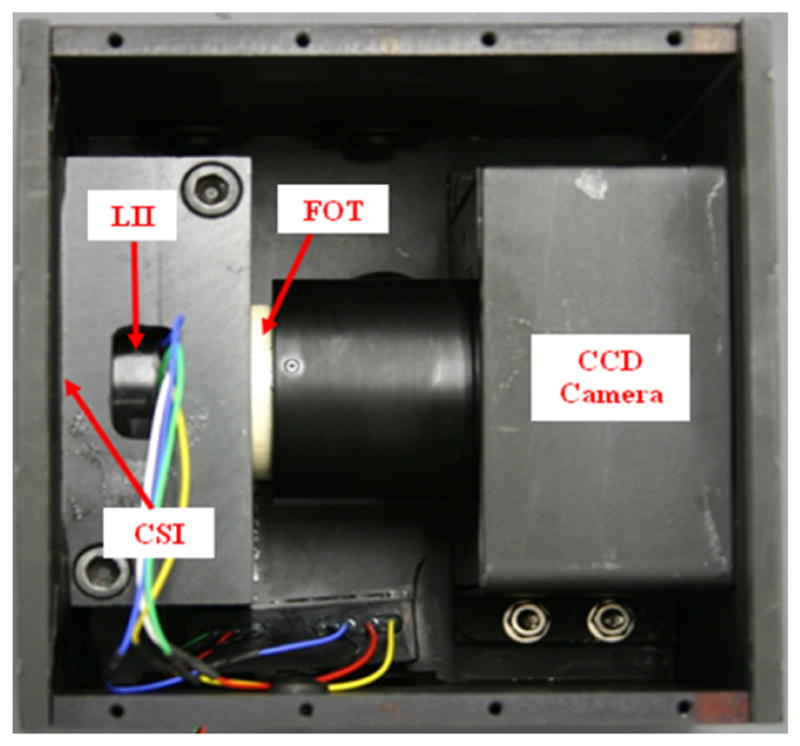
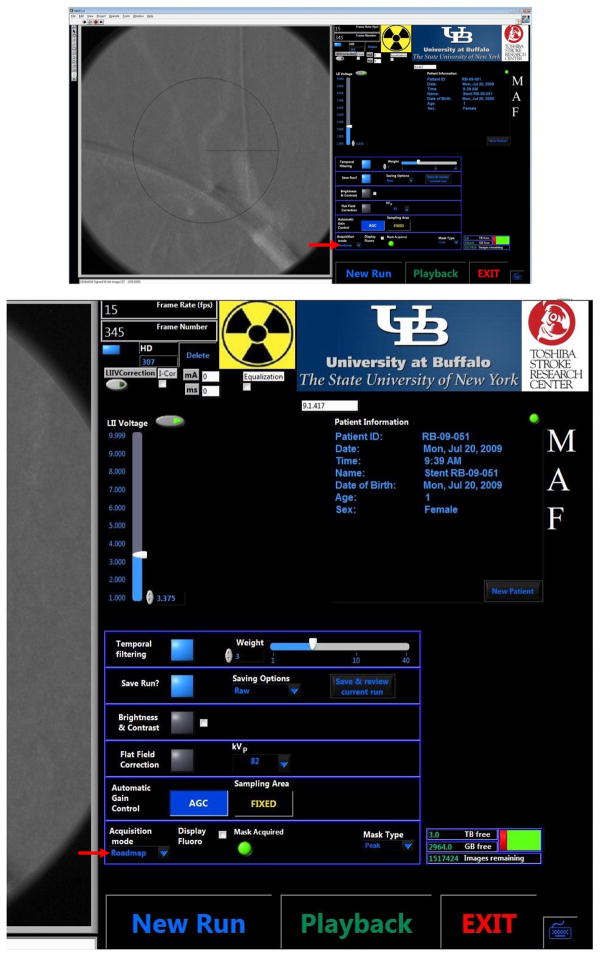
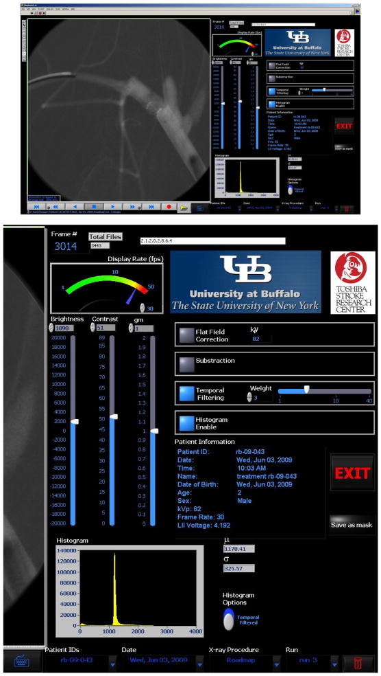
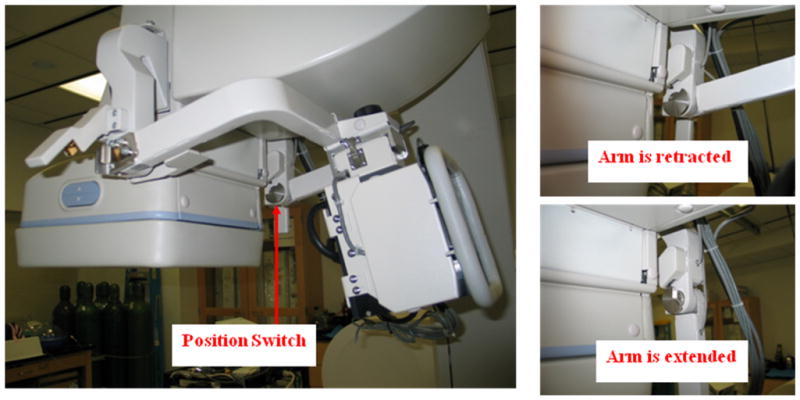
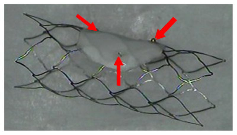
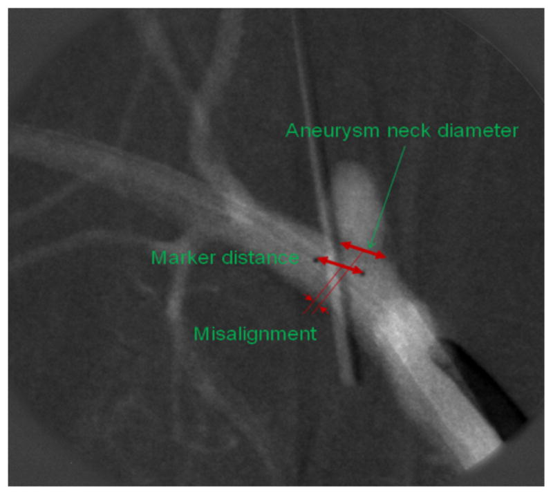
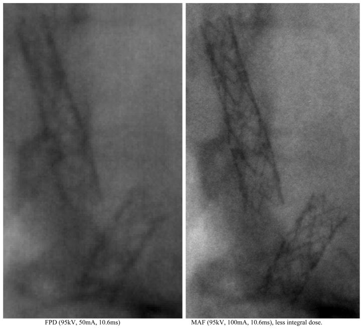
Due to the high-resolution needs of angiographic and interventional vascular imaging, a Micro-Angiographic Fluoroscope (MAF) detector with a Control, Acquisition, Processing, and Image Display System (CAPIDS) was installed on a detector changer which was attached to the C-arm of a clinical angiographic unit. The MAF detector provides high-resolution, high-sensitivity, and real-time imaging capabilities and consists of a 300 μm-thick CsI phosphor, a dual stage micro-channel plate light image intensifier (LII) coupled to a fiber optic taper (FOT), and a scientific grade frame-transfer CCD camera, providing an image matrix of 1024×1024 35 μm square pixels with 12 bit depth. The Solid-State X-Ray Image Intensifier (SSXII) is an EMCCD (Electron Multiplying charge-coupled device) based detector which provides an image matrix of 1k×1k 32 μm square pixels with 12 bit depth. The changer allows the MAF or a SSXII region-of-interest (ROI) detector to be inserted in front of the standard flat-panel detector (FPD) when higher resolution is needed during angiographic or interventional vascular imaging procedures. The CAPIDS was developed and implemented using LabVIEW software and provides a user-friendly interface that enables control of several clinical radiographic imaging modes of the MAF or SSXII including: fluoroscopy, roadmapping, radiography, and digital-subtraction-angiography (DSA). The total system has been used for image guidance during endovascular image-guided interventions (EIGI) using prototype self-expanding asymmetric vascular stents (SAVS) in over 10 rabbit aneurysm creation and treatment experiments which have demonstrated the system's potential benefits for future clinical use.
Figures


References
-
- Ionita CN, et al. Implementation of a high-sensitivity Micro-Angiographic Fluoroscope (HS-MAF) for in-vivo endovascular image guided interventions (EIGI) and region-of-interest computed tomography (ROI-CT) Proc Soc Photo Opt Instrum Eng. 2008. p. 69181I. [PMCID:2572822] http://www.ncbi.nlm.nih.gov/entrez/query.fcgi?cmd=Retrieve&db=PubMed&dop.... - PMC - PubMed
-
- Patel V, et al. Effect of projection angles used in multi-view reconstruction (MVR) using images from a microangiographic (MA) detector and an image-intensifier (II) system. Medical Physics. 2006;33(6):2229–2229.
-
- Keleshis C. AUTOMATED HIGH RESOLUTION, MICRO-ANGIOGRAPHIC FLUOROSCOPY MEDICAL SYSTEMS. 2009. http://hdl.handle.net/10465/449.
-
- Kuhls-Gilcrist A, et al. The Solid-State X-Ray Image Intensifier (SSXII): An EMCCD-Based X-Ray Detector. Proc Soc Photo Opt Instrum Eng. 2008. p. nihpa68284. [PMCID:2557100] http://www.ncbi.nlm.nih.gov/entrez/query.fcgi?cmd=Retrieve&db=PubMed&dop.... - PMC - PubMed
-
- Yadava GK, et al. Generalized Objective Performance Assessment of a New High-Sensitivity Microangiographic Fluoroscopic (HSMAF) Imaging System. Proc Soc Photo Opt Instrum Eng. 2008. p. nihpa68288. [PMCID:2557068] http://www.ncbi.nlm.nih.gov/entrez/query.fcgi?cmd=Retrieve&db=PubMed&dop.... - PMC - PubMed
Grants and funding
LinkOut - more resources
Full Text Sources