. 2011 Mar 22;50(11):1875-84.
doi: 10.1021/bi101863g. Epub 2011 Feb 4.

Slow conformational dynamics in the cystoviral RNA-directed RNA polymerase P2: influence of substrate nucleotides and template RNA

Affiliations

Slow conformational dynamics in the cystoviral RNA-directed RNA polymerase P2: influence of substrate nucleotides and template RNA

Zhen Ren et al. Biochemistry. .

Erratum in

  • Biochemistry. 2011 May 3;50(17):3578

Abstract

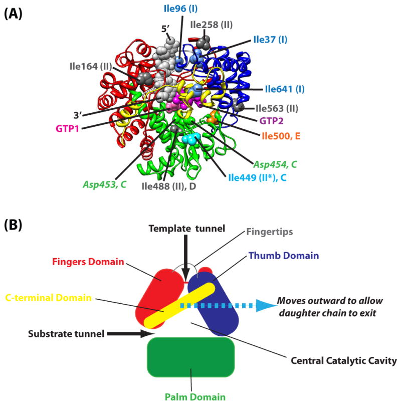
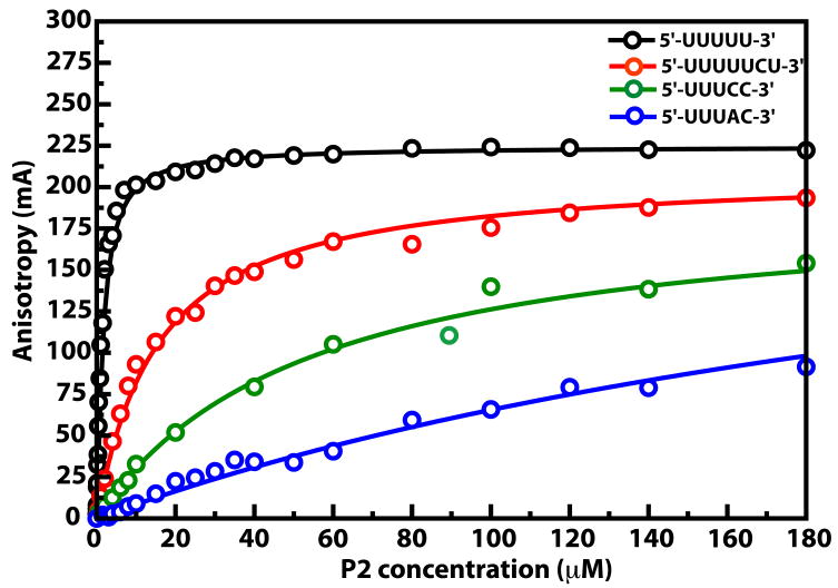
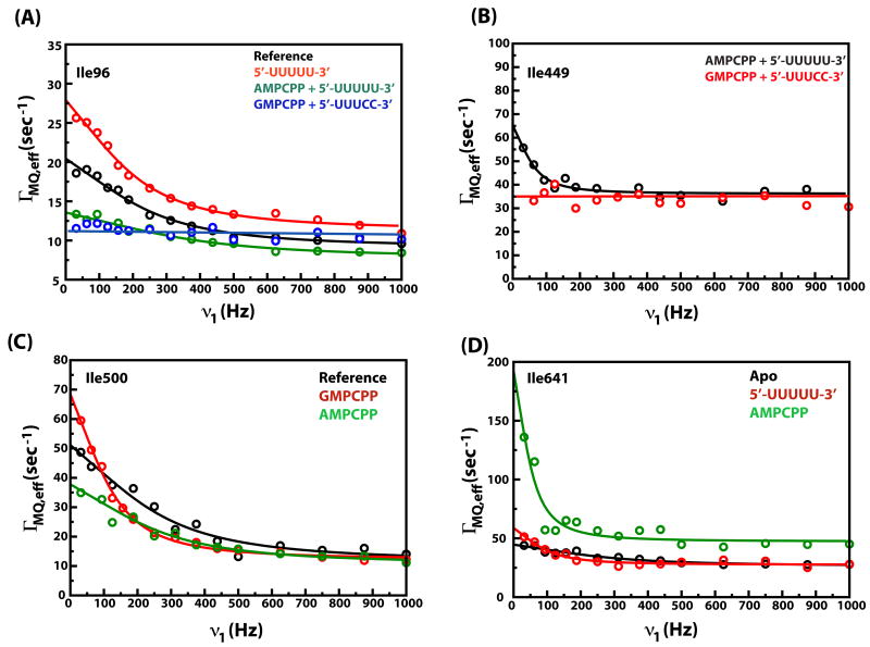
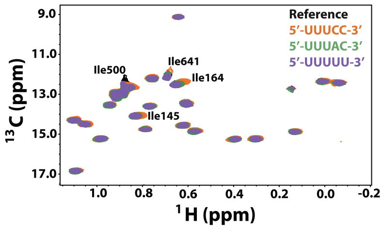
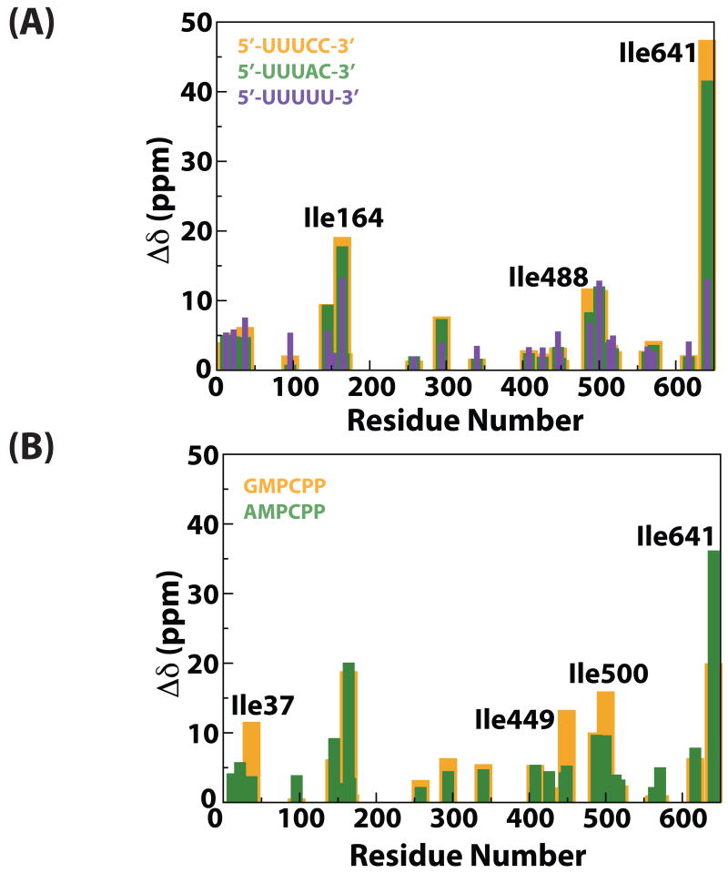
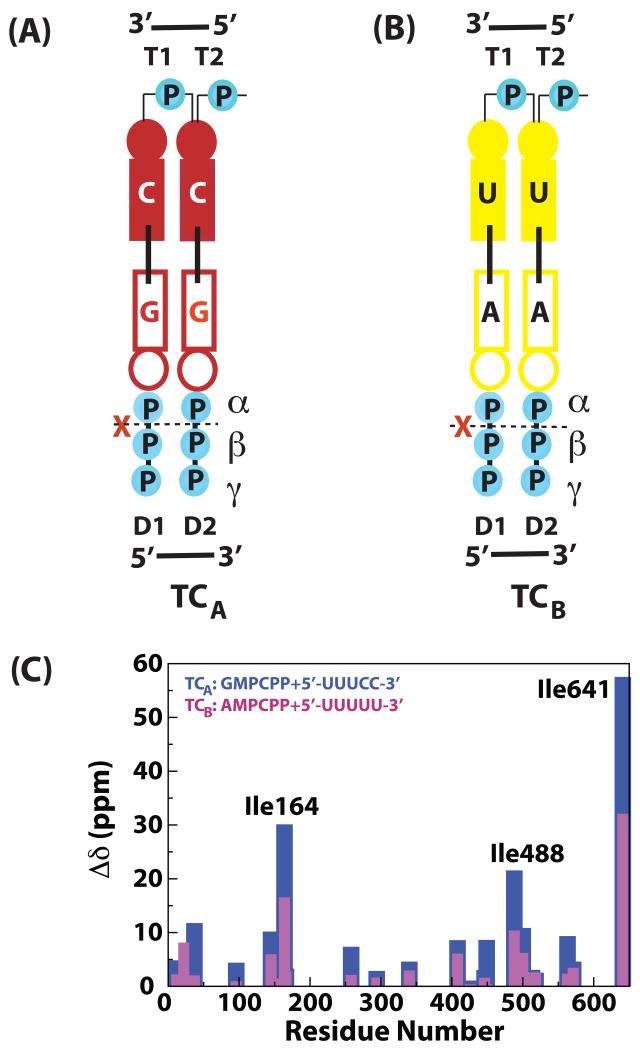
The RNA-directed RNA polymerase P2 from cystovirus ϕ6 catalyzes the de novo synthesis of positive and negative strands of the viral double-stranded RNA genome. P2 is mobile on the slow, microsecond to millisecond time scale with various motional modes, putatively assisting in RNA translocation and catalysis. Here we investigate the influence of the extreme 3'-end sequence of the single-stranded RNA templates and the nature of the substrate nucleotide triphosphates on these motional modes using multiple-quantum NMR spectroscopy. We find that P2, in the presence of templates bearing the proper genomic 3'-ends or the preferred initiation nucleotide, displays unique dynamic signatures that are different from those in the presence of nonphysiological templates or substrates. This suggests that dynamics may play a role in the fidelity of recognition of the correct substrates and template sequences to initiate RNA polymerization.


Figures

Figure 1
Figure 1
(A) φ6 P2 showing the conserved polymerase fold (4). The fingers domain (red; 1-30, 104-276, 333-397), thumb domain (blue; 37-91, 518-600), palm domain (green; 277-332, 398-517) and C-terminal domain (yellow; 601-664) are shown. Key Ile residues that are shown to undergo slow, μs-ms timescale dynamics in apo-P2 characterized previously (19) are labeled. Ile residues that undergo Group I and Group II are represented by blue and dark grey spheres respectively and labeled accordingly. Ile449 (motif C) undergoes slow dynamics (group II) only under certain circumstances (indicated by a “*”) and is depicted by a light blue sphere. Ile500 (motif E) that switches between the fast (group I) and slow motional (group II) modes is shown in orange. The two bound GTP molecules (pink and purple) are shown, as is the template single-stranded RNA (grey, 5′- and 3′-ends labeled). The conserved Asp residues, Asp453 and Asp454 (a part of the G/SDD motif found in DNA and RNA polymerases), that lie on motif C are represented by green spheres and labeled. (B) Schematic representation of P2 structure showing the structural domains and template and substrate channels.
Figure 2
Figure 2
Representative binding curves demonstrating the affinities of several ssRNA constructs towards φ6 P2, determined by fluorescence anisotropy measurements using 5′-carboxyfluorescein tagged ssRNA. Only a few binding curves are shown for ease in illustrating their differences.
Figure 3
Figure 3
Representative dispersion curves with experimental data depicted by circles and fits to Equation 4 by solid lines. (A) Data for the fingertips residue Ile96. (B) Data for the motif C residue Ile449. (C) Data for the motif E residue Ile500. (D) Data for Ile641 that lies in the C-terminal domain. The blue line in (A) and the red line in (B) are drawn to guide the eye and do not represent fits. These correspond to situations where the dispersion curves are flat i.e. ΓMQ,eff values are independent of ν1. Data for only a small number of states are shown for visual convenience.
Figure 4
Figure 4
Overlay of the reference spectra (φ6 P2 in the NMR buffer, black) with each of three templates C1 (5′-UUUCC-3′, 3′-ends of the genomic s- and m- strands, orange), C2 (5′-UUUAC-3′, 3′-end of the genomic l-, green) and C5 (5′-UUUUU-3′, purple).
Figure 5
Figure 5
(A) Scaled chemical shift changes shown for each of the three templates (reference – black, C1 – yellow, C2 – green and C5 – purple). (B) Scaled chemical shift changes for the substrate analogs (GMPCPP – yellow, AMPCPP – green). Scaled chemical shift changes, with respect to the reference, for (A) and (B) were calculated using Equation 1.
Figure 6
Figure 6
(A) Ternary complex TCA forms between 5′-UUUCC-3′ (template C1), the non-hydrolysable GTP-analog GMPCPP and φ6 P2. The template bases T1 and T2 (numbered 3′ to 5′) base pair with the substrate bases D1 and D2 (numbered 5′ to 3′) forming a stable initiation complex (based on base-pairing considerations alone). The first phosphodiester bond forms between D1 and D2 creating the daughter chain and releasing pyrophosphate. In TCA, D2 (and D1) is the substrate GMPCPP that cannot be hydrolyzed between the α– and β–phosphates and the reaction cannot proceed, though the initiation complex forms. TCA is physiologically relevant since template 1 corresponds to the 3′-ends of the s- and m- genomic segments. (B) TCB the ternary complex between template C5 (5′-UUUUU-3′) and the non-hydrolysable ATP-analog AMPCPP, also allows proper base-pairing between T1:D1 and T2:D2, as in TCA above, but C5 does not correspond to 3′-ends of any of the φ6 genomic segments. (C) Scaled chemical shift changes (with respect to the reference state) shown for the ternary complexes (TCA – blue, TCB – pink).

Similar articles

Cited by

References

    1. Poranen MM, Tuma R. Self-assembly of double-stranded RNA bacteriophages. Virus Res. 2004;101:93–100. - PubMed
    1. Ferrer-Orta C, Arias A, Escarmis C, Verdaguer N. A comparison of viral RNA-dependent RNA polymerases. Curr Opin Struct Biol. 2006;16:27–34. - PubMed
    1. Bruenn JA. A structural and primary sequence comparison of the viral RNA-dependent RNA polymerases. Nucleic Acids Res. 2003;31:1821–1829. - PMC - PubMed
    1. Butcher SJ, Grimes JM, Makeyev EV, Bamford DH, Stuart DI. A mechanism for initiating RNA-dependent RNA polymerization. Nature. 2001;410:235–240. - PubMed
    1. Makeyev EV, Bamford DH. Replicase activity of purified recombinant protein P2 of double-stranded RNA bacteriophage phi6. EMBO J. 2000;19:124–133. - PMC - PubMed

Publication types

MeSH terms