. 2011 Jan 19;100(2):284-93.
doi: 10.1016/j.bpj.2010.11.082.

Elucidating the role of matrix stiffness in 3D cell migration and remodeling

Affiliations

Elucidating the role of matrix stiffness in 3D cell migration and remodeling

M Ehrbar et al. Biophys J. .

Abstract

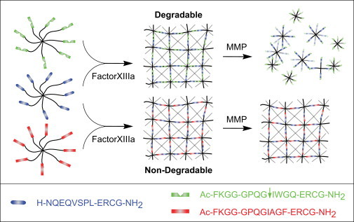
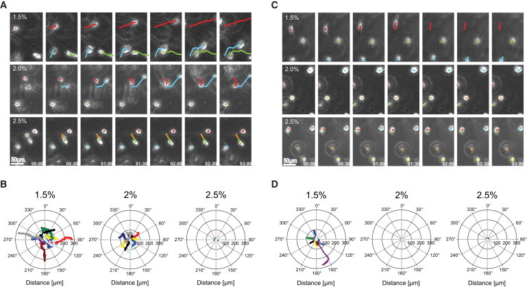
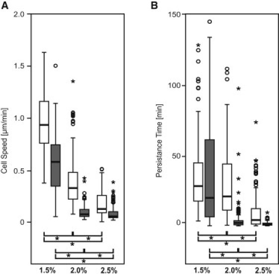
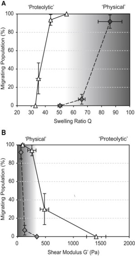
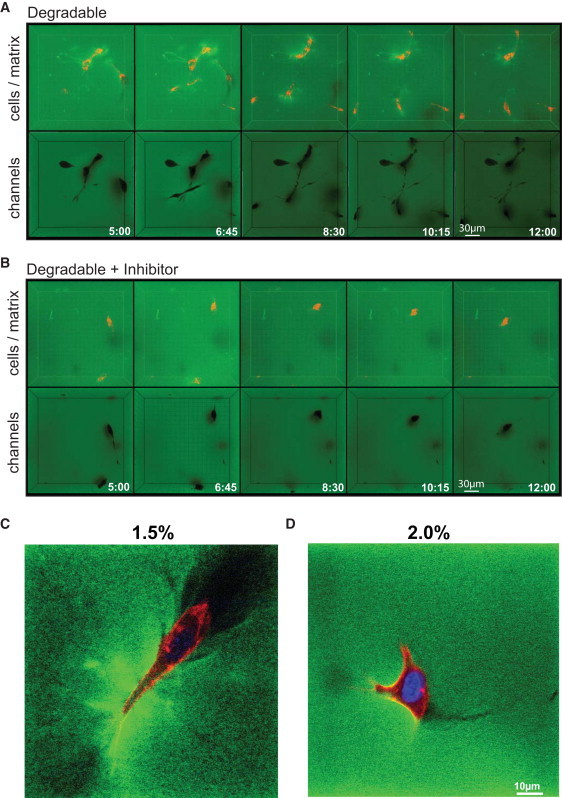
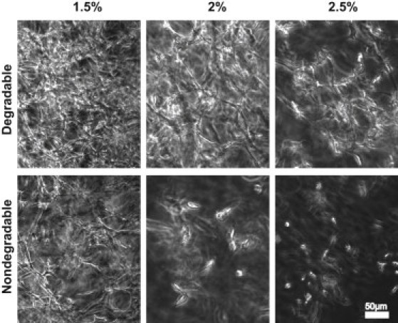
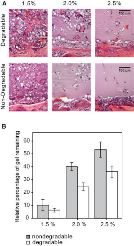
Reductionist in vitro model systems which mimic specific extracellular matrix functions in a highly controlled manner, termed artificial extracellular matrices (aECM), have increasingly been used to elucidate the role of cell-ECM interactions in regulating cell fate. To better understand the interplay of biophysical and biochemical effectors in controlling three-dimensional cell migration, a poly(ethylene glycol)-based aECM platform was used in this study to explore the influence of matrix cross-linking density, represented here by stiffness, on cell migration in vitro and in vivo. In vitro, the migration behavior of single preosteoblastic cells within hydrogels of varying stiffness and susceptibilities to degradation by matrix metalloproteases was assessed by time-lapse microscopy. Migration behavior was seen to be strongly dependent on matrix stiffness, with two regimes identified: a nonproteolytic migration mode dominating at relatively low matrix stiffness and proteolytic migration at higher stiffness. Subsequent in vivo experiments revealed a similar stiffness dependence of matrix remodeling, albeit less sensitive to the matrix metalloprotease sensitivity. Therefore, our aECM model system is well suited to unveil the role of biophysical and biochemical determinants of physiologically relevant cell migration phenomena.


Figures

Figure 1
Figure 1
Scheme of the modular design of PEG-based aECMs. Stoichiometrically balanced ([Lys]/[Gln] = 1) 8-arm PEG macromers in a buffer solution are enzymatically cross-linked via their pending glutamine acceptor [Gln] and lysine-donor [Lys] FXIIIa substrate sequences to form a hydrogel. By variation of the linker sequence and the initial precursor concentration, aECMs with different stiffness and MMP sensitivities in presence of constant RGD concentrations can be generated.
Figure 2
Figure 2
Time-lapse images and representative track plots of MC3T3-E1 cells in three-dimensional culture. (A and C Typical time-lapse images of cells in three-dimensional culture (time interval t = 30 min). The track of three individual cells is indicated by the sequentially determined center of the cell. (A) Efficient migration in soft MMP-sensitive hydrogels was observed that is reduced with increasing stiffness. (C) In soft MMP-insensitive hydrogels, cells also migrated, whereas in intermediate or stiff gels, cells remained round. (B and D) The projection of 10 migrating cells was acquired and overlaid with a common starting point. The tracks representing a 14.5 h period (58 × 15 min) do not show a preferred orientation.
Figure 3
Figure 3
Three-dimensional single cell migration as a function of gel stiffness and MMP-sensitivity (degradable gels depicted in white, nondegradable gels in gray). (A) Mean cell speed and (B) persistence time of all cells of a group were analyzed from time-lapse images of at least 24 consecutive time points. Values are displayed as box plots ranging from 25th to 75th percentile including the whiskers from the 10th to the 90th percentile. (A) The average cell speed and (B) the average persistence time (() p < 0.05) decreased significantly with increasing stiffness of the matrix (() p < 0.05).
Figure 4
Figure 4
(A) The high swelling ratio Q results in a large population of migrating cells which, in MMP-sensitive gels (white triangles), is due to mostly proteolytic matrix remodeling and in MMP-insensitive gels (gray diamonds) due to physically controlled cell migration. With decreasing swelling ratios, the migrating population decreases. (B) In soft matrices, almost all the cells migrate in MMP-sensitive and MMP-insensitive gels. By increasing the stiffness, migration ceases and only cells in MMP-sensitive substrates can migrate.
Figure 5
Figure 5
Visualization of cell-induced matrix deformation and macroscopic cavities. (A and B) Migrating PKH26-labeled cells were encapsulated in FITC-conjugated, soft (1.5%) matrices in the absence (A) or presence (B) of GM6001 and followed by four-dimensional time-lapse confocal microscopy for 8 h. In both cases, three-dimensional reconstruction of z stacks over time in maximum intensity projection mode (cells in red, matrix in green) revealed an increase in matrix intensity (green), indicating localized matrix deformation around migrating cells. Three-dimensional reconstruction of stack from the same time points performed in minimum intensity projection mode (matrix in green) revealed the presence of a network of interconnected macroscopic cavities and small cracks indicated the cell migrating paths. (C and D) High-resolution z section of encapsulated cells within soft and intermediate degradable gels after one day in culture (fixed samples).
Figure 6
Figure 6
Matrix stiffness and MMP-sensitivity influence the formation of cellular networks. Starting from single cells, dense and evenly distributed cellular networks are formed in soft MMP-sensitive gels within three weeks. With increasing stiffness the cell distribution becomes less homogenous and less dense. The formation of cellular networks in MMP-insensitive soft hydrogels is largely reduced, and in intermediate or stiff hydrogels is almost absent.
Figure 7
Figure 7
In vivo matrix remodeling is controlled by gel properties. (A) Critical-sized calvarial defects in rats were treated with soft, intermediate, and stiff MMP-sensitive and -insensitive hydrogels containing 1 μg BMP-2. Within two weeks, endogenous cells invade, degrade, and remodel provisional MMP-sensitive matrices in a stiffness-dependent manner. Cell invasion is homogenous in soft gels and becomes less regular in more dense gels. MMP-insensitive gels are less efficiently invaded by cells (n = 5). (B) The stiffness and MMP-dependent degradation rate of the aECM was confirmed by histomorphometrical quantification of the gel leftovers (mean ± SD, n = 5).

References

    1. Lutolf M.P., Hubbell J.A. Synthetic biomaterials as instructive extracellular microenvironments for morphogenesis in tissue engineering. Nat. Biotechnol. 2005;23:47–55. - PubMed
    1. Phelps E.A., García A.J. Engineering more than a cell: vascularization strategies in tissue engineering. Curr. Opin. Biotechnol. 2010;21:704–709. - PMC - PubMed
    1. Mooney D.J., Vandenburgh H. Cell delivery mechanisms for tissue repair. Cell Stem Cell. 2008;2:205–213. - PubMed
    1. Zaman M.H., Matsudaira P., Lauffenburger D.A. Understanding effects of matrix protease and matrix organization on directional persistence and translational speed in three-dimensional cell migration. Ann. Biomed. Eng. 2007;35:91–100. - PubMed
    1. Ilina O., Friedl P. Mechanisms of collective cell migration at a glance. J. Cell Sci. 2009;122:3203–3208. - PubMed

Publication types

LinkOut - more resources