. 2011 Jan 19;100(2):430-9.
doi: 10.1016/j.bpj.2010.11.045.

Detailed tuning of structure and intramolecular communication are dispensable for processive motion of myosin VI

Affiliations

Detailed tuning of structure and intramolecular communication are dispensable for processive motion of myosin VI

Mary Williard Elting et al. Biophys J. .

Abstract

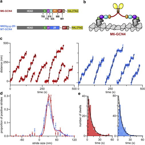
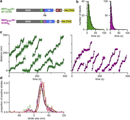
Dimeric myosin VI moves processively hand-over-hand along actin filaments. We have characterized the mechanism of this processive motion by measuring the impact of structural and chemical perturbations on single-molecule processivity. Processivity is maintained despite major alterations in lever arm structure, including replacement of light chain binding regions and elimination of the medial tail. We present kinetic models that can explain the ATP concentration-dependent processivities of myosin VI constructs containing either native or artificial lever arms. We conclude that detailed tuning of structure and intramolecular communication are dispensable for processive motion, and further show theoretically that one proposed type of nucleotide gating can be detrimental rather than beneficial for myosin processivity.


Figures

Figure 1
Figure 1
Single molecule stepping results for M6-GCN4 and M6DI816-2R∼MT-GCN4. (a) Schematics of constructs. (Gray, head of native myosin VI; green (C), converter domain; purple (UI), calmodulin binding unique insert; cyan (IQ), IQ domain; orange (PT), proximal tail domain; red (MT), medial tail domain; brown (G), GCN4; yellow, HaloTag; blue (2R), two spectrin repeats from α-actinin; and wavy line, (GSG)3 flexible linkers.) (b) Cartoon of M6-GCN4 structure, color-coded to match part a. (c) Example stepping traces for M6-GCN4 (red) and M6DI816-2R∼MT-GCN4 (blue). Steps fit to these traces (black). (d) Distributions of stride sizes for M6-GCN4 and M6DI816-2R∼MT-GCN4. Stride size histograms (markers) are shown with a fit to the distribution of positive stride sizes (solid). Error bars are calculated as the square-root of the number of strides in each bin, scaled to proportion of strides. Peak positive stride sizes are 33.5 ± 0.7 nm (N = 158) for M6-GCN4 (red) and 30.1 ± 0.6 nm (N = 252) for M6DI816-2R-MT-GCN4 (blue). (e) Dwell-time distributions for M6-GCN4 (red), mean dwell-time of 8.7 ± 0.7 s and M6DI816-2R∼MT-GCN4 (blue), mean dwell-time of 7.4 ± 0.4 s. Short dwells below a cutoff (dashed lines) were ignored to avoid undersampling near our time resolution. Curves (solid black) are exponential distributions with decay constants equal to the mean dwell-time, shifted by the undersampling cutoff.
Figure 2
Figure 2
Three-state model for myosin VI kinetic cycle. (a) Schematic of kinetic pathway (based on de la Cruz et al. (5)) showing which states are grouped together or ignored in the simplified kinetic model. (Green) States in which myosin is bound to actin in the absence of nucleotide. (Yellow) States in which myosin is detached or weakly bound to the actin filament. (Red) States in which myosin is bound to actin and ADP. The remaining states (gray) are assumed to have negligible contributions. (b) Cartoon of the modeled kinetic cycle of a single myosin head. States and transitions between states are colored to match panel a. The prestroke state (yellow box) is bound to ADP.Pi (green circle) and unbound from actin. At rate krebind, myosin rebinds to actin, releases phosphate, and strokes, bringing it to a poststroke state (red box) where it is bound to actin and ADP (red circle). Note that krebind may actually be limited by phosphate release or the weak to strong transition instead of rebinding. ADP is released at rate kADPoff to enter a state where the motor is in a poststroke conformation and bound to actin but not nucleotide (green box). The cycle is completed when myosin binds ATP at rate kATPon and releases from the actin filament. (c) Diagram of transitions in a dimeric motor, highlighting the paths that predominate with different mechanisms of gating. Motor begins in state A, and forward steps are directed toward the left (black arrow). Myosin heads are marked as containing ATP or ADP.Pi (green dot), ADP (red dot), or no nucleotide (no dot) at the nucleotide binding site. Transitions that are in competition with rebinding (thinner arrows) have low probabilities because the rebinding rate is the fastest rate in the cycle. Shading indicates the predominant pathways in the case of gating of ADP release (pink); in the case of gating of ATP binding (green shading, which includes the pink shaded region); and in the case of no gating (blue shading, which encompasses all states). Inhibition marks indicate rates that are slowed by gating of ADP release (red) or gating of ATP binding (green).
Figure 3
Figure 3
Velocity and run-length measurements for M6-GCN4 and M6DI816-2R∼MT-GCN4. (a) M6-GCN4 (red) and (b) M6DI816-2R∼MT-GCN4 (blue) at 2 mM ATP. Kaplan-Meier survivor functions, with compensation for runs terminating at filament ends (32,33), are shown (color). Exponential distributions based on the Kaplan-Meier estimate of the mean (0.556 ± 0.050 μm for M6-GCN4; 0.226 ± 0.016 μm for M6DI816-2R∼MT-GCN4) are shown (solid black). Runs shorter than 0.216 μm (two pixels) are truncated due to undersampling of runs close to our time resolution, and the mean estimator has been shifted accordingly (see Materials and Methods). (c) Velocity and (d) run-length as a function of ATP concentration for M6-GCN4 (red) and M6DI816-2R∼MT-GCN4 (blue). M6DI816-2R∼MT-GCN4 is slightly slower than M6-GCN4. If velocities for both constructs are scaled to an expected stepping rate (based on measured stepsize), the velocity difference is reduced, because the chimera has a slightly shorter stepsize (Fig. S7). M6DI816-2R∼MT-GCN4 shows reduced processivity compared to M6-GCN4, possibly as a result of damaged gating. (Error bars) Standard deviation of bootstrapped mean (for velocity) and Kaplan Meier estimate for standard deviation of the mean (run-length). (Solid lines) Fit to processivity model (see parameters in Table 1).
Figure 4
Figure 4
Comparison of processivity model using different mechanisms of gating: no gating (blue), 10-fold gating of ADP release from front head (red), and 10-fold gating of ATP binding to front head (green). Parameters input into this model are the same as the M6-GCN4 fit shown in Fig. 3. All parameters are kept constant when comparing the models; only the mechanism of gating is changed (see Table 1).
Figure 5
Figure 5
Single molecule stepping results for M6PI790-2R∼MT-GCN4 and M6DI816-2R∼GCN4IL at 5 μM ATP. (a) Schematic of constructs. (Gray, head of native myosin VI; green (C), converter domain; purple (UI), calmodulin binding unique insert or unique insert truncated at residue 790 (before calmodulin binding site); red (MT), medial tail domain; brown (G/IL), GCN4 or GCN4IL; yellow, HaloTag; blue (2R), two spectrin repeats from α-actinin; and wavy line, (GSG)3 flexible linkers.) (b) Dwell-time distributions at 5 μM ATP for M6PI790-2R∼MT-GCN4 (green), mean dwell-time of 11.7 ± 0.7 s and M6DI816-2R∼GCN4IL (magenta), mean dwell-time of 6.3 ± 0.3 s. (Dotted lines) Cutoff of short dwells from undersampling near our time resolution. Curves (solid black) are exponential distributions with time constants of the mean dwells, shifted by the undersampling cutoff. (c) Example stepping traces of M6PI790-2R∼MT-GCN4 (green) and M6DI816-2R∼GCN4IL (magenta). Modeled steps are shown (black). (d) Stride size distributions of chimeras with M6-GCN4 for comparison. Histograms are shown with fits as in Fig. 1. Peak positive stride sizes are 33.5 ± 0.7 nm (N = 158) for M6-GCN4 (red), 27.9 ± 0.6 nm (N = 238) for M6PI790-2R∼MT-GCN4 (green), and 26.2 ± 0.5 nm (N = 335) for M6DI816-2R∼GCN4IL (magenta).

References

    1. Sweeney H.L., Houdusse A. What can myosin VI do in cells? Curr. Opin. Cell Biol. 2007;19:57–66. - PubMed
    1. Spudich J.A., Sivaramakrishnan S. Myosin VI: an innovative motor that challenged the swinging lever arm hypothesis. Nat. Rev. Mol. Cell Biol. 2010;11:128–137. - PMC - PubMed
    1. Okten Z., Churchman L.S., Spudich J.A. Myosin VI walks hand-over-hand along actin. Nat. Struct. Mol. Biol. 2004;11:884–887. - PubMed
    1. Rock R.S., Rice S.E., Sweeney H.L. Myosin VI is a processive motor with a large step size. Proc. Natl. Acad. Sci. USA. 2001;98:13655–13659. - PMC - PubMed
    1. de la Cruz E.M., Ostap E.M., Sweeney H.L. Kinetic mechanism and regulation of myosin VI. J. Biol. Chem. 2001;276:32373–32381. - PubMed

Publication types

LinkOut - more resources