. 2011 Jan 19;100(2):498-506.
doi: 10.1016/j.bpj.2010.12.3691.

Separating instability from aggregation propensity in γS-crystallin variants

Affiliations

Separating instability from aggregation propensity in γS-crystallin variants

William D Brubaker et al. Biophys J. .

Abstract

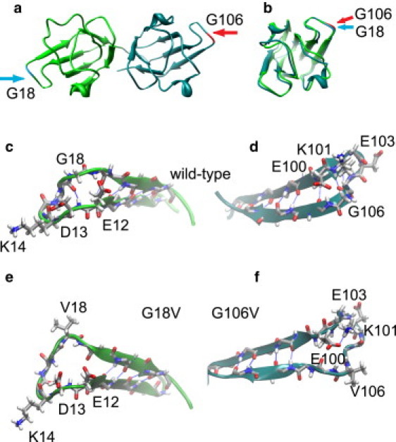
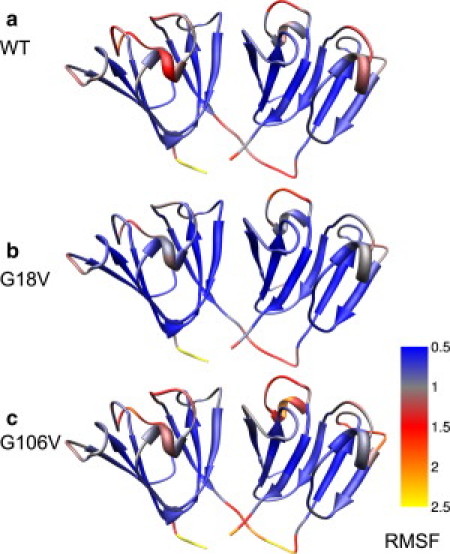
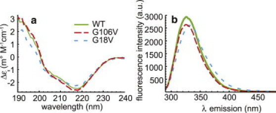
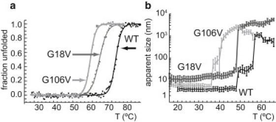
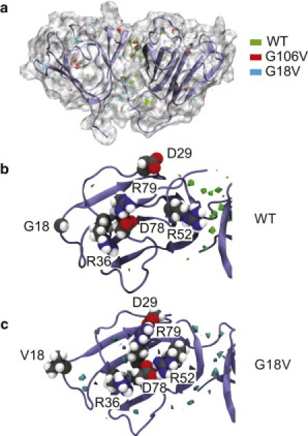
Molecular dynamics (MD) simulations, circular dichroism (CD), and dynamic light scattering (DLS) measurements were used to investigate the aggregation propensity of the eye-lens protein γS-crystallin. The wild-type protein was investigated along with the cataract-related G18V variant and the symmetry-related G106V variant. The MD simulations suggest that local sequence differences result in dramatic differences in dynamics and hydration between these two apparently similar point mutations. This finding is supported by the experimental measurements, which show that although both variants appear to be mostly folded at room temperature, both display increased aggregation propensity. Although the disease-related G18V variant is not the most strongly destabilized, it aggregates more readily than either the wild-type or the G106V variant. These results indicate that γS-crystallin provides an excellent model system for investigating the role of dynamics and hydration in aggregation by locally unfolded proteins.


Figures

Figure 1
Figure 1
(a) Human γS-crystallin homology model based on the murine γS NMR structure (35). The N- and C-terminal domains are shown overlaid with the mutated residues highlighted. This view shows the highly accessible positions of G18 and G106 and the symmetry of γS-crystallin domains. (b) Structural overlay of a backbone alignment of the N- and C-terminal domains of γS-crystallin. The backbone alignment between the two domains has an RMSD of only 1.08 Å. (c and d) Detail of the loops containing G18 and G106, respectively, from a configuration of the WT MD simulation. In both panels the Gly residues, the charged side chains in their neighborhood, and backbone atoms for the rest of the residue are shown in licorice representation. The backbone hydrogen-bond interactions are also highlighted. (e and f) Configuration snapshot for the same regions of the protein from the MD simulations of the G18V and G106V variants, respectively. In both variants, significant loss of secondary structure in the region occurs, along with opening of each loop. In each case the hydrophobic valine side chain is pointing out into solution because of unfavorable contacts with nearby charged residues.
Figure 2
Figure 2
Cα RMSFs mapped onto the γS-crystallin homology model. (a) WT. (b) G18V. (c) G106V. In both variants, changes in conformational dynamics with respect to the WT are not limited to the region around the mutations and are also found in the helices, connecting turns, and loops. Compared with the WT protein, G18V shows diminished thermal fluctuations in these regions. In contrast, G106V appears to be more flexible overall, with increased fluctuations in both surface regions and the β-sheet core.
Figure 3
Figure 3
(a) CD spectra of the WT, G18V, and G106V crystallins at a concentration of 0.125 mg/mL in a solution of 10 mM phosphate buffer (pH 6.9) at 20°C. All three display the negative ellipticity at 218 nm that is indicative of β-sheet secondary structure and common to γ-crystallins. (b) Fluorescence emission spectra of WT, G106V, and G18V, 0.25 mg/mL in 10 mM phosphate buffer pH 6.9 at 22°C, collected on a Hitachi F4500 fluorescence spectrophotometer. Both the WT and the G106V variant have a maximum at 326 nm, whereas in the G18V variant it is slightly shifted to 332 nm. Both of these methods indicate little structural difference between the WT and variant proteins at room temperature.
Figure 4
Figure 4
(a) Thermal unfolding curves of WT, G18V, and G106V crystallins measured by monitoring the CD signal at 218 nm, with best-fit unfolding curves. Unfolding measurements were obtained with a JASCO J-810 spectropolarimeter equipped with a thermal controller. The protein concentration was 0.25 mg/mL in the same buffer conditions as in Fig. 3a, but with 150 mM NaCl, and 1 mM DTT (reducing agent). Tm-values for WT, G18V, and G106V are 72.0°C, 65.6°C, and 59.0°C, respectively. All three variants exhibit behavior consistent with two-state equilibrium unfolding. (b) DLS measurement of thermally induced aggregation of WT, G18V, G106V γS. Although the CD unfolding curves show G106V to be the least thermodynamically stable, G18V is found to be the most aggregation-prone variant.
Figure 5
Figure 5
(a) Regions in the first coordination shell of the protein with water density of twice the bulk value are highlighted. The region between the two domains exhibits the largest number of water sites in all three proteins. A significant number of water sites are also observed in the N-terminal loop regions of the G18V variant. A configuration snapshot of the WT protein is shown in secondary structure representation and as a molecular surface. (b and c) Top view of the N-terminal domain in the WT and G18V variant, respectively. The water isodensity surfaces (twice bulk value) are shown in green (WT) and blue (G18V). Crevices are formed in the protein surface of the G18V variant in the region of the mutation and between the inner loops as a key charged pair (D78 and R79) form persistent salt-bridges with residue located in the outer loops. In the WT, in contrast, D78 alternates salt-bridge interactions with R79 and the neighboring R36. In both panels the protein is shown in secondary structure representation and the specific side chains are shown in CPK representation colored by atom (carbon, gray; nitrogen, blue; oxygen, red; hydrogen, white).

References

    1. Chiti F., Dobson C.M. Protein misfolding, functional amyloid, and human disease. Annu. Rev. Biochem. 2006;75:333–366. - PubMed
    1. Booth D.R., Sunde M., Pepys M.B. Instability, unfolding and aggregation of human lysozyme variants underlying amyloid fibrillogenesis. Nature. 1997;385:787–793. - PubMed
    1. Pepys M.B., Hawkins P.N., Hsuan J.J. Human lysozyme gene mutations cause hereditary systemic amyloidosis. Nature. 1993;362:553–557. - PubMed
    1. Liemann S., Glockshuber R. Influence of amino acid substitutions related to inherited human prion diseases on the thermodynamic stability of the cellular prion protein. Biochemistry. 1999;38:3258–3267. - PubMed
    1. Caughey B., Lansbury P.T. Protofibrils, pores, fibrils, and neurodegeneration: separating the responsible protein aggregates from the innocent bystanders. Annu. Rev. Neurosci. 2003;26:267–298. - PubMed

Publication types

LinkOut - more resources